DNA Detection of
Schistosoma japonicum
:
Diagnostic Validity of a LAMP Assay for
Low-Intensity Infection and Effects of
Chemotherapy in Humans
Jing Xu1☯, Zhi-Xun Guan1☯, Bo Zhao1, Yan-Yan Wang1, Yun Cao1, Hui-Qin Zhang1, Xing-Quan Zhu2, Yong-Kang He3, Chao-Ming Xia1*
1Department of Parasitology, Medical College of Soochow University, Suzhou, Jiangsu Province, China, 2State Key Laboratory of Veterinary Etiological Biology, Key Laboratory of Veterinary Parasitology of Gansu Province, Lanzhou Veterinary Research Institute, Chinese Academy of Agricultural Sciences, Lanzhou, Gansu Province, China,3Hunan Provincial Institute of Schistosomiasis Control, Yueyang, Hunan Province, China
☯These authors contributed equally to this work. *xiachaoming@suda.edu.cn, xcm624@hotmail.com
Abstract
Background
Schistosomiasis has decreased significantly in prevalence and intensity of infection in China, thus more accurate and sensitive methods are desperately needed for the further control of schistosomiasis. The present work aimed to assess the utility of the loop-mediated isothermal amplification (LAMP) for detection of light intensity infection or false-negative pa-tients and papa-tients post-treatment, targeting the highly repetitive retrotransposon SjR2 of Schistosoma japonicum.
Methodology/ Principal Findings
LAMP was first assessed in rabbits with low intensity infection (EPG<10). Then 110 patient sera from Hunan Province, China, and 47 sera after treatment by praziquantel were used to evaluate the diagnostic validity of LAMP. Meanwhile, 42 sera from healthy individuals in a non-endemic area, and 60 sera from "healthy”residents who were identified as being nega-tive for feces examination and immuno-methods in an endemic area were also examined. The results showed that LAMP could detectS.japonicumDNA in sera from rabbits at 3rd day post-infection. Following administration of praziquantel, theS.japonicumDNA in rabbit sera became negative at 10 weeks post-treatment. Of 110 sera from patients, LAMP showed 95.5% sensitivity, and even for 41 patients with less than 10 EPG, the sensitivity of LAMP still reached to 95.1%. For 47 patients after treatment, the negative conversion rate ofS.japonicumDNA in patient sera increased from 23.4%, 61.7% to 83.0% at 3 months, 6 months and 9 months post-treatment, respectively. No false-positive result was obtained for 42 human sera from non-endemic area, while for the 60“healthy”individuals from endemic OPEN ACCESS
Citation:Xu J, Guan Z-X, Zhao B, Wang Y-Y, Cao Y, Zhang H-Q, et al. (2015) DNA Detection of
Schistosoma japonicum: Diagnostic Validity of a LAMP Assay for Low-Intensity Infection and Effects of Chemotherapy in Humans. PLoS Negl Trop Dis 9(4): e0003668. doi:10.1371/journal.pntd.0003668
Editor:Aaron R. Jex, University of Melbourne, AUSTRALIA
Received:October 7, 2014
Accepted:March 2, 2015
Published:April 13, 2015
Copyright:© 2015 Xu et al. This is an open access
article distributed under the terms of theCreative
Commons Attribution License, which permits unrestricted use, distribution, and reproduction in any medium, provided the original author and source are credited.
Data Availability Statement:All relevant data are within the paper.
area, 10 (16.7%) individuals were positive by LAMP, which suggested that these individuals might be false-negative patients.
Conclusions/ Significance
The present study demonstrated that the LAMP assay is sensitive, specific, and affordable, which would help reduce schistosomiasis transmission through targeted treatment of indi-viduals, particularly for those with negative stool examinations who may yet remain infected. The LAMP assay may provide a potential tool to support schistosomiasis control and elimination strategies.
Author Summary
Accurate diagnostic tests play a key role in patient management and control of schistoso-miasis, especially in China where the prevalence and intensity ofSchistosoma japonicum
infection is low in recent years. The present study aimed to assess the utility of the loop-mediated isothermal amplification (LAMP) assay for detection of light intensity infection or false-negative patients and for the evaluation of chemotherapy effectiveness in patients, targeting the highly repetitive retrotransposon SjR2 ofS.japonicum, using 110 serum sam-ples of schistosomiasis patients. The results showed that the LAMP assay had high sensi-tivity of 95.1% for the diagnosis ofS.japonicuminfection with the lowest intensity (EPG<10). For the assessment of efficacy after treatment with praziquantel, the negative
conversion rate increased from 23.4%, 61.7% to 83.0% at 3 months, 6 months and 9 months post-treatment by LAMP, whereas for ELISA and IHA, the negative conversion rate remains at a low level (25.5% by ELISA and 31.9% by IHA) even at 9 months after treatment. Our results demonstrated that the LAMP assay may provide a valuable tool for the diagnosis ofSchistosomainfection, especially for cases of light infection which is coin-cident with the current endemic situation.
Introduction
Schistosomiasis remains one of the most important chronic parasitic diseases in tropical re-gions and affects approximately 200 million people, despite the continued implementation of control measures [1]. Schistosomiasis japonica, caused by infection withSchistosoma japoni-cum, is mainly endemic in China. In areas where infection occurs, the prevalence and infection intensity is now low due to long-term and large-scale chemotherapy campaigns [2]. Methods that allow infections to be correctly diagnosed are a prerequisite for effective disease control. All present schistosomiasis control measures, including targeted treatment of all infected indi-viduals, especially those with low-intensity infections, and large-scale surveillance of disease transmission are strongly dependent on sensitive and accurate diagnostic assays [3]. Indeed, the move to identify and evaluate highly sensitive diagnostics has become increasingly neces-sary as the prevalence and intensity of schistosome infections continues to decline worldwide [4]. The Kato-Katz fecal smear technique is the most commonly used method to diagnose schistosomiasis. However, various studies have shown that its sensitivity is less appropriate for low endemic areas, post-treatment situations, and for determination of incidence [5,6,7]. Moreover, traditional stool examinations always underestimate the prevalence of schistosomia-sis due to their low diagnostic sensitivity [8]. Antibody detection methods generally have high
Competing Interests:The authors have declared
sensitivities, but the slow reduction of specific antibody levels after treatment and the inability to discriminate between active and pastS.japonicuminfection constituted great disadvantages of the antibody-based assays [9,10]. Moreover, the level of antibodies persisted after treatment, which could not indicate whether the patients were cured or not [11,12]. In recent years, detec-tion of circulating antigens, such as circulating anodic antigens (CAA) in serum or urine, seems a promising tool for diagnosis ofSchistosomainfection [7,13]. A field study done by van Dam et al. [7], using lateral-flow assay for determination of CAA in urine and serum, showed that the method was at least 6 times more sensitive than triplicate Kato-Katz thick smears. However, due to the lack of“golden”standard reference test, different diagnostic methods are not comparable, thus a more sensitive standard is still needed [14].
With the development of molecular techniques, polymerase chain reaction (PCR)-based methods have shown great sensitivity and specificity for detection ofSchistosomaDNA in a variety of samples [9,10,15–23]. However, the dependence on expensive apparatus and on spe-cialized training in molecular biology restricts their widespread applications for field condi-tions. As an alternative, the loop-mediated isothermal amplification (LAMP) may provide a potential tool for diagnosis of schistosomiasis. The LAMP assay, firstly reported by Notomi et al. [24], has been rapidly accepted for detection of various pathogens, includingPlasmodium falciparum,S.mansoni,S.haematobiumandS.japonicum, due to its simplicity and rapidity [25–32]. This technique does not require sophisticated equipment for DNA amplification or for amplicon detection [33], which is of great value for field use. Our previous study established a LAMP assay based on the sequence of highly repetitive retrotransposon SjR2, which is able to detect 0.08 fgS.japonicumDNA, and 104times more sensitive than conventional PCR [30].
To extend from our previous work, in this study, we assessed the utility of this LAMP assay for detection of light infection or false-negative patients of schistosomiasis and evaluation of chemotherapy efficacy, with the aim of providing a potential tool to support schistosomiasis control and elimination strategies.
Materials and Methods
Ethics statement
This study was funded by A Project Funded by the Priority Academic Program Development of Jiangsu Higher Education Institution(No.YX13400214); the National Basic Research Program of China(973 Program, Grant No. 2007CB513100); Key Laboratory of Control and Prevention of Parasitic Disease of Healthy Ministry, No. wk014-001). The funders had no role in study de-sign, data collection and analysis, decision to publish, or preparation of the manuscript.
Establishment of light-infection rabbit models and sample collections
This study was carried out in strict accordance with the recommendations in the Guide for the Care and Use of Laboratory Animals of the National Institutes of Health. The protocol was ap-proved by the Committee on the Ethics of Animal Experiments of the Soochow University (Permit Number: 2007–13). All surgery was performed under sodium pentobarbital anesthesia, and all efforts were made to minimize suffering.
Schistosoma japonicum-infected snails (Oncomelania hupensis) were obtained from Jiangsu In-stitute of Parasitic Diseases, China. The living snails were putting into a breaker filled with 4/5 vol-ume of water and then exposed to a light source to induce shedding of liveS.japonicumcercariae.
of group 2 and group 3 were given the same infective intensity as group1. Rabbits of group 4 administrated with saline were used as uninfected control. The faecal samples of rabbits for Kato-Katz egg examination were collected weekly from 1 week infection to 30 weeks post-infection. Rabbits in group 2 were treated with a single dose of 150 mg praziquantel per kilo-gram body weight on the 7th week post-infection. This dose was known to eliminateS. japoni-cumin our model [9,30]. Rabbits of group 3 remained as untreated control, and received no drug treatment. Blood samples from rabbits of group 2 to group 4 were collected on the 3rd day and then weekly until 30 weeks post-infection (Fig 1). Serum of each blood samples was separated by centrifugation (2,000 rpm for 10 min) after storage at 37°C for 2 h. All of the serum samples were stored at -20°C until use. On the 30th week post-infection, all of the rab-bits were sacrificed, and the portal system and liver of the rabrab-bits in group 2 were examined for evaluation of therapy efficacy.
Collection of field serum samples
One hundred and ten serum samples positive forS.japonicuminfection determined by tripli-cate Kato-Katz stool examinations were obtained from 30 females and 80 males living in en-demic areas in Hunan Province, China. Written consent letters were obtained from all participants. Ethical approval was obtained from the village (local government), county (anti-schistosomiasis office) and provincial ((anti-schistosomiasis headquarters) authorities. Five mL blood sample was collected from each participant, and about 2 mL serum was obtained from each blood sample. Then the serum samples were divided into 3 groups according to the eggs per gram of feces (EPG): 93 samples with<100 EPG, 13 samples with 100–400 EPG, 4 samples
with>400 EPG. According to the new classification in China, which sets 100 EPG as the lower
limit for a heavy infection; 40–99 EPG denotes a moderate infection, while 40 EPG is the upper limit for a light infection [34], the 93 samples with less than 100 EPG was divided again: 10 samples with 40–99 EPG, 42 samples with 10–39 EPG, 41 samples with<10 EPG (Table 1).
All the 110 cases were treated with praziquantel (single oral dose of 40 mg/kg), two stool sam-ples were obtained from each participant at the 3rd month, the 6th month and the 9th month post-treatment, respectively, and each sample was subjected to triplicate Kato-Katz thick
Fig 1. Schematic representation of rabbit infections and blood sample collections.
smears to evaluate the effectiveness of chemotherapy. Meanwhile, 2 mL serum samples were collected from the study population accordingly. However, only 47 serum samples from the participants had the complete data records. Forty-two serum samples from uninfected individ-uals collected from Wuxi, Jiangsu Province, China were used as negative control to evaluate the specificity of the LAMP assay. Meanwhile, 60 serum samples from individuals living in fishing villages in endemic areas, which were negative for fecal eggs by triplicate Kato-Katz thick smears, and also negative for serology diagnosis by triplicate IHA and ELISA tests, were used to evaluate the validity of the LAMP assay.
DNA extraction
DNA from all of the collected serum samples was extracted using the method described previ-ously [9]. Briefly, 200μl sera were dissolved in 400μl serum lysis buffer, incubated 20 min at
55°C, and then extracted with phenol–chloroform–isoamyl alcohol (25:24:1) and precipitated with dehydrated alcohol.
IgG-enzyme linked immunosorbent assay (IgG-ELISA)
Serum samples were tested for anti-Schistosomaantibodies by IgG-ELISA. Serology was per-formed according to the protocol described by Xia et al. [9]. Sera were considered positive when the OD value exceeded the mean±3 SD absorbance of sera from non-infected samples.
Indirect hemagglutination assay (IHA)
Indirect hemagglutination kit containing human erythrocytes coated with soluble egg antigen is commercially available from the Anhui Provincial Institute of Parasitic Diseases (Wuhu, China). The test procedure followed a previous study [2]. The test result was considered posi-tive when a posiposi-tive reaction appeared at a titer 1:10.
LAMP
The LAMP assay was based on a previously study targeting the highly repetitive retrotranspo-son SjR2 ofS.japonicum(GeneBank Accession no. AF412221) [9,30,35], with slight modifica-tions. The LAMP assay was carried out with a total of 25μl reaction mixture containing 2.5μl
10×Bst-DNA polymerase buffer, 6 mmol/L MgSO4, 1.4 mmol/L dNTP, 0.2mmol/L F3, B3, 1.6 mmol/L FIP, BIP, 0.8 mol/L betain, 8 U Bst-DNA polymerase, and 5μl template DNA. The
re-action mixture was incubated at 64°C for 90 min. The LAMP amplification results were identi-fied by adding 5μl 1:80 diluted 10000×SYBR Green I after incubation, positive reactions were
detected by an orange to green color change visible under normal light.
Table 1. General characteristics of the study population.
EPG No. male (mean age and range)
No. female (mean age and range)
Total population (mean age and range)
>400 4 (21.5,11–53) 0 4 (21.5,11–53)
100– 400
11 (51.8,27–70) 2 (35.0,23–47) 13 (49.2,23–70)
<100 65 (46.3,9–74) 28 56.0,39–70) 93 (49.5,11–53)
Data management and statistical analysis
Diagnostic sensitivity, specificity, PPV and NPV were calculated using the following formulae:
Sensitivity = True positives/(True positives+False negatives)
Specificity = True negatives/(True negatives+False positives)
Positive predictive value (PPV) = True positives/(True positives+False positives)
Negative predictive value (NPV) = True negatives/(True negatives+False negatives)
The SPSS 19.0 software was used for data analysis.
Results
Confirmation of real intensities of infection and therapy efficacy in rabbit
models
On average, 3 female and 10 male adult worms (3 pairs of worms) were collected in rabbits of Group 1, and the mean EPG of the rabbits infected with 30 cercariae was 16, which is very low intensity infection. In addition, for fecal examinations of the rabbits, the eggs could not be found untill 7 weeks post-infection from group 1 to group 3. No adult worms were found in the portal system and liver of the rabbits in group 2, indicating thoroughly treatment of the rabbits in group 2. While for the rabbits without praziquantel treatment, 3 female and 18 male adult worms were found, and egg-granulomas were clearly observed (Table 2).
Serum DNA detection in rabbit models with light infection of
S
.
japonicum
ELISA and IHA examination of rabbit serum samples gave positive results at 5 weeks and 4 weeks post-infection, respectively, and the antibody sustained at high level even at 23 weeks post-treatment (30 weeks post-infection,Table 2). Whereas theS.japonicumDNA was detect-able by LAMP at the 3rd day post-infection in serum of rabbit model with very low intensity infection (EPG = 16,Table 2,Fig 2). Following administration of praziquantel, the detection of
S.japonicumDNA in rabbit sera became negative at 10 weeks treatment (17 weeks post-infection,Table 2,Fig 3).
Validity of LAMP for diagnosis of schistosomiasis in humans
Of the 110 patient serum samples with confirmedS.japonicuminfection by stool examination, the sensitivity and specificity of LAMP assay was 95.5% and 100%, respectively. Whereas, the sensitivity and specificity of ELISA and IHA was 84.6% and 85.7%, and 91.8% and 88.1%, respectively. There was significant difference between the sensitivity of LAMP and ELISA (χ2= 7.273,P= 0.007).Table 3shows the PPV and NPV of the three detection methods, and the LAMP assay had the highest PPV (100%) and NPV (89.4%). Although all the three meth-ods showed 100% sensitivity for patients with high-intensity infections (EPG>400), LAMP
assay had higher sensitivity (95.7%) than ELISA (83.9%) and IHA (93.4%) for patients with
<100 EPG(χ2= 7.627,P= 0.022,Table 4). For 42 patients with 10–39 EPG, the detection
rate of LAMP achieved 97.6%, which was significantly higher than the two immunoassays (χ2= 6.157,P= 0.046,Table 5). Even for 41 patients excreting<10 EPG, which was very
After treatment with praziquantel, all the stool samples were negative for faecal eggs by trip-licate Kato-Katz method. However, the negative conversion rate ofS.japonicumspecific SjR2 DNA detected by LAMP assay increased remarkably from 23.4%, 61.7% to 83.0% at 3 months, 6 months and 9 months post-treatment, while for the two immunoassays, the negative conver-sion rate of antibodies sustained at a low level even after 9 months post-treatment (Table 6). Statistical analysis showed that there was significant difference between LAMP and the two
Table 2. Detection results of EPG, LAMP, IHA, ELISA and worm burden in rabbit experiments after infection ofSchistosoma japonicumcercaiea.
Post-infection Group 1* Group 2 Group 3
No.EPG (positive rate, %) No.EPG (positive rate, %) No.EPG (positive rate, %)
LAMP IHA ELISA LAMP IHA ELISA LAMP IHA ELISA
3d None** 50 0 0 None 50 0 0 None 50 0 0
1w None 50 0 0 None 50 0 0 None 50 0 0
2w None 100 0 0 None 100 0 0 None 50 0 0
3w None 100 0 0 None 100 0 0 None 50 0 0
4w None 100 0 0 None 100 0 0 None 100 0 0
5w None 100 100 0 None 100 100 0 None 100 0 0
6w None 100 100 100 None 100 100 100 None 100 100 100
7w 16 100 100 100 8 100 100 100 16 100 100 100
8w 8 100 100 100 16 100 100 100
9w None 100 100 100 8 100 100 100
10w None 100 100 100 None 100 100 100
11w None 100 100 100 8 100 100 100
12w None 100 100 100 8 100 100 100
13w None 100 100 100 None 100 100 100
14w None 100 100 100 None 50 100 100
15w None 50 100 100 None 100 100 100
16w None 50 100 100 None 100 100 100
17w None 0 100 100 None 100 100 100
18w None 0 100 100 None 100 100 100
19w None 0 100 100 None 50 100 100
20w None 0 100 100 None 50 100 100
21w None 0 100 100 None 100 100 100
22w None 0 100 100 None 100 100 100
23w None 0 100 100 None 100 100 100
24w None 0 100 100 None 100 100 100
25w None 0 100 100 None 100 100 100
26w None 0 100 100 None 50 100 100
27w None 0 100 100 None 100 100 100
28w None 0 100 100 None 100 100 100
29w None 0 100 100 None 50 100 100
30w None 0 100 100 None 100 100 100
Male worms 10 0 18
Female worms 3 0 3
*All of the rabbits in Group 1 were sacrificed on the 7th week post-infection.
**None means no egg was found by triplicate Kato-Katz thick smears in three fecal samples.
Fig 2. Detection ofSchistosoma japonicumDNA in sera of rabbits with light-infection by LAMP.Serum samples were collected at 3 days (3d) and 1–7 weeks (1w–7w) post-infection. Tube N, serum of non-infected rabbits; tube P, DNA ofS.japonicumadult worm (positive control).
doi:10.1371/journal.pntd.0003668.g002
Fig 3. Detection ofSchistosoma japonicumDNA in sera of rabbits after treatment with praziquantel by LAMP.Serum samples were collected from 15weeks to 20 weeks post-infection (8–13 weeks post-treatment). Tube N, serum of non-infected rabbits (negative control); tube P, DNA ofS.japonicum
adult worm (positive control).
doi:10.1371/journal.pntd.0003668.g003
Table 3. Results of ELISA, IHA and LAMP for diagnosis of 110Schsitosoma japonicumpositive cases by Kato-Katz method.
Methods No. examined cases Sensitivity (%) (95%CI) Specificity (%) (95%CI) PPV(95%CI) NPV(95%CI)
ELISA 110 84.6 (77.9–91.3) 85.7 (79.2–92.2) 93.9 (89.2–98.6) 67.9 (55.3–80.5)
IHA 110 91.8 (86.7–96.9) 88.1 (82.0–94.2) 95.3 (91.3–99.3) 80.4 (68.9–91.9)
LAMP* 110 95.5 (91.6–99.4) 100 100 89.4 (80.6–98.2)
*The sensitivity of LAMP assay and ELISA showed significant difference (χ2= 7.273,P= 0.007), and there was little difference between LAMP and IHA
(χ2= 1.221,P= 0.269), and between ELISA and IHA (χ2= 2.791P= 0.095). doi:10.1371/journal.pntd.0003668.t003
Table 4. Comparison of ELISA, IHA and LAMP for diagnosis of different infection intensities ofSchsitosoma japonicumin 110 samples.
No. positive (sensitivity, %, 95%CI)
Intensity of infection (EPG) No. examined cases ELISA IHA LAMP χ2 Pvalue
<100 93 78 (83.9) (76.4–91.4) 85 (93.4) (88.4–98.4) 89 (95.7) (91.6–99.8) 7.627 0.022
100–400 13 11 (84.6) 12 (92.3) 12 (92.3) / /
>400 4 4 (100) 4 (100) 4 (100) / /
immunodiagnostics for the negative conversion rate ofS.japonicaat 6 months and 9 months post-treatment (χ2= 17.63 and 37.43, bothPvalue<0.0001).
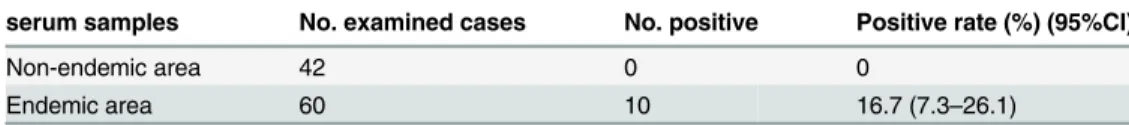
Additionally, 42 serum samples of individuals from non-endemic areas, and 60 serum sam-ples of residents in endemic areas who were identified with negative results for all the three commonly used diagnostic methods, including triplicate stool examinations, IHA and ELISA tests, were used to assess the validity of LAMP assay for field diagnosis of schistosomiasis. No positive result was observed for 42 uninfected human serum samples, indicating an excellent specificity of LAMP assay (Table 7). However, for the 60 serum samples from residents in en-demic areas, which were negative for faecal eggs and serological methods, 10 (16.7%) samples were positive diagnosed by LAMP assay (Table 7), which might be missed by the commonly used diagnostic methods, such as Kato-Katz method, IHA and ELISA.
Discussion
Diagnosis is central to control of schistosomiasis [36]. The prevention and control of the dis-ease need rapid and reliable diagnostic techniques to identify target population accurately for treatment [37]. However, the currently available diagnostic assays are not ideal, since the search for eggs in stools and detection of circulating antigens lack sensitivity in low prevalence and post-treatment situations, and antibody detection lacks specificity [38], and cannot distin-guish current and cured infections, which results in the difficulties in determining prevalence, identifying true infected individuals for selective chemotherapy and assessing the effectiveness of intervention including follow-up of chemotherapy [34,39,40,]. Our results further demon-strated the limitations of the direct parasitological technique (Kato-Katz method) and antibody detection assays (ELISA and IHA). In rabbit models infected withS.japonicumcercariea, we did not find faecal eggs until 7 weeks post-infection, and for the two immunoassays, the earliest positive detection result was obtained at 4 weeks post-infection. Our rabbit experiment results confirmed that both the stool examination and antibody detection methods could not give early diagnosis at the beginning ofS.japonicuminfection. Moreover, 4 egg-positive cases were negative for antibodies by ELISA and IHA, with ages ranging from 43 to 65, indicating that an-tibody detections were insufficient for diagnosis of schistosomiasis in patients of older ages, due to immune down-regulation in chronic infection stages [7,41]. However, DNA
Table 5. Comparison of ELISA, IHA and LAMP for diagnosis of 93 serum samples with various low-intensity infection ofSchistosoma japonicum.
Intensity of infection (EPG) No. examined cases No. positive (positive rate, %, 95%CI)
ELISA IHA LAMP χ2 Pvalue
<10 41 34 (82.9, 71.4–91.4) 39 (95.1, 88.5–100) 39 (95.1, 88.5–100) 4.657 0.097 10–39 42 35 (83.3, 72.0–94.6) 36 (85.7, 75.1–96.3) 41 (97.6, 93.0–100) 6.157 0.046
40–99 10 9 (90.0) 9 (90.0) 9 (90.0) / /
doi:10.1371/journal.pntd.0003668.t005
Table 6. Comparison of negative conversion rate of ELISA, IHA and LAMP assay in 47 cases infected withSchistosoma japonicumafter treatment with praziquantel.
Periods after treatment No. examined cases No. (negative conversion rate, %, 95%CI) χ2 Pvalue
ELISA IHA LAMP
3 months 47 8(17.0, 6.3–27.7) 11(23.4, 11.3–35.5) 11(23.4, 11.3–35.5) 0.76 0.683
6 months 47 9(19.1, 7.9–30.3) 20(42.5, 28.4–56.6) 29(61.7, 47.8–75.6) 17.63 <0.0001 9 months 47 12(25.5, 13.0–38.0) 15(31.9, 18.6–45.2) 39(83.0, 72.3–92.7) 37.43 <0.0001
amplification assays, which had identical diagnostic value with that of parasitological methods, may provide alternative approaches for sensitive and specific diagnosis of schistosomiasis. Lier et al. [21] reported a real-time PCR assay for detection of low intensityS.japonicuminfections in a pig model. Subsequently, this method was used in a clinical trial. This real-time PCR assay detected slightly more positive faecal samples than the microscopy method, but was consistent-ly negative in serum and urine samples [22]. To our knowledge, most of the PCR-based meth-ods were focused on the detection of specificSchistosomaDNA in faeces, all of these methods could be seen as improvements on the stool examination approach [7], thus the sensitivity was influenced by the large day-to-day egg fluctuations in infected individuals [42]. In general, serum and urine samples are easier to obtain and more accepted in many populations than fae-cal samples, and unlike eggs in faefae-cal samples, schistosome DNA would be equally distributed throughout the serum of the patient, resolving the restrictions of uneven distributions of eggs in stool samples [43]. Therefore, the detection ofSchistosomaDNA from serum samples would be more accurate in field conditions. In our previous study, we found that theS.japonicum
DNA in host serum primarily comes from the residual body of dead schistosomula in the first 4 weeks post-infection, while during the spawning stage of the female schistosome, the parasite DNA mainly comes from the disintegration of inactive eggs [44]. Furthermore, a 230-bp se-quence from the highly repetitive retrotransposon SjR2 was identified in our previous study, and showed high sensitivity and specificity for detectingS.japonicumDNA in sera of rabbit model and patients [9]. Although PCR-based methods have the potential for sensitive and spe-cific detection of schistosomiasis, the dependence on expensive apparatus restricts their wide application in the field.
Unlike PCR, LAMP assay does not require amplification cycles by thermocycling or ampli-con detection by electrophoresis. Given these features, LAMP is potentially useful for work in the field and has already used in rural laboratories in developing areas for the diagnosis of tu-berculosis [45]. Our previous study established a LAMP assay targetingS.japonicumSjR2, and the method was capable of detecting as little as 0.08 fgS.japonicumDNA, which was 104times more sensitive than common PCR assay. In particular, the LAMP assay was able to detect
S.japonicumDNA in rabbit sera at 1 week post-infection, and become negative at 12 weeks post-treatment [30]. However, the rabbit models used in our previous study were of high and moderate intensity infections, and the detection ofS.japonicumDNA in low-intensity infec-tions is more consistent with the current epidemiological situation.
In this study, the utility of LAMP assay was firstly assessed in rabbit models with very low grade intensities of infection (EPG = 16). It was able to detectS.japonicumDNA in serum at 3 days post-infection, and the detection results became negative at 10 weeks post-treatment, indi-cating that the LAMP method was useful for diagnosis of schistosomiasis, especially with low-intensity infection, and had potential for evaluation of chemotherapy effectiveness.
Then the field diagnostic value of the LAMP method, and its ability for evaluation of effectiveness of drug treatments was tested using 110 patient serum samples with confirmed
S.japonicuminfection by stool examination. Meanwhile, two of the most extensively used im-munoassays (ELISA and IHA) in the field were also used to assess the validity of the LAMP
Table 7. LAMP for detection of“healthy”human serum samples from endemic and non-endemic areas.
serum samples No. examined cases No. positive Positive rate (%) (95%CI)
Non-endemic area 42 0 0
Endemic area 60 10 16.7 (7.3–26.1)
assay for diagnosis of schistosomiasis. Our detection results showed that the LAMP assay per-formed better than the commonly used immunoassays in terms of higher sensitivity in patients with low-intensity infection (Table 4andTable 5). After treatment with praziquantel, the nega-tive seroconversion rate of IHA and ELISA sustained at low levels, while for the LAMP assay, the negative conversion rate ofS.japonicumDNA in serum increased from 23.4%, 61.7% to 83.0% after 3 months, 6 months and 9months post-treatment (Table 6). All of the results con-firmed that the LAMP assay was efficient for diagnosis of cases with low-intensity infections, and had potential for assessment of effectiveness of drug treatment.
Finally, 60 residents living in endemic areas with negative detection results of Kato-Katz, IHA and ELISA, were recognized as“healthy”residents, and were employed to assess the abili-ty of LAMP assay for accurate diagnosis of schistosomiasis. Of the 60 serum samples from
“healthy”individuals, 10 (16.7%) were diagnosed as positive by LAMP assay (Table 7), who might had been missed by parasitological methods, indicating that traditionally used methods lack sensitivity for diagnosis of individuals with low intensity infections. A field study done by Xu et al. [3] further confirmed our results. In this study, of 1371 enrolled residents, parasitolog-ical detection identified only 74 (5%) individuals as being egg-positive by Kato-Katz thick smears, of whom all the individuals were also positively diagnosed by LAMP detection of SjR2 DNA. More importantly, additional 368 (27%) individuals were positive for SjR2 DNA [3]. Professor Clive Shiff of Johns Hopkins Bloomberg School of Public Health, USA, commented on this paper as“New diagnostics reform infectious parasite epidemiology”[12]. The comment suggested that continuous surveillance to predict any resurgence of infection by accurate and sensitive measuring methods is highly recommended. It is very important to help reduce schis-tosomiasis transmission through targeted treatment of individuals, particularly those people who are presumed to be free of infection (false-negative) may actually remain infected and ca-pable of infecting snails when their faeces get into the water.
In conclusion, the LAMP assay with rapidity, simplicity, sensitivity and specificity is suitable not only for case detections, but also for disease surveillance in schistosomiasis-endemic areas. Application of this method may improve the identification of cases with low-intensity infec-tions and targeted treatment, which is of great significance for schistosomiasis control and elimination programmes.
Supporting Information
S1 Checklist. STARD checklist.
(DOC)
S1 Flowchart. STARD flowchart.
(PDF)
Acknowledgments
We are grateful to Professor Zhongdao Wu for excellent technical assistance and improvement of our manuscript.
Author Contributions
References
1. World Health Organisation (WHO). WHO fact sheet on Schistosomiasis. WHO Geneva, 2013.http:// www.who.int/mediacentre/factsheets/fs115/en/
2. Zhou XN, Guo JG, Wu XH, Jiang QW, Zheng J, et al. (2007) Epidemiology of schistosomiasis in the People's Republic of China, 2004. Emerg Infect Dis 13: 1470–1476. doi:10.3201/eid1310.061423 PMID:18257989
3. Xu XD, Zhang YB, Lin DD, Zhang JJ, Xu J, et al. (2014) Serodiagnosis ofSchistosoma japonicum infec-tion: genome-wide identification of a protein marker, and assessment of its diagnostic validity in a field study in China. Lancet Infect Dis 14: 489–497. doi:10.1016/S1473-3099(14)70067-2PMID:24656567 4. Bergquist R, Yang GJ, Knopp S, Utzinger J, Tanner M. (2015) Surveillance and response: Tools and
approaches for the elimination stage of neglected tropical diseases. Acta Trop 141(Pt B):229–234. http://dx.doi.org/10.1016/j.actatropica.2014.09.017doi:10.1016/j.actatropica.2014.09.017PMID: 25301340
5. Allam AF, Kader O, Zaki A, Shehab AY, Farag HF. (2009) Assessing the marginal error in diagnosis and cure ofSchistosoma mansoniin areas of low endemicity using Percoll and PCR techniques. Trop Med Int Health 14: 316–321. doi:10.1111/j.1365-3156.2009.02225.xPMID:19278527
6. Lin DD, Liu JX, Liu YM, Hu F, Zhang YY, et al. (2008) Routine Kato-Katz technique underestimates the prevalence ofSchistosoma japonicum: A case study in an endemic area of the People’s Republic of China. Parasitol Int 57: 281–286. doi:10.1016/j.parint.2008.04.005PMID:18485807
7. van Dam GJ, Xu J, Bergquist R, de Dood CJ, Utzinger J, et al. (2015) An ultra-sensitive assay targeting the circulating anodic antigen for the diagnosis ofSchistosoma japonicumin a low-endemic area, Peo-ple’s Republic of China. Acta Trop 141(Pt B):190–197.http://dx.doi.org/10.1016/j.actatropica.2014.08. 004doi:10.1016/j.actatropica.2014.08.004PMID:25128703
8. Gordon CA, Acosta LP, Gray DJ, Olveda RM, Jarilla B, et al. (2012) High Prevalence ofSchistosoma japonicumInfection in Carabao from Samar Province, the Philippines: Implications for Transmission and Control. PLoS Negl Trop Dis 6(9): e1778. doi:10.1371/journal.pntd.0001778PMID:23029571 9. Xia CM, Rong R, Lu ZX, Shi CJ, Xu J, et al. (2009)Schistosoma japonicum: A PCR assay for the early
detection and evaluation of treatment in a rabbit model. Exp Parasitol 121: 175–179. doi:10.1016/j. exppara.2008.10.017PMID:19027005
10. Pontes LA, Dias-Neto E, Rabello A. (2002) Detection by polymerase chain reaction ofSchistosoma mansoniDNA in human serum and feces. Am J Trop Med Hyg 66: 157–162. PMID:12135287 11. Zhu Y, Hua W, Xu M, He W, Wang X, et al. (2012) A novel immunodiagnostic assay to detect serum
an-tibody response against selected soluble egg antigen fractions fromSchistosoma japonicum. PloS one 7: e44032. doi:10.1371/journal.pone.0044032PMID:22952862
12. Shiff C. (2014) New diagnostics reform infectious parasite epidemiology. Lancet Infect Dis 14: 446–448. doi:10.1016/S1473-3099(14)70707-8PMID:24656566
13. van Dam GJ, de Dood CJ, Lewis M, Deelder AM, van Lieshout L, et al. (2013) A robust dry reagent lat-eral flow assay for diagnosis of active schistosomiasis by detection ofSchistosomacirculating anodic antigen. Exp Parasitol 135: 274–282. doi:10.1016/j.exppara.2013.06.017PMID:23850995 14. Utzinger J, N’Goran EK, Caffrey CR, Keiser J. (2011) From innovation to application: social-ecological
context, diagnostics, drugs and integrated control of schistosomiasis. Acta Trop 120 (Suppl 1): S121–S137. doi:10.1016/j.actatropica.2010.08.020PMID:20831855
15. Hamburger J, He-Na Abbasi I, Ramzy RM, Jourdane J, Ruppel A. (2001) Polymerase chain reaction assay based on a highly repeated sequence ofSchistosoma haematobium: a potential tool for monitor-ing schistosome infested water. Am J Trop Med Hyg 65: 907–911. PMID:11791997
16. Pontes LA, Oliverira MC, Katz N, Dlas-Neto E, Rabello A. (2003) Comparison of a polymerase chain re-action and the Kato-Katz technique for diagnosing infection withSchistosoma mansoni. Am J Trop Med Hyg. 68: 652–656. PMID:12887022
17. Sandoval N, Siles-Lucas M, Lopez Aban J, Perez-Arellano JL, Garate T, et al. (2006a)Schistosoma mansoni: a diagnostic approach to detect acute schistosomiasis infection in a murine model by PCR. Exp Parasitol. 114: 84–88. PMID:16571353
18. Sandoval N, Siles-Lucas M, Perez-Arellano JL, Carranza C, Puente S, et al. (2006b). A new PCR-based approach for the specific amplification of DNA from different Schistosoma species applicable to human urine samples. Parasitology 133: 581–587.
21. Lier T, Johansen MV, Hjelmevoll SO, Vennervald BJ, Simonsen GS. 2008. Real-time PCR for detection of low intensitySchistosoma japonicuminfections in a pig model. Acta Trop 105: 74–80. PMID: 18036505
22. Lier T, Simonsen GS, Wang T, Lu D, Haukland HH, et al. (2009) Real-time polymerase chain reaction for detection of low-intensitySchistosoma japonicuminfection in China. Am J Trop Med Hyg. 81: 428–432. PMID:19706908
23. ten Hove BJ, Verweij JJ, Vereecken K, Polman K, Dieye L, et al. (2008) Multiplex real-time PCR for the detection and quantification ofSchistosoma mansoniandS.haematobiuminfection in stool samples collected in northern Senegal. Trans R Soc Trop Med Hyg 102: 179–185. doi:10.1016/j.trstmh.2007. 10.011PMID:18177680
24. Notomi T, Okayama H, Masubuchi H, Yonekawa T, Watanabe K, et al. (2000) Loop-mediated isother-mal amplification of DNA. Nucleic Acids Res 28: E63. PMID:10871386
25. Poon LL, Wong BW, Ma EH, Chan KH, Chow LM, et al. (2006) Sensitive and inexpensive molecular test for falciparum malaria: detectingPlasmodium falciparumDNA directly from heat-treated blood by loop-mediated isothermal amplification. Clin Chem 52: 303–306. PMID:16339303
26. Poschl B, Waneesorn J, Thekisoe O, Chutipongvivate S, Karanis P. (2010) Comparative diagnosis of malaria infections by microscopy nested PCR, and LAMP in northern Thailand. Am J Trop Med Hyg 83: 56–60. doi:10.4269/ajtmh.2010.09-0630PMID:20595478
27. Lucchi NW, Demas A, Narayanan J, Sumari D, Kabanywanyi A, et al. (2010) Real-time fluorescence loop mediated isothermal amplification for the diagnosis of malaria. PLoS One 5: e13733. doi:10. 1371/journal.pone.0013733PMID:21060829
28. Abbasi I, King CH, Muchiri EM, Hamburger J. (2010) Detection ofSchistosoma mansoniand Schisto-soma haematobiumDNA by Loop-Mediated Isothermal Amplification: Identification of Infected Snails from Early Prepatency. Am J Trop Med Hyg 83: 427–432. doi:10.4269/ajtmh.2010.09-0764PMID: 20682894
29. Kumagai T, Furushima-Shimogawara R, Ohmae H, Wang TP, Lu S, et al. (2010) Detection of Early and Single Infections ofSchistosoma japonicumin the Intermediate Host Snail, Oncomelania hupensis, by PCR and Loop-Mediated Isothermal Amplification (LAMP) Assay. Am J Trop Med Hyg 83: 542–548. doi:10.4269/ajtmh.2010.10-0016PMID:20810818
30. Xu J, Rong R, Zhang HQ, Shi CJ, Zhu XQ, et al. (2010) Sensitive and rapid detection ofSchistosoma japonicumDNA by loop-mediated isothermal amplification (LAMP). Int J Parasitol 40: 327–331. doi: 10.1016/j.ijpara.2009.08.010PMID:19735662
31. Wang C, Chen L, Yin X, Hua W, Hou M, et al. (2011) Application of DNA-based diagnostics in detection of schistosomal DNA in early infection and after drug treatment. Parasit Vectors 4:164. doi:10.1186/ 1756-3305-4-164PMID:21864384
32. Fernandez-Soto P, Gandasegui Arahuetes J, Sanchez Hernandez A, Lopez Aban J, Vicente Santiago B, et al. (2014) A Loop-Mediated Isothermal Amplification (LAMP) Assay for Early Detection of Schisto-soma mansoni in Stool Samples: A Diagnostic Approach in a Murine Model. PLoS Negl Trop Dis 8: e3126. doi:10.1371/journal.pntd.0003126PMID:25187956
33. Mori Y, Nagamine K, Tomita N, Notomi T. (2001) Detection of loop-mediated isothermal amplification reaction by turbidity derived from magnesium pyrophosphate formation. Biochem Biophys Res Com-mun 289: 150–154. PMID:11708792
34. Utzinger J, Zhou XN, Chen MG, Bergquist R. (2005) Conquering schistosomiasis in China: the long march. Acta Trop 96: 69–96. PMID:16312039
35. Laha T, Brindley PJ, Smout MJ, Verity CK, McManus DP, et al. (2002) Reverse transcriptase activity and untranslated region sharing of a new RTE-like, non-long terminal repeat retrotransposon from the human blood fluke,Schistosoma japonicum. Int J Parasitol 32:1163–1174. PMID:12117499 36. Zhu YC. (2005) Immunodiagnosis and its role in schistosomiasis control in China: a review. Acta Trop
96: 130–136. PMID:16143288
37. Wang W, Li Y, Li H, Xing Y, Qu G, et al. (2012) Immunodiagnostic efficacy of detection ofSchistosoma japonicumhuman infections in China: a meta analysis. Asian Pac J Trop Med 5: 15–23. doi:10.1016/ S1995-7645(11)60238-1PMID:22182637
38. Rabello A, Pontes LA, Dias-Neto E. (2002) Recent advances in the diagnosis of Schistosoma infection: the detection of parasite DNA. Mem Inst Oswaldo Cruz 97 (Suppl. I): 171–172. PMID:12426615 39. Zhou YB, Zheng HM, Jiang QW. (2011) A diagnostic challenge for schistosomiasis japonica in China:
consequences on praziquantel-based morbidity control. Parasit Vectors 4: 194. doi: 10.1186/1756-3305-4-194PMID:21981948
41. van Lieshout L, Panday UG, De Jonge N, Krijger FW, Oostburg BF, et al. (1995) Immunodiagnosis of schistosomiasis mansoni in a low endemic area in Surinam by determination of the circulating antigens CAA and CCA. Acta Trop 59: 19–29. PMID:7785523
42. Engels D, Sinzinkayo E, Gryseels B. (1996) Day-to-day egg count fluctuation inSchistosoma mansoni
infection and its operational implications. Am J Trop Med Hyg 54: 319–324. PMID:8615440
43. Wichmann D, Panning M, Quack T, Kramme S, Burchard GD, et al. (2009) Diagnosing schistosomiasis by detection of cell-free parasite DNA in human plasma. PLoS Negl Trop Dis 3: e422. doi:10.1371/ journal.pntd.0000422PMID:19381285
44. Xu J, Liu AP, Guo JJ, Wang B, Qiu SJ, et al. (2013) The sources and metabolic dynamics of Schisto-soma japonicumDNA in serum of host. Parasitol Res 112:129–133. doi:10.1007/s00436-012-3115-3 PMID:22983220