Paraoxonase 3 Activity and The Ratio of Antioxidant
to Peroxidation in The Follicular Fluid of
Infertile Women
Mohammad-Reza Rashidi, Ph.D.1, Jalal Eisa-Khaje, M.Sc.1, Laya Farzadi, M.D.2, Masoud Darabi, Ph.D.3,
Alieh Gasemzadeh, M.D.2, Vahideh Shahnazi, M.Sc.2, Shabnam Fayezi, M.D.4, Amir Mehdizadeh, M.Sc.5,
Reza Haji Hosseini, Ph.D.6, Mohammad Nouri, Ph.D.7*
1. Research Center for Pharmaceutical Nanotechnology, Tabriz University of Medical Sciences, Tabriz, Iran 2. Women’s Reproductive Health Research Center, Alzahra Hospital, Tabriz University of Medical Sciences, Tabriz, Iran 3. Department of Biochemistry and Clinical Laboratories, School of Medicine, Tabriz University of Medical Sciences, Tabriz, Iran 4. Students Research Committee, Infertility and Reproductive Health Research Center, Shahid Beheshti University of
Medical Sciences, Tehran, Iran
5. Liver and Gastrointestinal Disease Research Center, Tabriz University of Medical Sciences, Tabriz, Iran 6. Department of Biology, Payame Noor University, Tehran, Iran
7. Umbilical Stem Cell Research Center, Tabriz University of Medical Sciences, Tabriz, Iran
Abstract
Background: Paraoxonase-3 (PON3), as a high density lipoprotein (HDL)-associated
lac-tonase, is capable of preventing the oxidative modiication of low density lipoprotein (LDL). PON3 activity in follicular luid (FF) is three times more than its activity in serum. However, the detailed role of PON3 in women’s fertility remains unknown. The aim of this study was to investigate the correlation between PON3 activity in the FF of women undergoing assisted repro
-ductive technique (ART), in vitro fertilization (IVF), or intra-cytoplasmic sperm injection (ICSI).
Materials and Methods: This cross-sectional study consisted of 50 women from couples with male factor infertility (MFI) or with female factor infertility (FFI). The FF samples were obtained during the ART intervention. PON3 activity, HDL cholesterol (HDL C), total antioxi -dant status (TAS) and the level of malondialdehyde (MDA) were determined. The morphology of the embryo was determined using embryo cell number (ECN) and embryo fragmentation score (EFS). In addition, fertilization rate (FR) was used an oocyte fertilization index.
Results:Of 50 women, 20 women belonged to FFI group and the remaining 30 women belonged to MFI group. PON3 activity in FF of women in FFI group was signiicantly lower (p<0.05) in comparison with corresponding value in MFI group. The value of PON3 activity/MDA in the FFI group was lower than that in MFI group. Moreover, MDA level in the FF of FFI group was signiicantly higher (p<0.05) than its concentra -tion in MFI group. Meanwhile, no signiicant difference was found in HDL-C concentra-tion and TAS of both groups. No signiicant correlaconcentra-tion was observed between the ECN and FF biochemical parameters. There was also a negative correlation between FR and MDA (r=-0.42, p=0.02), whereas a positive relation between FR with PON3 activity (r=0.59, p=0.004), HDL-C (r=0.35, p=0.04) and PON3/MDA (r=0.59, p=0.001).
Conclusion:According to the results of this study, PON3 activity level as a key compo-nent of antioxidant system in FF may directly be associated with the success rate of ART and fertilization rate in women.
Keywords: Infertility, PON3, Follicular Fluid, Peroxidation, In Vitro Fertilization
Citation: Rashidi MR, Eisa-Khaje J, Farzadi L, Darabi M, Gasemzadeh A, Shahnazi V, Fayezi Sh, Mehdizadeh A, Haji
Hosseini R, Nouri M. Paraoxonase 3 activity and the ratio of antioxidant to peroxidation in the follicular luid of infertile
women. Int J Fertil Steril. 2014; 8(1): 51-58.
Received: 19 Jan 2013, Accepted: 2 Jun 2013
* Corresponding Address: P.O. Box: 51666-15573, Umbilical Stem Cell Research Center, Tabriz University of Medical Sciences, Tabriz, Iran
Email: [email protected] Royan Institute
Introduction
Free radicals cause oxidative damages to the cell membrane lipid content (1). The role of the reactive oxygen species (ROS) in peroxidation of lipids and their interference with sperm func -tion, ovum func-tion, and human reproduction have been reported (2). Natural byproduct of metabolism is ROS which includes the superox -ide anion (O2• –) and the hydroxyl radical (OH). ROS can induce DNA fragmentation, protein oxidation, lipid per oxidation and cellular dam-age (3-5). Within a cell, ROS is neutralized by the antioxidants (6). Paraoxonase (PON) is one of the strong antioxidants in the serum and the follicular fluid (FF). PON1 and PON3, which are both associated in serum with high density lipoprotein (HDL) cholesterol (-C), protect the serum lipids from oxidation, probably through their ability to hydrolyze specific oxidized li-pids (7, 8). The PON gene family consists of three members of PON1 gene, PON2 gene and PON3 gene, encoding PON enzyme family (4). All three genes of PON have been preserved in mammals, and this fact indicates the important physiological role of this antioxidant enzyme (9). There is a higher PON enzymatic activity in FF compared with serum, which has been attributed to PON expression and secretion by granulosa cells in FF (10).
PON3 is synthesized in the liver and carried in the blood in association with HDL. PON3 is also able to prevent the oxidation of low density lipoproteins (LDL) (11). Plasma concentration of PON3 is 100 times less than that of PON1 (12, 13). In recent stud -ies, the activity of this enzyme in FF has been report -ed (14) and is calculat-ed to be three times more than its original concentration in serum (15). However, the role of PON3 in women’s fertility has not yet been fully studied. Considering the strong antioxi -dant property of PON3 and its high concentration in the FF, it is likely that this enzyme plays an important role in the oogenesis, eggs quality and fertilization. In the present study, PON3 activity and the ratio of antioxidant to peroxidation in the FF of women with male factor infertility (MFI) and with female factor infertility (FFI) were compared after ovarian stimu -lation, while their variation with respect to the num -ber of oocytes, embryo cell num-ber (ECN), embryo fragmentation score (EFS) and fertilization rate (FR) were statistically analyzed.
Materials and Methods
Study design and subjects
In this cross-sectional study, we gained the agreement of the Ethical Committee of Tabriz University of Medical Science, and all patients gave written informed consent. Fifty infertile cou -ples referred to Tabriz Alzahra Women’s Hospital, Tabriz, Iran, for infertility treatment using assist -ed reproductive technique (ART) were select-ed within a three-month period. Out of 50 couples, 30 women with MFI were used as the control group (MFI group), and the remaining 20 women with FFI were used as the test group (FFI group).
Among selected patients, 60% of infertile partner were male and 40% were female. Lack of infections and husband with no smoking hab -it were defined as including cr-iteria. The long protocol, gonadotropin-releasing hormone ago-nist (GnRHa) and human menopausal gonado-tropin (HMG) were used for all subjects as the treatment protocol for the stimulation of ovula-tion (16). On the 14th day of the menstrual cycle,
the follicles larger than 3 mm were punctured and the FF was extracted. After separating the oocytes from FF, the remaining was centrifuged and the supernatant was kept at -86˚C for furs-ther studies. The collected oocytes were incu -bated at 37˚C, 5% CO2 and 95% humidity for 4 hours, and then were used for in vitro fertili-zation (IVF). In couples with abnormal sperm parameters such as low sperm count, low sperm motility and morphological defects, intracyto-plasmaic sperm injection (ICSI) was used for oocytes fertilization, otherwise IVF was per -formed. Identification of zygotes was carried out 18 hours after insemination through the ap -pearance of two pronuclei (2PN). All embryos were recultured in standard culture medium (ISM1) at 37˚C, 5% CO2 and 95% humidity for 24 hours. On the second day of the culture, the morphology of the embryos was determined us -ing ECN and EFS (17, 18). In addition, FR was used as an oocyte fertilization index. FR was calculated as the number of fertilized oocytes/ number of mature oocytes ×100.
Determination of simvastatinase PON3 activity
PON3 activity was determined through monitor -ing of the conversion of simvastatin (SV) to β,δ-dihydroxyacid simvastatin (SVA) using reverse phase-high performance liquid chromatography (RP-HPLC) method as described before (20) with slight modiication as follows: SV (120 µM) was incubated with FF at 37˚C for 60 minutes, and the reaction was terminated by addition of acetoni -trile. The sample was centrifuged at 6000 rpm for 6 minutes, and the amount of SVA in the super -natant was analyzed using a high-performance liquid chromatography (HPLC) system (Waters Associates, Norwich, Cheshire, UK) which con -sisted of a Waters 515 pump, Waters 717 plus Au -tosampler, Waters 2487, and Dual λ Absorbance Detector. The mobile phase was a mixture of 100 mM monopotassium phosphate (pH=4.5)/ acetoni -trile (27/73 v/v). Separations were performed on a C18 column (Phenosphere-LUNA, 5 μM, 250×4.6 mm) with a C18 guard column (Perfectsil Target ODS-3, 5 μM, 10×4 mm). The efluent was moni -tored by ultraviolet (UV) detection at 239 nm at a low rate of 1 ml/minute.
Determination of MDA levels in FF
MDA levels in FF were determined by the thiobarbituric acid (TBA) method and expressed as nmol MDA formed/mL FF (20). Briefly, 0.5 ml FF was shaken with 2.5 ml of 20% trichlo -roacetic acid (TCA) in a 10 ml centrifuge tube. Then, 1ml of 0.67% TBA was added to the mix -ture, shaken, and heated in a boiling water bath for 60 minutes and it was cooled rapidly. MDA content in the serum was spectrophotometeri -cally determined at 532 nm. The calibration curve was plotted with 0.1 to 20 μmol/L tetra -ethoxypropane (TEP).
Measurement of plasma total antioxidant status (TAS) in FF
Total antioxidant status (TAS) was measured in FF using a commercial kit (Randox Laboratories, France). The assay was performed by incuba -tion of 2,2'-azino-di-(3-ethylbenzthiazoline sulphonate) (ABTS) with a peroxidase (meth -myoglobin) and hydrogen peroxide to develop a relatively stable blue-green color, which sub -sequently measured at 600 nm (21). Trolox, a traditional standard for TAS measurement, was used to calculate Trolox molar equivalent (22).
Determining the HDL-C levels
HDL-C level was measured after extraction of the particles from FF by means of phosphotungstic solution and centrifugation. The amount of cho -lesterol in the supernatant was determined spec -trophotometrically by cholesterol oxidase (Pars Azmon, Iran).
Statistical analysis
All data were expressed as mean ± SD. The normality test showed that all data were normal or nearly normally distributed. Statistical com -parisons were performed using t test. ANOVA was used to evaluate the differences between the means of more than two groups. A value of p<0.05 was considered statistically signiicant. Statistical analysis was carried out by Statistical Package for the Social Sciences (SPSS; version 16, SPSS Inc., Chicago, USA).
Results
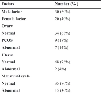
The general information of the population study is shown in table 1. In the test group, 68% of the pa -tients had normal ovaries, 18% had polycystic ova -ries and 14% had abnormal ova-ries (small ova-ries, ovaries which were operated upon, and no eggs were produced), 4% of the test subjects had abnormal uter -uses (small uterus and endometriosis) and 30% suf -fered from abnormal menstrual cycles.
Table 1: General information of the studied women
Number (% )
Factors
30 (60%) Male factor
20 (40%) Female factor
Ovary
34 (68%) Normal
9 (18%) PCOS
7 (14%) Abnormal
Uterus
48 (96%) Normal
2 (4%) Abnormal
Menstrual cycle
35 (70%) Normal
15 (30%) Abnormal
PON3 activity in the FF of the MFI group was found to be significantly (p<0.001) high -er than that in the women with FFI (4.7 ± 0.8 vs. 3.8 ± 0.7 µmol/min/ml). In contrast, the concentration of MDA, a lipid peroxidation indicator, in the FF of MFI group was 3.1 ± 1.4 nmol/ml compared to the value of 4.2 ± 1.7 nmol/ml measured in FF of women with FFI (p=0.024). Therefore, the ratio of antioxi -dant to peroxidation, which was evaluated as PON3/MDA value in the control group, was also higher than the corresponding value in
the women with FFI. No statistically signifi -cant difference was found in the HDL-C level in the FF of both groups (Table 2). Although there was no significant difference in the TAS levels in the women with MFI and FFI, the ra -tio of PON3 to TAS in two groups were statis -tically different (p= 0.013).
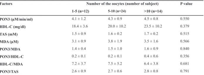
Biochemical factors measured in the FF based on the number of oocytes are displayed in table 3. No signiicant difference was observed in the studied factors with respect to the number of oo -cytes.
Table 2: Follicular luid biochemical parameters in the studied population
P value
FFI (Mean ± SD)
MFI (Mean ± SD) Chromosomal polymorphic variations
<0.001 3.8 ± 0.7
4.7 ± 0.8* PON3 (µM/min/ml)
0.556 20.0 ± 6.1
21.6 ± 11.4 HDL-C (mg/dl)
0.465 1.6 ± 0.2
1.6 ± 0.3 TAS (mM)
0.024 4.2 ± 1.7
3.1 ± 1.4 MDA (µM)
0.002 1.0 ± 0.6
1.8 ± 0.9 PON3/MDA
0.097 0.2 ± 0.1
0.4 ± 0.5 PON3/HDL-C
0.633 6.5 ± 4.7
7.1 ± 4.0 HDL-C/MDA
0.013 2.5 ± 0.5
3.0 ± 0.7 PON3/TAS
MFI; Male factor infertility, FFI; Female factor infertility and TAS; Total antioxidant status.
Table 3: Follicular luid biochemical parameters according to the oocyte numbers in studied women
P value Number of the oocytes (number of subject)
Factors
>10 (n=14)
5-10 (n=24)
1-5 (n=12)
0.550 4.5 ± 0.8
4.3 ± 0.9 4.1 ± 1.2
PON3 (µM/min/ml)
0.379 23.5 ± 10.2
20.0 ± 10.2 18.4 ± 3.6
HDL-C (mg/dl)
0.515 1.7 ± 0.2
1.6 ± 0.2 1.5 ± 0.9
TAS (mM)
0.566 3.5 ± 1.6
3.8 ± 1.9 3.1 ± 0.9
MDA (µM)
0.840 1.6 ± 0.9
1.5 ± 1.0 1.4 ± 0.4
PON3/MDA
0.356 0.4 ± 0.6
0.2 ± 0.1 0.2 ± 0.1
PON3/HDL-C
0.681 6.4 ± 3.8
7.5 ± 5.2 7.2 ± 3.7
HDL-C/MDA
0.791 2.8 ± 0.8
2.7 ± 0.6 2.6 ± 0.9
PON3/TAS
Comparison of number of the oocytes (t=1.32, p=0.13), EFS (t=0.81, p=0.41), ECN (t=0.89, p=0.32) and FR (t=1.52, p=0.11) between women with MFI and FFI by t test showed no signiicant difference between IVF and ICSI techniques. The relations between FF biochemical parameters with EFS, ECN and FR were assessed using regression analysis and the results have been summarized in table 4. A signiicant negative correlation was found between PON3 activity (r=-0.65, p=0.02) and PON3/MDA (r=-0.63, p=0.001) with EFS, whereas there was a positive correlation between EFS and MDA (r=0.55, p=0.002) which indicates
that an increase in antioxidant/peroxidation value is accompanied with an increase in the embryo quality. No signiicant correlation was found be -tween ECN and FF biochemical parameters. A negative correlation between FR and MDA (r=-0.42, p=0.02), while a positive relations between FR and PON3 activity (r=0.56, p=0.004), HDL-C (r=0.35, p=0.041) and PON3/MDA (r=0.59, p=0.001) could be indicative of the beneicial effects of antioxidant in the success of ART. The same pattern was also observed when correlations were examined separately for women with MFI and FFI.
Table 4: Correlation of follicular luid biochemical factors with embryo quality and fertilization rate
FR
ECN
EFS Chromosomal polymorphic variations
0.56 (0.004) 0.24 (0.140)
-0.65 (0.02) PON3 (µM/min/ml)
0.35 (0.041) 0.07 (0.821)
-0.25 (0.310) HDL-C (mg/dl)
0.14 (0.404) 0.18 (0.283)
-0.16 (0.425) TAS (mM)
-0.42 (0.020) -0.38 (0.029)
0.55 (0.002) PON3/MDA
0.59 (0.001) 0.22 (0.215)
-0.63 (0.001) PON3/HDL-C
0.22 (0.214) 0.15 (0.314)
-0.17 (0.406) HDL-C/MDA
0.25 (0.246) 0.09 (0.703)
-0.20 (0.374) PON3/TAS
EFS; Emberyo fragmentation score, ECN; Emberyo cell number, FR; Fertilization rate and TAS; Total antioxidant status. The numbers in brackets are p values.
Discussion
It has been shown that the concentration of li -pidic hydroperoxides and active substances of thiobarbituric acid in the FF is lower than serum in women who underwent IVF. This conirms the presence of a suitable antioxidant in oocyte’s en -vironment prior to ovulation (23). The concentra -tion of this enzyme in FF is much higher than its concentration in serum. The results of this study shows that PON3 activity in FFI is signiicantly lower in comparison with the MFI group. Accord -ing to a study by Closshey et al. (15) the level of PON3 activity in the FF in 14 infertile women who underwent IVF was higher in comparison with se -rum. Moreover, it was shown that there is a signii -cant positive correlation between PON3 and the rate of laboratory pregnancy and fertility. These
results match the results of this study. These ind -ings along with high ratio of PON3/TAS in MFI group indicate the important role of PON3 in FF and in oogenesis.
quality of oocyte during stimulation of ovulation (26). It has been shown that older ages are associ -ated with reduced amount of HDL apolipoprotein, which is accompanied by reduction of the number of mature oocytes in women (27). Although there are some reports verifying the role of HDL in oo-genesis, further studies are needed on this issue.
In our study, no signiicant difference was ob -served in the concentration of TAS in the two groups. In similar studies, no signiicant difference was found in the TAS levels in the women with FFI who suffered from endometriosis when com -pared with the TAS concentration of the MFI (28, 29). This is in line with the indings of this study. In present study, high level of PON3/TAS ratio in the control group (MFI group) and high level of MDA in FFI group indicate that PON3 plays an important role in the prevention of follicular oxi-dative stress. Furthermore, high level of MDA in FF of women with FFI could also be suggestive of the above-indings. This inding is similar to the indings of Yildrim et al. (30) who have shown that the lipidic peroxidation in the FF of the FFI group with polycyctic ovarian syndrome (PCOS) is much higher than that of the MFI group.
The results obtained in the present study show that the PON3/MDA ratio in the women with FFI is signiicantly lower than the corresponding val -ue in the women with MFI (p=0.002). It could be stated that the ratio of antioxidant to peroxidation in the FF is a suitable factor for assessing the oxi -dative stress in the follicles.
PON3 is a strong antioxidant in FF. Closshey et al. (15) have reported that high PON3 activity inside follicle could probably be due to being pro-duced locally in follicle. According to Browne et al. (26), the origin of the enzyme is granula-gener -ating cells. In this study, for the irst time, it was shown that PON3 activity in the FF of the women with FFI is lower than that in women with MFI. Now, it is known that PON3 is able to prevent LDL oxidation (10). This enzyme can utilize the products of lipids oxidation as substrates, and thus, reduces the severity of oxidative stress in the cell (31, 32). Therefore, it is more likely that PON3 have some important roles in the growth and matu -ration of the oocytes.
On the other hands, we were not able to ind any signiicant difference between PON3 activity and
the number of oocytes. Plachot et al. (33) have stated that the rate of fertilized oocytes is associat-ed with not only the number but also the quality of oocytes. Thus, high level of PON3 activity during the growth period, maturity and quality of oocytes plays a vital role. Moreover, current study showed that the ratio of PON3 to MDA, as an indicator of antioxidant to peroxidation, in the FF of FFI group is lower than that of MFI group. Therefore, the an -tioxidant and peroxidation status in FF could be correlated with female infertility.
A signiicant negative relation between PON3 and PON3/MDA with EFS, and a positive relation between these parameters with FR may indicate that PON3 plays an important role in fertilization and the quality of embryo. A high level of PON3 in FF could be indicative of its speciic role in de -velopment and maturation of good oocyte which, in turn, can lead to a healthy embryo.
The role of PON3 in fertility has not received enough attention. Browne et al. (34) have reported a signiicant negative association between HDL-C and EFS; however, they could not ind any signii -cant relation between EFS and PON3 activity in FF. Although the negative relation observed in our study between HDL-C and EFS was not statisti -cally signiicant, we were able to show a signii -cant positive relation between FR and HDL-C in FF. This may indicate the importance of HDL in fertilization which has also been reported by oth -ers (35). It has been shown that HDL and the pro -teins present in the structure of HDL could have a cytoprotective effects on oocyte and surrounding granulosa cells (36). As PON is one of the impor -tant antioxidant components of HDL and PON3 concentration in FF is much higher than its level in blood, it is likely that the local role of PON3 is much more dominant. It should be noted that studied variables including embryo quality and fertilization rate may be affected differentially in IVF and ICSI patients. However, because of our limited number of patients, it was not possible to perform two separate analyses for IVF and ICSI groups.
Conclusion
growth. Thus, PON3 could be a valuable therapeu -tic target to improve the success rate of ART.
Acknowledgements
This study was supported by a grant from the Research Center for Pharmaceutical Nanotechnol -ogy, Tabriz University of Medical Sciences. The researchers gratefully thank all the couples who participated in the study, Umbilical Stem Cell Research Center at Tabriz University of Medical Sciences, Tabriz, Iran and the coworkers of the Alzahra Hospital. There is no conlict of interest in this study.
References
1. Bezrukova GA. Free radical oxidation of red blood cell
membrane lipid structures as a trigger mechanism of an increase in red blood cell membrane permeability during blood coagulation in vitro. Gematol Transfuziol. 1991; 36(11): 7-9.
2. Gupta S, MalhotraN, Sharma D, Chandra A, Ashok A.
Oxi-dative stress and its role in female infertility and assisted reproduction: clinical implications. Int J Fertil Steril. 2009; 2 (4): 147-164.
3. Cemeli E, Anderson D. Mechanistic investigation of
ROS-induced DNA damage by oestrogenic compounds in lym-phocytes and sperm using the comet assay. Int J Mol Sci. 2011; 12 (5): 2783-2796
4. Bedaiwy MA, Elnashar SA, Goldberg JM, Sharma R,
Mascha EJ, Arrigain S, et al. Effect of follicular luid oxida-tive stress parameters on intracytoplasmic sperm injection outcome. Gynecol Endocrinol. 2012; 28(1): 51-55.
5. Lopes AS, Lane M, Thompson JG. Oxygen consumption
and ROS production are increased at the time of fertiliza-tion and cell cleavage in bovine zygotes. Hum Reprod. 2010; 25(11): 2762-2773.
6. Rizzo AM, Berselli P, Zava S, Montorfano G, Negroni M,
Corsetto P, et al. Endogenous antioxidants and radical scavengers. Adv Exp Med Biol. 2011; 698: 52-67.
7. Zhang C, Peng W, Wang M, Zhu J, Zang Y, Shi W, et al.
Studies on protective effects of human paraoxonases 1 and 3 on atherosclerosis in apolipoprotein E knockout mice. Gene Ther. 2010; 17(5): 626-633.
8. Browne RW, Shelly WB, Bloom MS, Ocque AJ, Sandler
JR, Huddleston HG, et al. Distributions of high-density lipoprotein particle components in human follicular luid and sera and their associations with embryo morphology parameters during IVF. Hum Reprod. 2008; 23(8): 1884-1894.
9. Primo-Parmo SL, Sorenson RC, Teiber J, La Du BN. The
human serum paraoxonase/arylesterase gene (PON1) is one member of a multigene family. Genomics.1996; 33(3): 498-507.
10. Aharoni A, Gaidukov L, Yagur S, Toker L, Silman I, Taw-ik DS. Directed evolution of mammalian paraoxonases PON1 and PON3 for bacterial expression and catalytic specialization. Proc Natl Acad Sci USA. 2004; 101(2): 482-487.
11. Draganov DI, Stetson PL, Watson CE, Billecke SS, La Du BN. Rabbit serum paraoxonase 3 (PON3) is a high density lipoprotein-associated lactonase and protects low density lipoprotein against oxidation. J Biol Chem. 2000;
275(43): 33435-33442.
12. Draganov DI. Human PON3, effects beyond the HDL: clues from human PON3 transgenic mice. Circ Res. 2007; 100(8): 1104-1105.
13. Reddy ST, Wadleigh DJ, Grijalva V, Ng C, Hama S, Gangopadhyay A, et al. Human paraoxonase-3 is an HDL-associated enzyme with biological activity similar to paraoxonase-1 protein but is not regulated by oxi-dized lipids. Arterioscler Thromb Vasc Biol. 2001; 21(4): 542-547.
14. Angelucci S, Ciavardelli D, Di Giuseppe F, Eleuterio E, Sulpizio M, Tiboni GM, et al. Proteome analysis of human follicular luid. Biochim Biophys Acta. 2006; 1764(11): 1775-1785.
15. Closshey WB, Browne RW, Huddleston HG, Sandler JR, Schisterman EF, Fujimoto VY. High activity of paraoxo-nase 3 (PON3), a known potent antioxidant, identiied in follicular luid. Fertil Steril. 2007; 88: 304-305.
16. Ata B, Yakin K, Balaban B, Urman B. GnRH agonist pro-tocol administration in the luteal phase in ICSI–ET cycles stimulated with the long GnRH agonist protocol: a rand-omized, controlled double blind study. Hum Reprod. 2008; 23(3): 668-673.
17. Steer CV, Mills CL, Tan SL, Campbell S, Edwards RG. The cumulative embryo score: a predictive embryo scor-ing technique to select the optimal number of embryos to transfer in an in-vitro fertilization and embryo transfer pro-gramme. Hum Reprod. 1992; 7(1): 117-119.
18. Puissant F, Van Rysselberge M, Barlow P, Deweze J, Leroy F. Embryo scoring as a prognostic tool in IVF treat-ment. Hum Reprod. 1987; 2(8): 705-708.
19. Draganov DI, Tiber JF, Speelman A, Osava Y, Sunahara R, La Du BN. Human paraoxonases (PON1, PON2 and PON3) are lactonase with overlapping and distinct sub-strate specifcities. J Lipid Res. 2005; 46(6): 1239-1247. 20. Suchocka Z, Swotowaka j, Pacheeka J, Suchocki P.
RP-HPLC determination of paraoxonase-3 activity in human serum. J Pharm Biomed Anal. 2006; 42: 113-119. 21. Conti M, Morand PC, Levillain P, Lemonnier A. Improved
luorimetric determination of malondialdehyde. Clin Chem. 1991; 37: 1273-1275.
22. Miller NJ, Rice-Evans C, Davies MJ. A new method for measuring antioxidant activity. Biochem Soc Trans. 1993; 21(2): 95-97.
23. Jowzik M, Wolczynski S, Jozwik M, Szamatowicz M. Oxi-dative stress markers in preovulatory follicular luid in hu -man. Molecular Human Reprod. 1999; 5: 409-413. 24. Becker S, von Otte S, Robenek H, Diedrich K, Nofer JR.
Follicular luid high-density lipoprotein-associated sphin -gosine 1-phosphate (S1P) promotes human granulosa lutein cell migration via S1P receptor type 3 and small G-protein RAC1. Biol Reprod. 2011; 84(3): 604-612. 25. Mehdizadeh A, Rahimipour A, Farzadi L, Darabi M,
Shah-nazi V, Shaaker M, Vatankhah AM. Correlation between the level of cholesteryl ester transfer protein in follicular luid with fertilization rates in IVF/ ICSI cycles. Iran J Re -prod Med. 2011; 9: 193-198.
26. Browne RW, Shelly WB, Bloom MS, Ocque AJ, Sandler JR, Huddleston HG, et al. Distributions of high-density lipoprotein particle components in human follicular luid and sera and their associations with embryo morphology parameters during IVF. Hum Reprod. 2008; 23(8): 1884-1894.
28. Hong-Nerng Ho1, Ming-Yih Wu, Shee-Uan Chen, Kuang-Han Chao, Chin-Der Chen, Yu-Shih Yang. Total antioxi-dant status and nitric oxide do not increase in peritoneal luids from women with endometriosis. Human Reproduc -tion. 1997; 12(12): 2810-2815.
29. Bedaiwy M, Agarwal A, Said TM, Goldberg JM, Sharma RK, Worley S, et al. Role of total antioxidant capacity in the differential growth of human embryos in vitro. Fertil Steril. 2006; 86(2): 304-309.
30. Yildirim B, Demir S, Temur I, Erdemir R, Kaleli B. Lipid per-oxidation in follicular luid of women with polycystic ovary syndrome during assisted reproduction cycles. J Reprod Med. 2007; 52(8): 722-726.
31. Aviram M, Kaplan M, Rosenblat M, Fuhrman B. Dietary antioxidants and paraoxonases against LDL oxidation and atherosclerosis development. Handb Exp Pharmacol. 2005; 170: 263-300.
32. Draganov DI, Stetson PL, Watson CE, Billecke SS, La Du BN. Rabbit serum paraoxonase 3 (PON3) is a high density lipoprotein-associated lactonase and protects low density lipoprotein against oxidation. J Biol Chem. 2000;
275(43): 33435-33442.
33. Plachot M, Belaisch-Allart J, Chouraqui A, Tesquir A, Serkine AM, Agaheyrached F. Oocyte and embryo qual-ity in poly cystic ovary syndrome. Gynecol Obstet Fertil. 2003; 31: 350-354.
34. Browne RW, Shelly WB, Bloom MS, Ocque AJ, Sandler JR, Huddleston HG, et al. Distributions of high-density lipopro-tein particle components in human follicular luid and sera and their associations with embryo morphology parameters during IVF. Hum Reprod. 2008; 23(8): 1884-1894.
35. Browne RW, Bloom MS, Shelly WB, Ocque AJ, Huddles-ton HG, Fujimoto VY. Follicular luid high density lipopro -tein-associated micronutrient levels are associated with embryo fragmentation during IVF. J Assist Reprod Genet. 2009; 26(11-12): 557-560.