This work is licensed under a Creative Commons Attribution 4.0 International License. O R I G I N A L S CI E N T I F I C P A P E R
Croat. Chem. Acta 2016, 89(2), 197–202 Published online: September 26, 2016
DOI: 10.5562/cca2872
Can Crystal Symmetry and Packing Influence the
Active Site Conformation of Homohexameric Purine
Nucleoside Phosphorylases?
Marija Luić,# Zoran Štefanić*Division of Physical Chemistry, Ruđer Bošković Institute, POB 180, HR-10002 Zagreb, Croatia * Corresponding author’s e-mail address: zoran.stefanic@irb.hr
# Author’s e-mail address: marija.luic@irb.hr
RECEIVED: March 22, 2016 REVISED: June 17, 2016 ACCEPTED: June 26, 2016
THIS PAPER IS DEDICATED TO THE LOVING MEMORY OF IVANA WEYGAND-ĐURAŠEVIĆ (1952–2014)
Abstract: It is generaly believed that enzymes retain most of their functionality in the crystal form due to the large solvent content of protein crystals. This is facilitated by the fact that their natural environment in solution is not too far from the one found in the crystal form. Nevertheless, if the nature of the enzyme is such to require conformational changes, overcoming of the crystal packing constraints may prove to be too difficult. Such conformational change is present in one class of enzymes (purine nucleoside phosphorylases), that is the subject of our scientific interest for many years. The influence of crystal symmetry and crystal packing on the conformation of the active sites in the case of homohexameric purine nucleoside phosphorylases is presented and analysed.
Keywords: crystal symmetry, crystal packing, active site conformation, enzyme reaction, purine nucleoside phosphorylase.
INTRODUCTION
ATALYTICAL mechanism of homohexameric purine nucleoside phosphorylases (PNPs), key enzymes in the purine salvage pathway, is the subject of our sci-entific interest for many years. PNPs catalyse the phos-phorolytic cleavage of the glycosidic bond of purine (2'-deoxy)nucleosides, generating the corresponding free base and (2'-deoxy)ribose-1-phosphate. The biologically active form of this enzyme is a homohexamer (Figure 1) that can be described as a trimer of dimers.
Allosteric regulation and cooperativity of phosphate and nucleoside binding to PNPs is very complex and still poorly understood process. In Escherichia coli PNP phos-phate binding induces a segmentation of the helix located at the active site pocket border, leading to a structural change in part of the active sites. In this way, the active site conformation changes from so called open to closed one[1] (Figure 2).
In the open conformation, where the helix (H8) is continuous, the entry into the active site is widely open and
C
198 M. LUIĆ, Z. ŠTEFANIĆ: Can Crystal Symmetry Influence the Active Site Conformation?
Croat. Chem. Acta 2016, 89(2), 197–202 DOI: 10.5562/cca2872
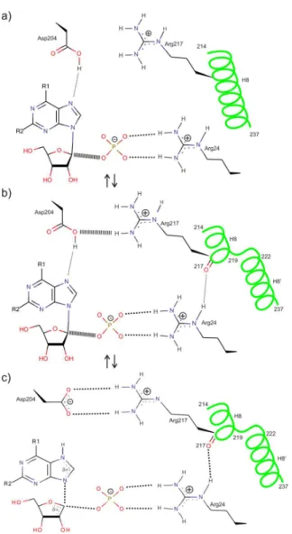
the ligands are bound only loosely. By segmentation of the H8, the entry into the active site pocket partially closes and the ligands are bound more tightly. We suppose that from two substrates, phosphate binds first while the active site is in the open conformation. Phosphate binding stabilizes Arg24 (conserved in all hexameric PNPs) and favours break-ing of H8 into two segments. Before the bindbreak-ing of the sec-ond substrate (nucleoside), catalytically important amino acid Asp204 has to be protonated (Figure 3a).
Catalytic action occurs while the active site is closed. As a consequence of the helix segmentation, two important protein-substrate contacts are established: the H-bond be-tween Arg24 and a main-chain carbonyl oxygen of Arg217 and guanidinium group of Arg217 moves into hydrogen bond distance to Asp204 (Figure 3b). After proton transfer from Asp204 to N7 of purine base, a transition state is formed (Figure 3c).
The two possible conformations of the active sites revealed by X-ray crystallography are in line with solution studies of E. coli PNP which also observe strong and weak binding sites for phosphate and nucleoside inhibitor.
In the crystal structures of E. coli PNP complexed with its ligands[1–14] the following distributions of the closed and open active sites can be found: 3 open + 3 closed,[1] 4 open + 2 closed sites[2–6] and all six open (see for example reference).[6] It is interesting to stress out, that in the case with 3 open + 3 closed active site conformations they alter-nate regularly, while in the case of 4 open and 2 closed ac-tive sites, the closed sites are always next to each other and belong to two different dimers of one homohexamer.
Recently we focused our interest on PNP from path-ogen bacteria Helicobacter pylori (HP). Although HP PNP
has 50 % identity and 70 % similarity with E. coli PNP, it seems that there are significant differences in their enzy-matic activity. In some HP PNP ternary structures we have found unexpected distribution of 5 open and 1 closed ac-tive site conformation (data not published). To the best of our knowledge, this is the first such case among homohex-americ PNP enzymes.
Figure 2. Superimposed open and closed active site conformations in the structure of the E. coli purine nucleo-side phosphorylase in ternary complex (PDB code 1K9S) with phosphate ion and formycin B (FMC).
M. LUIĆ, Z. ŠTEFANIĆ: Can Crystal Symmetry Influence the Active Site Conformation? 199
DOI: 10.5562/cca2872 Croat. Chem. Acta 2016, 89(2), 197–202
In order to facilitate the analysis of the distribution of open and closed active site conformations in ever-grow-ing number of PNP crystal structures available in the Pro-tein data bank (PDB), and to insure that all available structures will be taken into account by such an analysis, a custom made Python script was written, which automated the task of recognizing active site conformations. In addi-tion to that, the script extracts the informaaddi-tion about crys-tal contacts between specified parts of the monomers, in particular those that are in proximity of the active site. In this way we tried to identify whether a correlation between the active site conformation on one side, and crystal symmetry and/or crystal packing factors on the other side exists.
Such an analysis would contribute to better under-standing of the enzyme mechanism(s) of these complicated two-substrate two-product oligomeric proteins, by identi-fying a possible sequence of events in ligands binding to dis-tinct subunits of the homohexameric enzyme. At the same time, such an analysis could help us to reveal if there are limitations imposed by crystal symmetry and/or crystal packing which could mask features of the enzyme function when analysed by X-ray crystallographic methods.
EXPERIMENTAL SECTION
The set of protein structures, which are similar in sequence to the referent strain of H. pylori (strain 26695) purine nu-cleoside phosphorylase (HP PNP), was extracted from PDB by sequence alignment. The parameters for sequence alignment were chosen to yield „Significant“ entries which corresponded to E value of 0.01.[15] This resulted in 172 structures out of which 149 were hexameric proteins and the rest were dimeric and were not included in further analysis.
The resulting 149 PDB structures were analysed with custom-made Python script (https://www.python.org/). The script uses excellent cctbx library[16] to process a PDB file, and automatically identify whether monomers are in open or closed active site conformation. Due to the differ-ence in numbering and different chain lengths in different PNPs, it was not possible to automate the process of de-tecting the open and closed conformations by for instance measuring some distances on certain amino acid numbers. Instead, the segmentation of the helix was followed by parsing a secondary structure record and extracting the in-formation on the helix H8 (Figure 2). Namely, this helix is segmented in two helices in the case of closed active site conformation. If another α-helix was found not further than 5 amino acids away from the beginning of H8 helix than the conformation was assigned as closed.
In addition, this script was used to identify all the crystal contacts in the crystal structure. For the crystal
contacts, all distances between protein atoms (excluding waters) from different monomers shorter than 3.5 Å were counted. More specifically, the script could narrow down this list of contacts taking into account any protein region of interest. This was used to identify crystal contacts in the vicinity of the closing region of the helix H8. In order to see if there is any correlation between closed conformation of the active site and crystal contacts the following analysis was performed. For the 22 structures that contained at least one closed monomer, the number of crystal contacts of every monomer in that structure was calculated, taking into account only the region close to the part of H8 helix that closes. Due to differences in numbering of amino acids, this could not have been done by simply taking some amino acid range. To overcome this, the number of amino acids which marks the beginning of the helix H8 was identified from the secondary structure. Then the range of ten amino acids forward and five amino acids back from that amino acid was taken into account. Taking this amino acid region insured that part of the protein that undergoes conforma-tional change was taken into account.
RESULTS AND DISCUSSION
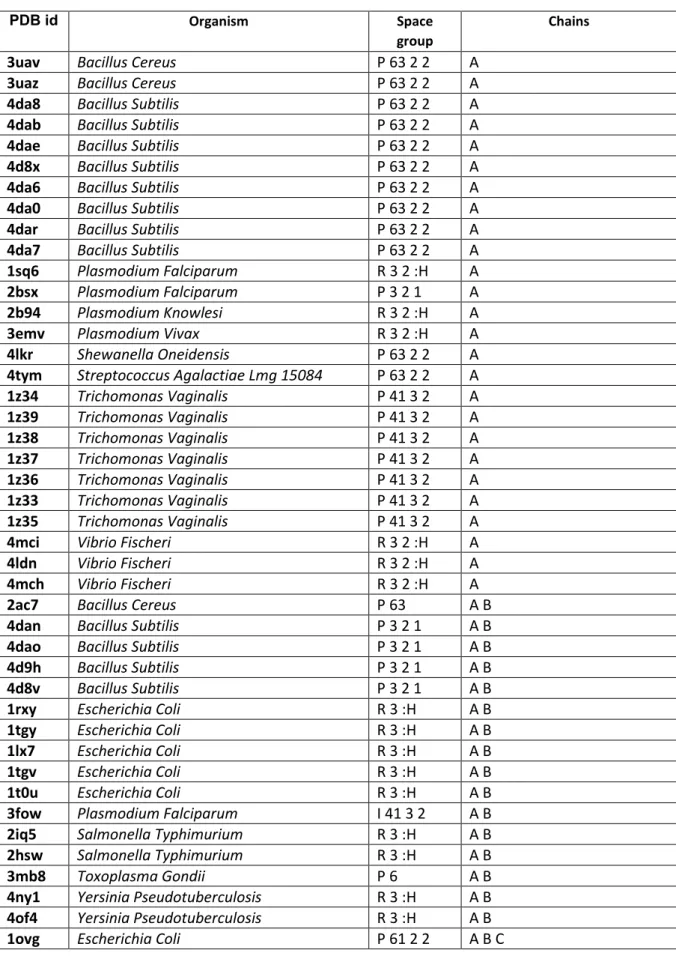
The 149 structures retrieved from Protein data bank (PDB) by sequence alignment of HP PNP referent strain (26695) originated from 23 different bacteria. In 127 bacterial PNPs our custom made script identified no active sites in closed conformation (Supplementary materials, Table S1) while in 22 of them (from 7 different bacteria) the closed confor-mation of the active site was detected (Table 1).
The distribution of the open and closed active site conformations is as follows:
a) The most common distribution between open and closed active site conformations is 4+2, respectively (Table 1) and it occurs in 13 cases. It is interesting to mention that in all of them two closed sites are located close to each other and that they belong to two differ-ent dimers forming the same homohexamer. This ar-rangement is realized in four different space groups (P6122, P212121, P62 and P21) indicating that crystal packing probably does not influence such a distribu-tion. Also, this arrangement is found almost exclu-sively in E. coli with only one exception the PNP structure from T. vaginalis.
200 M. LUIĆ, Z. ŠTEFANIĆ: Can Crystal Symmetry Influence the Active Site Conformation?
Croat. Chem. Acta 2016, 89(2), 197–202 DOI: 10.5562/cca2872
that, comparing with E. coli PNP, have more closed the critical part of the active site (residues 207–217). The remainig two structures (PDB codes 3OCC and 3OF3) are the result of Protein structure initiative and are not published yet.
c) In three crystal structures the distribution between closed and open active sites is 3+3. One is of the ter-nary complex of homohexameric PNP from Echerichia coli with formycin A derivatives and phosphate or sul-phate ions.[1] This is a very special case of PNP's
ternary complex. 6-methyl formycin A is the best know inhibitor of E. coli PNP with Ki = 0.3 µM at neutral pH. In aqueous solution this compound undergoes rear-rangement to four different formycin derivatives. The main product of hydrolysis is N7-methlyformycin A. Detailed inspection of the active sites of the homohex-amer present in the asymmetric unit revealed unam-biguously that 6-methylformycin was located in three monomers (closed conformation) and in other three N7-methylformycin (open conformations). Open and closed conformations in the homohexamer alternate
Table 1. The structures of purine nucleoside phosphorylases from the Protein Data Bank that contain at least one active site in closed conformation. Structures can be classified in three groups: 4+2, 0+6 and 3+3
PDB code Organism Space group (closed marked with *) Chains Open +closed In active site 1PK9 Escherichia coli P6122(a) A B C* 4+2 2FA(b)+PO4
1PR1 Escherichia coli P6122 A B C* 4+2 FMB+PO4
1PR5 Escherichia coli P6122 A B C* 4+2 TBN+PO4
2I4T Trichomonas vaginalis P62 A B* C 4+2 UA2+PO4
1A69 Escherichia coli P6122 A* B C 4+2 FMB+SO4
3UT6 Escherichia coli P6122 A* B C 4+2 FMC+2PO4
4TS9 Escherichia coli P6122 A* B C 4+2 FMC+PO4
3OOE Escherichia coli P212121 A* B C D E F* 4+2 PO4
4TS3 Escherichia coli P212121 A* B C D E F* 4+2 FMC+PO4 in closed PO4 in open
4TTA Escherichia coli P212121 A* B C D E F* 4+2
SO4 in closed FMC+PO4 in 2 open PO4 in other 2 open
4TTI Escherichia coli P212121 A* B C D E F* 4+2
FMC+PO4 in closed FMC+PO4 in 2 open PO4 in other 2 open
4TTJ Escherichia coli P6122 A* B D 4+2 FMC+PO4
3OOH Escherichia coli P21 A* B C D* E F* G* H I J K L* M* N O P Q R* (4+2)x3 PO4
3UAW Bacillus cereus P6322 A* 0+6 ADN+SO4
3UAX Bacillus cereus P6322 A* 0+6 NOS+SO4
3UAY Bacillus cereus P6322 A* 0+6 ADN+SO4
3UAZ Bacillus cereus P6322 A* 0+6 ADN+SO4
3OCC pseudotuberculosis Yersinia P212121 A* B* C* D* E* F* 0+6 DIH+PO4
3OF3 Vibrio cholerae P212121 A* B* C* D* E* F* G* H* I*
J* K* L* (0+6)x2 DIH+PO4
4D98 Bacillus subtilis R32 A B* 3+3 SO4
4M7W Leptotrichia buccalis P32 A B* C (3+3)+(6+0) PO4
1K9S Escherichia coli P41212 A* B* C* D E F 3+3 FM2+PO4 (a) In the space groups P6122, P62 and P32 one half of the hexamer is present in the asymmetric unit. In the space group R32 two monomers and in the P6322
M. LUIĆ, Z. ŠTEFANIĆ: Can Crystal Symmetry Influence the Active Site Conformation? 201
DOI: 10.5562/cca2872 Croat. Chem. Acta 2016, 89(2), 197–202
regularly. In all six active sites one phosphate or sul-phate ion (it is not possible to distinguish between them in the electron density maps calculated at the given resolution) is bound. Such a distribution was in agreement with half-the-sites binding for 6-methyl-formycin determined in solution.[1] The same alternat-ing arrangement is found in the crystal structure of
Bacillus subtilis (PDB id 4D98), the only structure with one dimer in the asymmetric unit.[17] One other very interesting case in this group is the structure of PNP from Leptotrichia buccalis (PDB code 4M7W). Namely, in the crystal packing of this protein there is an equal number of hexamers with 3+3 and 6+0 arrangement.
There is a substantial variability of the space groups found among the members belonging to the same distribu-tion of active sites (for example, 4+2 distribudistribu-tion is found in four different space groups, 0+6 distribution is found in two space groups and one of the space groups has two varia-tions). Furthermore, the same space group alone does not impose similar packing and therefore similar crystal con-tacts. For this to be true unit cell axis need to be similar too.
Therefore, the appearance of the same open and closed site distribution (such as 4+2) in different crystal symme-tries implies, that this distribution is not influenced by crys-tal packing. Although the number of structures with closed active sites is arguably not high enough to make good sta-tistics, this set of structures does not display any regularity which would indicate any correlation between open and closed active site conformations and crystal-lographic symmetry.
Very recently we have determined several crystal structures of PNP from the pathogen bacteria H. pylori
(data not published). To our surprise, distribution with five open and one closed conformation was found, which is not present in any structure available in PDB.
The results of detailed analysis of crystal contacts in the crystal structures which contained at least one active site in closed conformation (Table 1) are summarized in Figure 4. It turns out that active sites in closed conformation have, in most of the cases less crystal contacts than those in open conformation. There are quite a few open active sites that have from 15–21 crystal contacts in this region,
Figure 4. This figure displays the number of crystal contacts that particular monomers have in proximity of the active site in 22 PNP structures that have at least one active site in closed conformation. Each bordered cell in the table contains the data for one PDB code given in the top shaded area. Letters indicate the chain identifiers. In each bordered cell the two left columns represent results for the monomers (if any) in open and two right columns for the monomers in closed conformation. The red bars are graphical help to compare values accross the cells.
B 1 A 9 A 0 B 21 A 4 B 0 A 0
C 2 D 1 B 0 C 0 F 4 C 0 F 1
E 3 F 3 C 0 D 0 D 0
H 1 G 0 D 1 E 0 E 0
I 1 L 1 E 0
J 0 M 0 F 1 A 1 B 5 A 0 C 0
K 0 R 9 G 0 C 0 B 20
N 4 H 0
O 3 I 1 B 6 A 8 A 0 C 0
P 3 J 1 C 0 F 4 B 18
Q 7 K 0 D 0
L 1 E 10 A 14 B 7
B 15 A 8
C 0 F 3 B 0 A 0 D 3 A 2 A 1
D 0 C 19 E 0 B 2
E 6 F 0 C 1 B 0 A 0
B 0 A 0 C 17
B 7 A 5 D 14 A 1
C 3 F 4 B 0 A 0
D 0 A 2 B 0 A 0 C 4 C 16
E 1 C 6 B 13
A 8 A 3
4ttj 3uax 4ts3 3uaz 4ts9 4d98 3uaw 3ut6
4m7w 1pr5 1a69
2i4t 1pk9 1pr1 4tta 1k9s 3uay 3of3 4tti 3ooe 3ooe
open closed open closed open closed
open closed
202 M. LUIĆ, Z. ŠTEFANIĆ: Can Crystal Symmetry Influence the Active Site Conformation?
Croat. Chem. Acta 2016, 89(2), 197–202 DOI: 10.5562/cca2872
while the number of contacts in closed active sites is never higher than 9. This could perhaps be explained by the fact that in open active sites helix H8 is slightly more distant from the central region, and therefore more available for crystal contacts. In any case, it does not support our first hy-pothesis that closing of the active sites could be influenced by neighbouring homohexamers in the crystal packing (somehow pressing them and thereby forcing them to close). Therefore, based on our analyses presented here, the straightforward influence of the crystal symmetry and/or crystal contacts on the conformation of the active sites in homohexameric purine nucleoside phosphorylases cannot be inferred.
Acknowledgment. This work has been fully supported by the Croatian Science Foundation under the project number 7423.
Supplementary Information. Supporting information to the paper is enclosed to the electronic version of the article at: http://dx.doi.org/10.5562/cca2872.
REFERENCES
[1] G. Koellner, A. Bzowska, B. Wielgus-Kutrowska, M. Luić, T. Steiner, W. Saenger, J. Stępiński, J. Mol. Biol. 2002, 315, 351.
[2] G. Koellner, M. Luić, D. Shugar, W. Saenger, A. Bzowska, J. Mol. Biol.1998, 280, 153.
[3] E. M. Bennett, C. Li, P. W. Allan, W. B. Parker, S. E. Ea-lick, J. Biol. Chem.2003, 278, 47110.
[4] G. Mikleušević, Z. Štefanić, M. Narczyk, B. Wielgus-Kutrowska, A. Bzowska, M. Luić, Biochimie2011,
93, 1610.
[5] Z. Štefanić, M. Narczyk, G. Mikleušević, B. Wielgus-Kutrowska, A. Bzowska, M. Luić, FEBS Letters 2012,
586, 967.
[6] Z. Štefanić, G. Mikleušević, M. Narczyk, B. Wielgus-Kutrowska, A. Bzowska, M. Luić, Croat. Chem. Acta 2013, 86, 117.
[7] D. Paul, S. E. O'Leary, K. Rajashankar, W. Bu, A. Toms, E. C. Settembre., J. M. Sanders, T. P. Begle, S. E. Ealick, Biochemistry2010, 49, 3499.
[8] W. Bu, E. C. Settembre, M. H. el Kouni, S. E. Ealick, Acta Crystallogr., Sect. D: Biol. Crystallogr.2005, 61, 863. [9] T. T. Caradoc-Davies, S. M. Cutfield, I. L. Lamont, J. F.
Cutfield, J. Mol. Biol.2004, 337, 337.
[10] E. M. Bennett, R. Anand, P. W. Allan, A. E. Hassan, J. S. Hong, D. N. Levasseur, D. T. McPherson, W. B. Parker, J. A. Secrist, E. J. Sorscher, T. M. Townes, W. R. Waud, S. E. Ealick, Chem. Biol.2003, 10, 1173. [11] F. T. Burling, R. Kniewel, J. A. Buglino, T. Chadha, A.
Beckwith, C. D. Lima, Acta Crystallogr., Sect. D: Biol. Crystallogr.2003, 59, 73.
[12] C. Mao, W. J. Cook, M. Zhou, G. W. Koszalka, T. A. Krenitsky, S. E. Ealick, Structure 1997, 5, 1373. [13] E. Y. Morgunova, A. M. Mikhailov, A. N. Popov, E. V.
Blagova, E. A. Smirnova, B. K. Vainshtein, C. Mao, Sh. R. Armstrong, S. E. Ealick, A. A. Komissarov, FEBS Lett.1995, 367, 183.
[14] S. F. Altschul, W. Gish, W. Miller, E. W. Myers, D. J. Lipman, J. Mol. Biol.1990, 215, 403.
[15] R. W. Grosse-Kunstleve, N. K. Sauter, N. W. Moriarty, P. D. Adams, J. Appl. Cryst.2002, 35, 126.
Supplementary materials
Table S1. The structures of purine nucleoside phosphorylases that contain no closed chains in the
Protein Data Bank.
PDB id Organism Space
group Chains
3uav
Bacillus Cereus
P 63 2 2
A
3uaz
Bacillus Cereus
P 63 2 2
A
4da8
Bacillus Subtilis
P 63 2 2
A
4dab
Bacillus Subtilis
P 63 2 2
A
4dae
Bacillus Subtilis
P 63 2 2
A
4d8x
Bacillus Subtilis
P 63 2 2
A
4da6
Bacillus Subtilis
P 63 2 2
A
4da0
Bacillus Subtilis
P 63 2 2
A
4dar
Bacillus Subtilis
P 63 2 2
A
4da7
Bacillus Subtilis
P 63 2 2
A
1sq6
Plasmodium Falciparum
R 3 2 :H
A
2bsx
Plasmodium Falciparum
P 3 2 1
A
2b94
Plasmodium Knowlesi
R 3 2 :H
A
3emv
Plasmodium Vivax
R 3 2 :H
A
4lkr
Shewanella Oneidensis
P 63 2 2
A
4tym
Streptococcus Agalactiae Lmg 15084
P 63 2 2
A
1z34
Trichomonas Vaginalis
P 41 3 2
A
1z39
Trichomonas Vaginalis
P 41 3 2
A
1z38
Trichomonas Vaginalis
P 41 3 2
A
1z37
Trichomonas Vaginalis
P 41 3 2
A
1z36
Trichomonas Vaginalis
P 41 3 2
A
1z33
Trichomonas Vaginalis
P 41 3 2
A
1z35
Trichomonas Vaginalis
P 41 3 2
A
4mci
Vibrio Fischeri
R 3 2 :H
A
4ldn
Vibrio Fischeri
R 3 2 :H
A
4mch
Vibrio Fischeri
R 3 2 :H
A
2ac7
Bacillus Cereus
P 63
A B
4dan
Bacillus Subtilis
P 3 2 1
A B
4dao
Bacillus Subtilis
P 3 2 1
A B
4d9h
Bacillus Subtilis
P 3 2 1
A B
4d8v
Bacillus Subtilis
P 3 2 1
A B
1rxy
Escherichia Coli
R 3 :H
A B
1tgy
Escherichia Coli
R 3 :H
A B
1lx7
Escherichia Coli
R 3 :H
A B
1tgv
Escherichia Coli
R 3 :H
A B
1t0u
Escherichia Coli
R 3 :H
A B
3fow
Plasmodium Falciparum
I 41 3 2
A B
2iq5
Salmonella Typhimurium
R 3 :H
A B
2hsw
Salmonella Typhimurium
R 3 :H
A B
3mb8
Toxoplasma Gondii
P 6
A B
4ny1
Yersinia Pseudotuberculosis
R 3 :H
A B
4of4
Yersinia Pseudotuberculosis
R 3 :H
A B
1oum
Escherichia Coli
P 61 2 2
A B C
3onv
Escherichia Coli
P 61 2 2
A B C
1otx
Escherichia Coli
P 61 2 2
A B C
1ov6
Escherichia Coli
P 61 2 2
A B C
1oty
Escherichia Coli
P 61 2 2
A B C
1ou4
Escherichia Coli
P 61 2 2
A B C
1pk7
Escherichia Coli, Escherichia Coli O157:H7
P 61 2 2
A B C
1pw7
Escherichia Coli, Escherichia Coli O157:H7
P 61 2 2
A B C
1pr0
Escherichia Coli, Escherichia Coli O157:H7
P 61 2 2
A B C
1pr6
Escherichia Coli, Escherichia Coli O157:H7
P 61 2 2
A B C
1pr2
Escherichia Coli, Escherichia Coli O157:H7
P 61 2 2
A B C
1pr4
Escherichia Coli, Escherichia Coli O157:H7
P 61 2 2
A B C
1pke
Escherichia Coli, Escherichia Coli O157:H7
P 61 2 2
A B C
4mar
Meiothermus Ruber
C 2 2 21
A B C
4m3n
Meiothermus Ruber Dsm 1279
C 2 2 21
A B C
1je0
Sulfolobus Solfataricus
C 2 2 21
A B C
1jdt
Sulfolobus Solfataricus
C 2 2 21
A B C
1jdu
Sulfolobus Solfataricus
C 2 2 21
A B C
1jdz
Sulfolobus Solfataricus
C 2 2 21
A B C
1jp7
Sulfolobus Solfataricus
C 2 2 21
A B C
1jpv
Sulfolobus Solfataricus
C 2 2 21
A B C
1k3f
Escherichia Coli
P 1 21 1
A B C D E F
4ogk
Salmonella Enterica Subsp. Enterica
Serovar Typhimurium
P 61
A B C D E F
2hrd
Salmonella Typhimurium
P 21 21 21 A B C D E F
2i8a
Salmonella Typhimurium
P 21 21 21 A B C D E F
1sj9
Salmonella Typhimurium
P 61
A B C D E F
2rj3
Salmonella Typhimurium
P 21 21 21 A B C D E F
4oeh
Vibrio Cholerae O1 Biovar El Tor
P 1
A B C D E F
4r31
Actinobacillus Succinogenes
P 21 21 21 A B C D E F
1xe3
Bacillus Anthracis
P 21 21 21 A B C D E F
4d8y
Bacillus Subtilis
P 21 21 21 A B C D E F
1u1c
Escherichia Coli
P 21 21 21 A B C D E F
1ecp
Escherichia Coli
P 1 21 1
A B C D E F
1u1f
Escherichia Coli
P 21 21 21 A B C D E F
1u1e
Escherichia Coli
P 21 21 21 A B C D E F
3kvv
Escherichia Coli
P 32
A B C D E F
4rj2
Escherichia Coli
P 1 21 1
A B C D E F
1u1g
Escherichia Coli
P 21 21 21 A B C D E F
1u1d
Escherichia Coli
P 21 21 21 A B C D E F
3enz
Plasmodium Falciparum
I 41 2 2
A B C D E F
1q1g
Plasmodium Falciparum
P 21 21 21 A B C D E F
3phc
Plasmodium Falciparum
P 1
A B C D E F
1y1q
Salmonella Typhimurium
P 21 21 21 A B C D E F
3fwp
Salmonella Typhimurium
P 21 21 21 A B C D E F
2oec
Salmonella Typhimurium
P 21 21 21 A B C D E F
2pga
Salmonella Typhimurium
P 21 21 21 A B C D E F
1zl2
Salmonella Typhimurium
P 21 21 21 A B C D E F
2hwu
Salmonella Typhimurium
P 21 21 21 A B C D E F
2qdk
Salmonella Typhimurium
P 21 21 21 A B C D E F
3c74
Salmonella Typhimurium
P 21 21 21 A B C D E F
1y1r
Salmonella Typhimurium
P 21 21 21 A B C D E F
2hn9
Salmonella Typhimurium
P 21 21 21 A B C D E F
1y1s
Salmonella Typhimurium Lt2
P 21 21 21 A B C D E F
4r2x
Shewanella Oneidensis
P 1 21 1
A B C D E F
4yjk
Shewanella Oneidensis (Strain Mr-1)
P 1 21 1
A B C D E F
4r2w
Shewanella Oneidensis Mr-1
P 1 21 1
A B C D E F
1jds
Sulfolobus Solfataricus
P 1 21 1
A B C D E F
1je1
Sulfolobus Solfataricus
P 1 21 1
A B C D E F
1jdv
Sulfolobus Solfataricus
P 1 21 1
A B C D E F
2isc
Trichomonas Vaginalis
P 21 21 21 A B C D E F
4u2k
Vibrio Cholerae
P 31
A B C D E F
4h1t
Vibrio Cholerae
P 1
A B C D E F
4g8j
Vibrio Cholerae
P 1 21 1
A B C D E F
4k6o
Vibrio Cholerae
P 1 21 1
A B C D E F
4lzw
Vibrio Cholerae
P 1 21 1
A B C D E F
4ip0
Vibrio Cholerae
P 1
A B C D E F
1vhj
Vibrio Cholerae
P 1 21 1
A B C D E F
1vhw
Vibrio Cholerae
P 1 21 1
A B C D E F
4ogl
Vibrio Cholerae O1 Biovar El Tor
P 1 21 1
A B C D E F
4jp5
Yersinia Pestis
P 32
A B C D E F
4i2v
Yersinia Pseudotuberculosis
P 3
A B C D E F
4e1v
Salmonella Enterica Subsp. Enterica
Serovar Typhimurium
C 1 2 1
A B C D E F G H I
1rxc
Escherichia Coli
P 1 21 1
A B C D E F G H I J K L
3opv
Escherichia Coli
C 1 2 1
A B C D E F G H I J K L
1rxu
Escherichia Coli
P 1 21 1
A B C D E F G H I J K L M N O P
Q R
3qpb
Streptococcus Pyogenes Serotype M6
P 1
A B C D E F G H I J K L M N O P
Q R
1rxs
Escherichia Coli
P 1 21 1
A B C D E F G H I J K L M N O P
Q R a b c d e h i j k l m o
2oxf
Salmonella Typhimurium
R 3 :H
A F
3dps
Salmonella Typhimurium
R 3 :H
A F
1y1t
Salmonella Typhimurium Lt2
R 3 :H
A F
1odk
Thermus Thermophilus
P 43 21 2
A? B? C? D? E? F?
1odi
Thermus Thermophilus
P 43 21 2
A? B? C? D? E? F?
1odl
Thermus Thermophilus
P 43 21 2
A? B? C? D? E? F?
Python script that was used in crystal contacts calculations
#!/bin/env cctbx.python_dev-546 import sys
import pickle import iotbx.pdb import glob
from mmtbx.lattice import *
from scitbx.array_family import flex
organisms = pickle.load(open('organisms.pkl','rb'))
def open_or_closed(f):
inpdb = iotbx.pdb.input(f) h1 = inpdb.construct_hierarchy() chains = [c.id for c in h1.chains()] res = ""
for chain in sorted(set(chains)):
res += chain_open_closed(inpdb,h1,chain) + " " return res
def open_and_closed_chains(f): inpdb = iotbx.pdb.input(f) h1 = inpdb.construct_hierarchy() chains = [c.id for c in h1.chains()] opn, clsd= [],[]
for chain in sorted(set(chains)):
if '*' in chain_open_closed(inpdb,h1,chain): clsd.append(chain)
else:
opn.append(chain) return opn, clsd
def average_chain_length(f): from numpy import array inpdb = iotbx.pdb.input(f) h1 = inpdb.construct_hierarchy() chains = list(h1.chains())
l = [len(chain.as_sequence()) for chain in chains] l = array(l)
return int(l[l>5].mean())
def chain_open_closed(inpdb,h,chain):
"""This tries to extract the secondary structure and see if the helix is open or closed""" try:
secstr = inpdb.extract_secondary_structure() except:
return chain + '?'
h8 = filter(lambda h: h.start_chain_id == chain, secstr.helices)[-1] h7 = filter(lambda h: h.start_chain_id == chain, secstr.helices)[-2] start_h8 = h8.get_start_resseq_as_int()
end_h7 = h7.get_end_resseq_as_int() if start_h8 - end_h7 < 5:
oc = "*" else: oc = "" return chain+oc
def get_resseq(f,chain):
"""Calculates resseq around helix is open or closed""" return 210,225
inpdb = iotbx.pdb.input(f) try:
return 210,225
h8 = filter(lambda h: h.start_chain_id == chain, secstr.helices)[-1] start_h8 = h8.get_start_resseq_as_int()
return start_h8 - 10, start_h8 + 5
def crystal_contacts_from_selection(pdb,selection="all",distance_cutoff=3.5): inpdb = iotbx.pdb.input(pdb)
h = inpdb.construct_hierarchy() x = inpdb.xray_structure_simple() #x.show_summary()
atoms = list(h.atoms_with_labels())
sc = h.atom_selection_cache() selected = sc.iselection(selection)
sites = x.sites_frac() unit_cell = x.unit_cell()
pat = x.pair_asu_table(distance_cutoff) pst = pat.extract_pair_sym_table() contacts = []
selected_sites = list(selected)
pairs = {}
for i, psd in enumerate(pst): for j in psd.keys(): pairs[(i,j)] = psd[j]
selected_pairs = sorted(filter(lambda p: p[0] in selected_sites or p[1] in selected_sites, pairs.keys()))
for pair in selected_pairs: # (i_seq, j_seq) i_seq, j_seq = pair
site_i = sites[i_seq] atom_i = atoms[i_seq]
chain_i = atom_i.parent().parent().parent() res_i = atom_i.parent()
site_j = sites[j_seq] atom_j = atoms[j_seq]
chain_j = atom_j.parent().parent().parent() res_j = atom_j.parent()
sym_op = pairs[pair][0]
if res_j.resname == "HOH" or res_i.resname == "HOH" : continue
if not sym_op.is_unit_mx(): site_ji = sym_op * site_j
distance = unit_cell.distance(site_i, site_ji) contacts.append((i_seq, j_seq, sym_op, distance))
#print "%s %s %-30s %8.4f" % (res_i.id_str(), res_j.id_str(), sym_op, distance) #print "%s %s %-30s %8.4f" % (chain_i.id, chain_j.id, sym_op, distance)
#print 'Number of contacts: %d' % len(contacts) return contacts
def get_chains(pdb):
h = iotbx.pdb.input(pdb).construct_hierarchy() return set(sorted([c.id for c in list(h.chains())]))
def get_xray_structures(): pdbs = glob.glob('*.pdb') xray_structures = {} for pdb in pdbs:
xray_structures[pdb] = iotbx.pdb.input(pdb).xray_structure_simple() return xray_structures
oc = open_and_closed_chains(f) print f
print 'Open' for c in oc[0]:
start,stop = get_resseq(f,c)
ncont = crystal_contacts_from_selection(f,'chain %s and resseq %d:%d' %(c,start,stop)) print c, len(ncont)
print 'Closed' for c in oc[1]:
start,stop = get_resseq(f,c)
ncont = crystal_contacts_from_selection(f,'chain %s and resseq %d:%d' %(c,start,stop)) print c, len(ncont)
print '-'*80
def print_table():
xray_structures = get_xray_structures() def similars(x):
res = []
for k, v in xray_structures.items(): if x.is_similar_symmetry(v): res.append(k)
return res
for k, v in xray_structures.items():
#print '%s\t%-50s\t%-20s\t%-20s\t%20s%5s\t%s' %(k, v.unit_cell(), v.space_group().info(),
v.space_group().crystal_system(),open_or_closed(k),average_chain_length(k),similars(v)) #print '%s\t%-50s\t%-10s\t%30s\t%s' %(k[:4], organisms[k.upper()[:4]],
v.space_group().info(),open_or_closed(k),similars(v))
print '%s\t%-50s\t%-10s\t%-30s\t%5.2f' %(k[:4], organisms[k.upper()[:4]],
v.space_group().info(),open_or_closed(k),len(crystal_contacts_from_selection(k))/len(get_chain s(k)))
def run1():
f = sys.argv[1] print f
selection = sys.argv[2]
contacts = crystal_contacts_from_selection(f,selection) if not contacts:
print 'No contacts' print open_or_closed(f) print "-"*80
if __name__ == "__main__": run1()