(Annals of the Brazilian Academy of Sciences)
Printed version ISSN 0001-3765 / Online version ISSN 1678-2690 www.scielo.br/aabc
Purification and identification of metabolites produced by
Bacillus cereus
and
B. subtilis
active against
Meloidogyne exigua
, and their
in silico
interaction
with a putative phosphoribosyltransferase from
M. incognita
DENILSON F. OLIVEIRA1,HELVÉCIO M. DOS SANTOS JÚNIOR1, ALEXANDRO S. NUNES1, VICENTE P. CAMPOS2, RENATA S.C. DE PINHO2 and GIOVANNA C. GAJO1
1Universidade Federal de Lavras, Departamento de Química, Caixa Postal, 3037, 37200-000 Lavras, MG, Brasil 2
Universidade Federal de Lavras, Departamento de Fitopatologia, Caixa Postal 3037, 37200-000 Lavras, MG, Brasil
Manuscript received on August 16, 2012; accepted for publication on May 20, 2013
ABSTRACT
To contribute to the development of products to control Meloidogyne exigua, the bacteria Bacillus cereus and B. subtilis were cultivated in liquid medium to produce metabolites active against this plant-parasitic nematode. Fractionation of the crude dichloromethane extracts obtained from the cultures afforded uracil, 9H-purine and dihydrouracil. All compounds were active against M. exigua, the latter being the most efficient. This substance presented a LC50 of 204 µg/mL against the nematode, while a LC50 of 260 µg/mL was observed for the commercial nematicide carbofuran. A search for protein-ligand complexes in which the ligands were structurally similar to dihydrouracil resulted in the selection of phosphoribosyltransferases, the sequences of which were used in an in silico search in the genome of M. incognita for a similar sequence of amino acids. The resulting sequence was modelled and dihydrouracil and 9H-purine were inserted in the active site of this putative phosphoribosyltransferase resulting in protein-ligand complexes that underwent molecular dynamics simulations. Calculation of the binding free-energies of these complexes revealed that the dissociation constant of dihydrouracil and 9H-purine to this protein is around 8.3 x 10⁻⁷
and 1.6 x 10⁻⁶ M, respectively. Consequently, these substances and the putative phosphoribosyltransferase are promising for the development of new products to control M. exigua.
Key words: 9H-purine, dihydrouracil, molecular modelling, nematicidal activity, root-knot nematode, uracil.
Correspondence to: Denilson Ferreira E-mail: [email protected]
INTRODUCTION
Plant-parasitic nematodes are a constant source of
problems for Brazilian farmers due to the negative
impact these animals cause to the production of
several crops in Brazil (Dias-Arieira et al. 2010).
For example, the production of coffee is severely
affected by species of Meloidogyne (Campos
and Villain 2005). In the State of Minas Gerais,
where ~50% of this commodity in Brazil is
produced (Ministério da Agricultura, Pecuária e
Abastecimento - Mapa 2012), Meloidogyne exigua
Goeldi occurs in 22% of the coffee farms in the
southern region of this state (Castro et al. 2008).
Although exclusion methods and plant resistance
to nematodes should be emphasised, synthetic
nematicide application is the most often used method
to control M. exigua on coffee farms (Campos and
Silva 2008), which increases production costs and
contaminates humans and the environment with
harmful substances (Chitwood 2002). A possible
alternative to circumvent such problems comprises
the use of rhizobacteria, which colonise plant roots
and can reduce the population of plant-parasitic
nematodes. For example,
Pseudomonas fluorescens
(Flügge) Migula and Pseudomonas putida Trevisan
can reduce the population of Meloidogyne
spp.
and
Radopholus similis (Cobb) Thorne in tomato
and banana plants (Aalten et al. 1998). The use of
a combination of Bacillus thuringiensis Berliner
and
Lysinibacillus sphaericus (Meyer and Neide)
Ahmed et al. to control plant-parasitic nematodes
was protected by a patent (B'Chir 2000).
One of the mechanisms by which
rhizo-bacteria can act against nematodes consists of the
production of nematicidal substances, for example
the production of 2,4-diacetylphloroglucinol
by
P. fluorescens
that can control Globodera
rostochiensis (Wollenweber) Behrens in potato
plants (Cronin et al. 1997). Also worthy of mention
is the ability of Bacillus amyloliquefaciens
(Fukumoto) Priest et al.
to produce a cyclic
peptide active against nematodes. The use of this
substance as a nematicide was protected by a patent
some years ago (Bendzko et al. 1998). Thus, in
order to select rhizobacteria for the development
of new products to control plant-parasitic
nematodes and to elucidate their mechanism of
action, in a previous work several rhizobacterial
strains were screened for the ability to produce
substances active against M. exigua (Oliveira et
al. 2007). Then, the most active crude metabolites
underwent a fractionation process monitored
by
in vitro assays with M. exigua second-stage
juveniles (J2), which resulted in the identification
of common amino acids (Oliveira et al. 2009) that
were probably formed in the culture medium by
hydrolysis of proteins or peptides that may be used
as nutrients by rhizobacteria. Although the low
nematicidal properties of amino acids preclude
such substances from being commercially used
to control M. exigua
, they are sufficiently active
to cause false positive results in a screening
program aimed to detect bacterial metabolites
with nematicidal activities. Consequently, in a
preliminary work they were removed from crude
bacterial metabolites by solvent extraction before
the screening process. To continue such work, the
most active amino-acid-free crude metabolites,
produced by Bacillus subtilis Cohn and Bacillus
cereus Frankland and Frankland, underwent
fractionations in the present study to isolate
and identify those substances with nematicidal
properties against M. exigua. Furthermore, these
compounds were also submitted to a computational
study to elucidate their mode of action.
MATERIALS AND METHODS
BACTERIA
PRODUCTION OF BACTERIAL METABOLITES
Each bacterium was grown in 2.5 L of tryptic
soy broth (TSB, Merck) for seven days, at 28°C,
under constant stirring (100 rpm), in the dark. After
bacterial cell removal by centrifugation (10,000 g,
15 min), the supernatant liquids were freeze-dried
and washed with dichloromethane (4 x 1.0 L).
The resulting dichloromethane-soluble fractions
were combined and concentrated to dryness in a
rotary evaporator to afford the crude extracts.
An aliquot (0.5%) of each extract was dissolved in
2.0 mL of an aqueous 0.01 g/mL Tween 80 solution
to be tested in vitro for activity against M. exigua J2.
IN VITROASSAY
The test was performed as described by Amaral et
al. (2002). Briefly,
M. exigua eggs were extracted
from coffee (Coffea arabica L.) roots infected with
the nematode in accordance with the Hussey and
Barker (1973) technique, modified by Boneti and
Ferraz (1981). J2 were hatched from the eggs and
collected to be employed in the in vitro assays.
Only less than two-day old J2 were used. Into each
300 μL well of a 96-well polypropylene plate, 20 μL
aqueous suspension containing approximately 25 J2
and 100 μL of samples dissolved in aqueous 0.01 g/
mL Tween 80 solution, was poured. To evaluate the
crude extracts, 30 μL of 3.0 mg/mL Pentabiótico
(a mixture of antibacterial substances produced by
Fort Dodge, Brazil) suspension was also poured
into each well to prevent bacterial growth. After
48 h at 28°C, one drop of an aqueous 1.0 M
NaOH solution was added and J2 which changed
their body shape from straight to curled or
hook-shaped within 3 min were considered to be alive,
whereas the nematodes not responding to the
addition of NaOH were considered dead. This
experiment was performed with four replicates
per treatment, employing aqueous 0.01 g/mL
Tween 80 solution and aldicarb
[(2-methyl-2-(methylthio)propanal
O-(N-methylcarbamoyl)
oxime] (50 μg/mL) as negative and positive
controls, respectively. For the aldicarb solution
preparation, 8.6 g of Temik 150 (150 g of aldicarb/
kg), from Rhône-Poulenc AgroBrasil Ltda, was
suspended in water and filtered through filter paper.
The resulting solution was diluted with water to the
desired concentration. All values of dead J2 were
converted to percentage before analysis of variance
(ANOVA) and means separation according to the
Scott and Knott (1974) test (P
≤ 0.05), which were
performed using the SISVAR 5.1 software (Sistema
para Análises Estatísticas, UFLA, Lavras, 2006).
FRACTIONATION OF BACTERIAL EXTRACTS
Dichloromethane-soluble crude extracts, obtained
as described above, were successively eluted
through a silica gel column (3 x 15 cm; 40-63 µm,
Merck) with hexane (400 mL), hexane/ethyl acetate
(1:1, 400 mL), ethyl acetate (400 mL), methanol
(800 mL), distilled water (1600 mL) and 0.1 M
hydrochloric acid (1600 mL). All resulting fractions
were concentrated in a rotary evaporator and
freeze-dried. The analytical and semi-preparative
high performance liquid chromatography (HPLC)
analyses and fractionations were carried out on a
Shimadzu instrument (Tokyo, Japan) equipped with
a photodiode array detector, model SPD-M20A, set
at 190-400 nm; pumps LC-6AD; injector Reodyne
7725i or auto-injector SIL-10AF; degasser DGU –
20A
3; and CBM-20A (SCL-10Avp) interface. Data
were processed on LC-SOLUTION 1.21 software
(Shimadzu). Only those fractions eluted with
methanol were submitted to HPLC analyses with a
Gemini Si-C18 column (4.6 mm x 250 mm x 5 µm,
Phenomenex). A gradient elution at a flow rate of
1.0 mL/min was carried out employing water (10
min), water/methanol (0 to 100% methanol, 10-40
min) and methanol (40-50 min), as mobile phases.
(0-100%, 10-40 min) and methanol (40-50 min)
as mobile phases, at a flow rate of 15.0 mL/min.
All eluents contained 0.1% (v/v) acetic acid. All
fractions were concentrated in a rotary evaporator,
freeze-dried and analysed by HPLC. Only fraction
2 (8.5–9.1 min; uracil: 3.3 mg) was submitted to the
identification process.
Similarly, the methanol-eluted fraction of B.
subtilis underwent fractionation by HPLC to afford
seven new fractions, among which those numbered
1 (4.6–8.9 min) and 3 (16.5–19.2 min) were
further purified by a new elution through the
semi-preparative Gemini column with an aqueous 0.1%
(v/v) acetic acid solution at flow rates of 2.3 mL/min
and 13.0 mL/min, respectively. After concentration
to dryness in a rotary evaporator and freeze-drying,
dihydrouracil (51.4–54.8 min; 19.1mg) and uracil
(57.1–60.2 min; 1.2 mg) were obtained as white
solids from fraction 1, while fraction 3 afforded
9H-purine (24.2–25.7 min; 3.2 mg).
IDENTIFICATION OF ISOLATED SUBSTANCES
To obtain mass spectra, ~0.5 mg of each substance
was dissolved in 0.5 mL water/methanol (1:1)
solution and 20 μL of the resulting solutions were
directly infused at a flow rate of 5.0 μL/min into
an Agilent 1100 LC/MS Trap mass spectrometer
equipped with an electrospray ionization source
in positive and negative ion modes. Probe and
cone were maintained at ±3.5 kV and ±25 V,
respectively. Nitrogen at 250°C was used as nebuliser
(200 L/h) and gas drier (20 L/h), while selected ions
underwent fragmentation by collisions with helium
at 6 x 10
-6bar. Substances were also introduced
into a Shimadzu mass spectrometer model PQ5050
through a probe, which was heated from 60°C to
300°C for 20 min. Ionization was carried out by
electron impact at 70 eV.
Substances were also dissolved in 0.8 mL
of hexadeuterated dimethyl sulphoxide to obtain
hydrogen (
1H) and carbon-13 (
13C) nuclear magnetic
resonance (NMR) spectra on a Bruker AVANCE
DPX 200 spectrometer (
1H at 200 MHz and
13C at
50 MHz). Solvent peak was used as reference.
CONCENTRATION LETHAL TO 50% OF THE INDIVIDUALS (LC50)
Solutions of carbofuran (Sigma-Aldrich, 98%)
and dihydrouracil in aqueous 0.01 g/mL Tween 80
solution were prepared and used in the in vitro assay
with
M. exigua J2 as described above, employing
the Tween 80 solution as control. The final
concentrations inside the wells were 224, 244, 265
and 296 µg/mL for carbofuran, and 171, 192, 213
and 233 µg/mL for dihydrouracil. Average values of
dead J2 were converted to a percentage, corrected
{correct value = 100 [(value - control value)/100 -
control value]} and submitted to probit analyses on
POLO-PC software (LeOra Software 1987).
PROTEIN MODELLING
(Silva et al. 2004b), 1QB7 (Phillips et al. 1999),
2H3D (Wang et al. 2006a), 1VQU (Joint Center for
Structural Genomics, unpublished data) and 3LAR
(Kang et al. 2011). The sequences with the highest
scores (prot: Minc06801, contig: MiV1ctg193;
prot: Minc10020, contig: MiV1ctg367; and prot:
Minc03376, contig: MiV1ctg69) obtained when
the amino acid sequence of 1BZY was searched
against the MincV1A1.fas database, were
combined to form the sequence of the putative
phosphoribosyltransferase from M. incognita
(PPTM_A). Then, using the SWISS-MODEL
Workplace (Arnold et al. 2006), this sequence
was submitted to the SWISS-MODEL automated
protein homology-modelling service (Schwede et
al. 2003), which used chain A of the homotetramer
1BZY (1BZY_A) as the template. Employing
Swiss PDBViewer 4.0.4 (Guex and Peitsch 1997),
the generated tridimensional structure was aligned
with each one of the four chains of 1BZY to generate
a tetramer (PPTM). Multiple sequence alignments
of the putative phosphoribosyltransferase from
Meloidogyne incognita (PPTM_A), the amino
acids sequences of the genome from this nematode
(prot: Minc06801, contig: MiV1ctg193; prot:
Minc10020, contig: MiV1ctg367; and prot:
Minc03376, contig: MiV1ctg69), chain A of 1BZY
and another putative phosphoribosyltransferase
from
M. incognita that was described in the
literature (0186631, Kloek et al. 2005) were
performed using ClustalW 2 (Larkin et al. 2007)
in Seaview 4.3.3 software (Gouy et al. 2010) and
plotted by Jalview 2.7 software (Waterhouse et al.
2009) (Figure 2).
OPTIMISATION OF THE PROTEIN STRUCTURE CONTAINING BACTERIAL METABOLITES
Initially, both dihydrouracil and 9H-purine
underwent optimisation with the Hamiltonian
RM1, using the software MOPAC 2009 (James
J. P. Stewart, Stewart Computational Chemistry,
Version 11.052W). In this step, water was implicitly
considered using the conductor-like screening
model (COSMO). Four molecules of each optimised
structure were aligned with four molecules of the
ligand {phosphoric acid
mono-[5-(2-amino-4-oxo-
4,5-dihydro-3h-pyrrolo[3,2-d]pyrimidin-7-yl)-3,4-dihydroxy-pyrrolidin-2-ylmethyl] ester} that was
complexed to 1BZY. Using Swiss PDBViewer
4.0.4 (Guex and Peitsch 1997), each chain of
PPTM was aligned with the corresponding chain
in 1BZY and merged with the optimised structures,
resulting in two complexes containing four
molecules of each ligand: PPTM-dihydrouracil
and PPTM-9H-purine. The optimised structures
of dihydrouracil and 9H-purine were also used for
the generation of Charmm topology and parameter
files based on the Charmm General Force Field
(top_all36_cgenff.rtf and par_all36_cgenff.prm, v.
2a5) by using MATCH software (Yesselman et al.
2012). The PPTM-dihydrouracil and
PPTM-9H-purine complexes underwent optimisation of the
protein side-chains by SCWRL4 software (Krivov
et al. 2009) and hydrogen atoms were added to
the protein by the autoPSF plugin of VMD 1.9.1
software (Humphrey et al. 1996), which employed
the generated topology files for the ligands and
the Charmm topology file top_all27_prot_lipid.rtf
for protein. Finally, the resulting complexes were
minimised by NAMD 2.8 software (Phillips et al.
2005) using the generated parameter files for the
ligands and the Charmm parameter file par_all27_
prot_lipid.prm for protein. Solvent was implicitly
considered in this step (Generalized Born), which
comprised 1000 iterations.
MOLECULAR DYNAMICS SIMULATIONS
underwent minimisation by NAMD 2.8 with the
Charmm force field (5000 iterations), followed by
NPT (isothermal-isobaric ensemble) molecular
dynamics simulations for 3 ns, at 300 K, employing
2 fs time step, Particle Mesh Ewald (PME) algorithm
to take electrostatic interactions into account,
constant temperature control by Langevin dynamics,
constant pressure dynamics by Nosé-Hoover
Langevin piston and periodic boundary conditions.
NAMDPlot (a plugin of VMD) was used to extract
data from the output files generated by NAMD,
while trajectory was analysed employing RMSD
Trajectory Tool (another plugin of VMD), which
aligned all frames to the starting structure before
calculating root-mean square deviation (RMSD).
LIGAND BINDING AFFINITIES
A frame with the smallest total energy in the last
300 ps of each of the above-mentioned molecular
dynamics simulation was taken, and water as well
as Na
+ions were removed. Both resulting pdb
files containing only the protein-ligand complexes
were manually altered and finally corrected by
Swiss PDBViewer 4.0.4 in order to get them to the
appropriate format for calculation of the binding
free-energy by the use of PEARLS software (Han
et al. 2006).
Using VMD, the last 200 ps of the dcd files
generated by NAMD were converted to dcd files
containing only protein and ligands, which were
transformed into Amber trj trajectory files by the use
of Simulaid software (Mezei 2010). The last frame
of each simulation was converted by VMD to a pdb
file containing only protein and the ligand. Then, the
resulting pdb files were transformed into Amber pdb
files by Simulaid software, to be used in the generation
of Amber prmtop files by Chimera 1.6.1 software
(Pettersen et al. 2004), which employed Amber Force
Field ff99SB for protein and General Amber Force
Field (GAFF; Wang et al. 2004) for ligands. Charges
of ligands were computed through the AM1-BCC
method, using the Antechamber (Wang et al. 2006b)
module of Chimera. Both trj and prmtop files were
used by Sietraj-20-03-2012 software (Cui et al. 2008,
Naïm et al. 2007) to carry out solvated interaction
energy (SIE) calculations in order to obtain the
binding free-energies of ligands to protein.
RESULTS AND DISCUSSION
The phylogenetic analysis of the partial 16S
ribosomal DNA sequences revealed that the
bacterial isolates corresponded to B. cereus and B.
subtilis, which seems reasonable since species in
the genera Bacillus
are among the most beneficial
rhizobacteria described in the literature (Benizri et
al. 2001). B. cereus is widely distributed in nature,
being commonly found in soil (Oka et al. 1993),
while
B. subtilis is described as a plant
growth-promoting rhizobacteria (Gupta et al. 2000, Ongena
et al. 2005).
According to Bunch et al. (2003), amino acids
present low solubility in liquids like dichloromethane.
Thus, this was the solvent of choice to eliminate
these substances, since they could produce false
positives during the in vitro assay with M. exigua
J2 (Oliveira et al. 2009). When submitted to the
previously mentioned assay,
dichloromethane-soluble metabolites produced by both B. cereus and
B. subtilis presented activity against the nematode
(Table I). This result seems reasonable since these
bacteria can reduce the population of root-knot
nematodes (Meloidogyne spp.) in plant roots (Araújo
and Marchesi 2009, Xiao et al. 2012). According
to the literature, this activity may be a result of
the production of macromolecules active against
nematodes by these bacteria (Germida et al. 2000,
Sela et al. 1998).
The methanol fractions (Table II) were still
very complex according to HPLC analyses. Thus,
during the semi-preparative HPLC process, the
main goal was the purification of substances
present in larger amounts that could not be detected
in the dichloromethane-soluble fraction of TSB not
exposed to the bacteria. Such a process resulted
in four fractions that underwent NMR and mass
spectrometry analyses to elucidate their chemical
structures. Interpretation and comparison of
data with those reported in the literature allowed
the identification of three substances: uracil,
dihydrouracil and 9H-purine (Figure 1). The
metabolite produced by B. cereus
that was purified
and identified was identical to one of the substances
obtained from B. subtilis (see Experimental
procedures: Fractionation of bacterial metabolites).
Chemical shifts observed (Table III) during
the NMR analyses are in perfect agreement with
NMR data published for dihydrouracil (Roberts
TABLE I
Effect of dichloromethane-soluble metabolites
produced by Bacillus cereus and B. subtillis on
Meloidogyne exigua second-stage juveniles (J2).
Dichloromethane-soluble metabolites Dead J2 (%)a
B. cereus at 1.22 mg/mL 51b
B. subtilis at 0.72 mg/mL 60c Tween 80 at 0.01 g/mL (negative control) 6a
Aldicarb at 50 μg/mL (positive control) 83d
a Means followed by the same letter do not differ significantly
(P ≤ 0.05).
TABLE II
Fractions of dichloromethane-soluble metabolites
produced by Bacillus cereus and B. subtilis, obtained
after elution through a silica gel column.
Fractions Amount (mg)
B. cereus B. subtilis
Hexane 1.5 4.6
Hexane/ethyl acetate (50:50) 1.9 7.5
Ethyl acetate - 10.1
Methanol 108.0 129.0
Water -
-0.1 M hydrochloric acid -
-"-" Less than 0.1 mg.
and Poulter 1978), uracil (Roberts and Poulter
1978) and 9H-purine (Therese et al. 1975). Such
results were corroborated by the mass spectrometry
analyses, since 9H-purine afforded peaks at m/z
(mass-to-charge ratio) 121 [M+H]
+and 119 [M-H]
+during the positive and negative ion mode analyses,
respectively, performed by direct infusion of the
substance into the electrospray ionization source.
Regarding dihydrouracil and uracil, peaks at m/z
114 (M
+˙) and 112 (M
+˙) were respectively obtained
after the electron impact ionization process.
When tested for in vitro activity against M.
exigua
J2, all the isolated substances significantly
increased nematode mortality (Table IV). For
dihydrouracil, which was the most active structure,
a LC
50of 204 µg/mL was obtained while the
commercial nematicide carbofuran
(2,2-dimethyl-2,3-dihydro-1-benzofuran-7-yl methylcarbamate)
presented a LC
50of 260 µg/mL under the same
conditions, suggesting that dihydrouracil could
be a more efficient nematicide than carbofuran.
Although various biological properties have
already been described for dihydrouracil (Roberts
and Poulter 1978), uracil (Roberts and Poulter
1978) and 9H-purine (Therese et al. 1975), this is
the first time that their nematicidal properties have
been demonstrated. Thus, in order to understand
their mode of action, an in silico study was carried
out to identify the nematode enzyme that these
substances may be affecting.
This part of the work initiated with a search
for protein-ligand systems in which the ligand was
Figure 1 - Metabolites produced by Bacillus cereus and
structurally similar or identical to dihydrouracil.
The best-matched system, with a 3Dscore of
0.80 (Kinnings and Jackson 2011), comprised an
enzyme for telomere protection. As no relation
with nematodes for this protein could be found in
the literature, it was discarded. The next system
in the rank (3Dscore = 0.72) consisted of a
phosphoribosyltransferase complexed to xanthine
(PDB code: 1A95; Parry et al. 1998), which was
interesting since most parasites are unable to
synthesise purine bases by de novo pathways.
Thus, enzymes in the salvage pathways of
pre-formed bases, like the phosphoribosyltransferases,
are potential targets for the development of new
products to control parasites (Craig III and Eakin
2000), especially those of the Meloidogyne genus
(Kloek et al. 2005, Liu et al. 2006). Consequently,
a search for analogous sequences of amino acids
produced by M. exigua was carried out, but no
good match could be found. Since the genomic
information about Meloidogyne incognita (Kofoid
and White) Chitwood
is more comprehensive,
it was used instead of M. exigua, resulting in
some sequences that could correspond to a
phosphoribosyltransferase. However, the scores for
these sequences were very low (28 bits; Altschull
et al. 1997). Therefore, the sequences of other
phosphoribosyltransferases deposited in the Protein
Data Bank (http://www.rcsb.org) were also used in
this search and the best result was obtained for the
PDB code 1BZY (Shi et al. 1999), which is a human
homotetrameric enzyme. Scores of 143, 142 and
140 bits were obtained for the Minc06801 (contig:
MiV1ctg193), Minc10020 (contig: MiV1ctg367)
and Minc03376 (contig: MiV1ctg69) amino acids
sequences, respectively, when the search was
carried out in the MincV1A1.fas database. As these
sequences were very similar to each other, they
were combined to form the sequence of the putative
phosphoribosyltransferase from M. incongita
(PPTM_A; Figure 2). The score of the alignment
TABLE III
Hydrogen (1H) and carbon-13 (13C) chemical shifts (ppm), number of
hydrogens (H), multiplicity (d: doublet; s: singlet; t: triplet; dt: double triplet; br s: broad singlet) and coupling constant (J) in Hz, obtained by nuclear
magnetic resonance analyses of dihydrouracil, uracil and 9H-purine.
Positiona Uracil Dihydrouracil 9H-purine
13C 1H 13C 1H 13C 1H
1 - - - 7.39 NH br s -
-2 151.5 - 154.0 - 151.9 9.06 1H s
3 - - - 9.84 NH br s -
-4 164.3 - 171.1 - 154.8
-5 100.2 5.45 1H d J 7.6 30.4 2.35 2H t J 6.7 130.5 -6 142.2 7.40 1H d J 7.6 35.4 3.1 2H dt J 2.4/6.7 145.4 9.11 1H s
7 - - -
-8 - - - - 146.4 8.59 1H s
9 - - -
-a
See Figure 1. "–" Without absorption.
Substance Dead J2 (%)a
Dihydrouracil 100d
Uracil 38c
9H-purine 21b
Tween 80 (0.01 g/mL) 2a
TABLE IV
Effect of isolated bacterial metabolites at 500 µg/mL
on Meloidogyne exigua second-stage juveniles (J2).
a
of PPTM_A with 1BZY was 36.0%, while for
another putative phosphoribosyltransferase from
M. incognita (0186631, Kloek et al. 2005) the
scores of the alignments with PPTM_A and 1BZY
were 8.0 and 10.0%, respectively, suggesting the
production by M. incognita of enzymes with similar
functions, but with large differences in their amino
acid sequences.
Figure 2 - Amino acid sequences from: the putative phosphoribosiltransferase from Meloidogyne incognita
(PPTM_A); the genome from this nematode (prot: Minc06801, contig: MiV1ctg193; prot: Minc10020, contig: MiV1ctg367; and prot: Minc03376, contig: MiV1ctg69); chain A of 1BZY (1BZY_A), which is a human enzyme of the same class; and another putative phosphoribosyltransferase from M. incognita that was described in the literature (0186631, Kloek et al. 2005). Sequences were aligned by ClustalW 2 (Larkin et al. 2007) in SeaView 4.3.3 software (Gouy et al. 2010) and ploted by Jalview 2.7 software (Waterhouse et al. 2009).
In the following step, PPTM_A underwent
homology modelling, which was carried out using
chain A of 1BZY (1BZY_A) as the template. This
process resulted in a three-dimensional structure
(PPTM) with a Z-score of -2.217 according to
the Qmean4 global score (SWISS-MODEL
Workplace; Benkert et al. 2011). As 1BZY is a
tetramer, four chains of PPTM were used to build
the complete protein complexed to the bacterial
metabolites. In this study, only dihydrouracil
and 9H-purine were used. To reduce any clashes
or torsion problems in the protein structure, the
complexes dihydrouracil and
PPTM-9H-purine had the positions of their side chains
corrected and both systems were minimised
before the molecular dynamics simulation
process, which was carried out until stabilisation
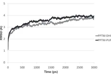
of their RMSD (Figure 3).
Frames with the lowest total energy in the last
300 ps of each simulation (Figure 4) were used
for the free-energy calculation with PEARLS
(Han et al. 2006), which is a force-field-based
scoring function. The calculated binding
free-energy for dihydrouracil (Table V) corresponded
to a dissociation constant (K
d) of ~1.3 x 10
-5M [ln
(1/K
d) = -ΔGo/2476.38; where ΔG
o: binding Gibbs
Figure 3 - Root mean square deviation (RMSD) of the putative phosphoribosyltransferase from Meloidogyne incognita, during the molecular dynamics simulation of the complex of this enzyme with dihydrouracil (PPTM-DHU) and 9H-purine (PPTM-PUR).
Figure 4 - Structures of the complexes PPTM-dihydrouracil (right side) and PPTM-9H-purine (left side), in which the protein is represented by Tube format and ligands are represented by the VDW format. This image was created by VMD 1.9.1 software.
In an attempt to obtain more reliable values
for the binding affinities of dihydrouracil and
9H-purine to PPTM, each snapshot of the last
200 ps of the molecular dynamics simulation of
the complexes dihydrouracil and
PPTM-9H-purine underwent calculation of SIE by rigid
obtained by the authors of the software employed
in this calculation (Cui et al. 2008, Naïm et al.
2007). The binding free-energy of dihydrouracil to
PPTM (Sietraj, Table V) corresponded to a K
dof
~8.3 x 10
-7M, which suggests that this substance
is a better inhibitor of PPTM than 9H-purine, for
which the K
dwas ~1.6 x 10
-6M.
In summary, this study has demonstrated that
uracil produced in vitro by B. cereus and B. subtilis,
as well as dihydrouracil and 9H-purine produced by
the latter rhyzobacterium, were active against M.
exigua
J2. Specifically for dihydrouracil, the LC50
against
M. exigua was less than that observed for
the commercial nematicide carbofuran, suggesting
that dihydrouracil is a promising substance for the
control of plant-parasitic nematodes. According
to the in silico study, these substances may be
acting against M. exigua through inhibition of its
phosphoribosyltransferase, which may be a promising
enzyme for the development of new products to
control Meloidogyne spp., since this macromolecule
appears to be important for the salvage pathways of
preformed bases in these pathogens.
ACKNOWLEDGMENTS
The authors express their sincere thanks to Dr.
José Dias de Souza Filho and Dr. Ivana Silva Lula
(Departamento de Química, Universidade Federal
de Minas Gerais - UFMG, Brazil) for providing
access to nuclear magnetic resonance facilities
and Dr. Flaviano Oliveira Silvério for mass
spectrometry spectra (Departamento de Química,
Universidade Federal de Viçosa - UFV, Brazil). The
authors also gratefully acknowledge: Coordenação
de Aperfeiçoamento de Pessoal de Nível Superior
(CAPES), Conselho Nacional de Desenvolvimento
Científico e Tecnológico (CNPq) and Fundação
de Amparo à Pesquisa do Estado de Minas Gerais
(FAPEMIG) for financial support and fellowships;
Centro Nacional de Supercomputação (CESUP)
at the Universidade Federal do Rio Grande do
Sul (UFRGS), where part of the present work was
developed; the Theoretical and Computational
Biophysics Group in the Beckman Institute for
Advanced Science and Technology at the University
of Illinois at Urbana-Champaign, who developed
NAMD and VMD; and the developers of Chimera:
Resource for Biocomputing, Visualization, and
Informatics at the University of California, San
Francisco, with support from the National Institutes
of Health (National Center for Research Resources
grant 2P41RR001081, National Institute of General
Medical Sciences grant 9P41GM103311).
RESUMO
Com o objetivo de contribuir para o desenvolvimento de produtos para o controle de Meloidogyne exigua, as bactérias Bacillus cereus e B. subtilis foram cultivadas em meio líquido de cultura para produzirem metabólitos ativos contra este nematoide parasita de plantas. Os fracionamentos dos extratos em diclorometano dos meios de cultura produziram uracila, 9H-purina e di-idrouracila. Todos os compostos foram ativos contra M. exigua, sendo o último o mais eficiente. Ele apresentou CL50de 204 µg/mL contra o nematoide, enquanto uma CL50 de 260 µg/mL foi observada para o nematicida comercial carbofuran. Uma busca por complexos proteína-ligante nos quais o proteína-ligante fosse estruturalmente similar à di-idrouracila resultou na seleção de fosforibosiltransferases, cujas sequências foram Method Binding affinity (kcal/mol)
Dihydrouracil 9H-purine
PEARLSa -6.64 -6.08
Sietrajb -8.29±0.59 -8.01±0.56
TABLE V
Binding affinities of dihydrouracil and 9H-purine
to the putative phosphoribosyltransferase (PPTM) from Meloidogyne incognita.
a
Values correspond to the average of the binding free-energy of the four ligand molecules, in the frame with the smaller energy in the last 300 ps of the molecular dynamics simulation trajectory. b
utilizadas em uma busca in silico no genoma de M. incognita por sequência de aminoácidos semelhante. A sequência resultante foi modelada e di-idrouracila e 9H-purina foram inseridos nos sítios ativos desta provável fosforibosiltransferase, resultando em complexos proteína-ligante que foram submetidos a simulações por dinâmica molecular. Cálculos das energias livres de ligação destes complexos revelaram que a constante de dissociação de di-idrouracila e 9H-purina da enzima é da ordem de 8,3 x 10⁻⁷
e 1,6 x 10⁻⁶
M, respectivamente. Consequentemente, estas substâncias e a provável fosforibosiltransferase podem ser de grande utilidade para o desenvolvimento de novos produtos para o controle de M. exigua.
Palavras-chave: 9H-purina, di-idrouracila, modelagem molecular, atividade nematicida, nematoide das galhas, uracila.
REFERENCES
AALTEN PM, VITOUR D, BLANVILLAIN D, GOWEN SR AND SUTRA L. 1998. Effect of rhizosphere fluorescent
Pseudomonas strains on plant-parasitic nematodes
Radopholus similis and Meloidogyne spp. Lett Appl Microbiol27: 357-361.
ALTSCHULL SF, MADDEN TL, SCHÄFFER AA, ZHANG J, ZHANG Z, MILLER W AND LIPMAN DJ. 1997. Gapped BLAST and PSI-BLAST: a new generation of protein database search programs. Nucleic Acids Res 25: 3389-3402.
AMARAL DR, OLIVEIRA DF, CAMPOS VP AND CARVALHO DA. 2002. Efeito de alguns extratos vegetais na mobilidade, mortalidade e patogenicidade de Meloidogyne exigua do cafeeiro. Nematologia Brasileira 26: 43-48.
ARAÚJO FF AND MARCHESI GVP. 2009. Uso de Bacillus subtilis no controle da meloidoginose e na promoção do crescimento do tomateiro. Cienc Rural 39: 1558-1561. ARNOLD K, BORDOLI L, KOPP J AND SCHWEDE T. 2006. The
Swiss-Model Workspace: A web-based environment for protein structure homology modeling. Bioinformatics 22: 195-201.
AUSUBEL FM, BRENT R, KINGSTON RE, MOORE DD, SEIDMAN JG, SMITH JA AND STRUHL K. 1997. Phenol/SDS method for plant RNA preparation. In: AUSUBEL ET AL. (Eds), Short protocols in molecular biology: a compendium of methods from current protocols in molecular biology. New York: J Wiley & Sons, p. 4-7.
B'CHIR MM. 2000. Bionematicide with efficient ovicide activity against plant-parasitic nematodes. Patent EP0981277A1, 01 March 2000.
BENDZKO P, ETZEL W, HOEDING B, KREBS B, MAXIMOV J AND OCKARDT A. 1998. Cyclic peptide(s) from Bacillus amyloliquefaciens – useful as antimycotics, antivirals, fungicides, nematocides, etc. US Patent DE19641213-A1, 16 April 1998.
BENIZRI E, BAUDOIN E AND GUCKERT A. 2001. Root colonization by inoculated plant growth-promoting rhizobacteria. Biocontrol Sci Technol 11: 557-574. BENKERT P, BIASINI M AND SCHWEDE T. 2011. Toward the
estimation of the absolute quality of individual protein structure models. Bioinformatics 27: 343-350.
BONETI JIS AND FERRAZ S. 1981. Modificação do método de Hussey e Barker para extração de ovos de Meloidogyne exigua de raízes de cafeeiro. Fitopatol Bras 6: 553. BUNCH ASR, MEUNIER D, RODIERB CLC, RAULINA F AND
VIDAL-MADJARC C. 2003. Extraction of organic molecules of exobiological interest for in situ analysis of the Martian soil. J Chromatogr A 99: 165-174.
CAMPOS VP AND SILVA JRC. 2008. Management of Meloidogyne
spp. in coffee plantations. In: SOUZA RM (Ed), Plant para-sitic nematodes of coffee, São Paulo: Springer, p. 149-164. CAMPOS VP AND VILLAIN L. 2005. Nematode parasites of coffee
and cocoa. In: LUC M et al. (Eds), Plant parasitic nematodes in subtropical and tropical agriculture, Wallingford UK: CAB Internacional, p. 529-579.
CASTRO JMC, CAMPOS VP, POZZA EA, NAVES RL, ANDRADE JÚNIOR WC, DUTRA MR, COIMBRA JL, MAXIMINIANO C AND SILVA JRC. 2008. Levantamento de fitonematóides em cafezais do sul de Minas Gerais. Nematologia Brasileira 32: 56-64.
CHITWOOD DJ. 2002. Phytochemical based strategies for nematode control. Annu Rev Phytopathol 40: 221-249. CHRISTOFFERSEN S, KADZIOLA A, JOHANSSON E, RASMUSSEN
M, WILLEMOES M AND JENSEN KF. 2009. Structural and kinetic studies of the allosteric transition in Sulfolobus solfataricus uracil phosphoribosyltransferase: Permanent activation by engineering of the C-Terminus. J Mol Biol 393: 464-477.
COLE JR ET AL. 2009. The ribosomal database project: Improved alignments and new tools for rRNA analysis. Nucleic Acids Res 37: 141-145.
CRAIG III SP AND EAKIN EA. 2000. Purine Phosphori-bosyltransferases. J Biol Chem 275: 20231-20234. CRONIN D, MOËNNE-LOCCOZ Y, FENTON A, DUNNE C, DOWLING
DN AND O’GARA F. 1997. Role of 2,4-diacetylphloroglucinol in the interactions of the biocontrol pseudomonad strain F113 with the potato cyst nematode Globodera rostochiensis. Appl Environ Microb 63: 1357-1361.
CUI Q, SULEA T, SCHRAG JD, MUNGER C, HUNG M-N, NAÏM M, CYGLER M AND PURISIMA EO. 2008. Molecular dynamics and solvated interaction energy studies of protein-protein interactions: the MP1-p14 scaffolding complex. J Mol Biol 379: 787-802.
FOCIA PJ, CRAIG IIISP AND EAKIN AE. 1998. Approaching the transition state in the crystal structure of a phosphoribosyltransferase. Biochemistry37: 17120-17127. GERMIDA JJ, HEINS SD, MANKER DC, JIMENEZ DR AND
MARRONE PG. 2000. Bacillus subtilis strain for controlling insect and nematode pests. US Patent 6015553, 01 January 2000.
GOUY M, GUINDON S AND GASCUEL O. 2010. SeaView version 4: A multiplatform graphical user interface for sequence alignment and phylogenetic tree building. Mol Biol Evol 27: 221-224.
GUEX N AND PEITSCH MC. 1997. SWISS-MODEL and the Swiss-PdbViewer: An environment for comparative protein modeling.Electrophoresis 18: 2714-2723.
GUPTA VP, BOCHOW H, DOLEJ S AND FISCHER I. 2000. Plant growth promoting Bacillus subtilis strain as potential inducer of systemic resistance in tomato against
Fusarium wilt. Zeitschrift für Pflanzenkrankheiten und Pflanzenschutz 107: 145-154.
HAN LY, LIN HH, LI ZR, ZHENG CJ, CAO ZW, XIE B AND CHEN YZ. 2006. PEARLS: program for energetic analysis of receptor-ligand system. J Chem Inf Model 46: 445-450. HUMPHREY W, DALKE A AND SCHULTEN K. 1996. VMD
-Visual Molecular Dynamics. J Mol Graphics 14: 33-38. HUSSEY RS AND BARKER KR. 1973. A compararison of methods
of collecting inocula of Meloidogyne spp., including a new technique. Plant Dis Reporter 57: 1025-1028. KANG GB, KIM MK, YOUN HS, AN JY, LEE JG, PARK KR,
LEE SH, KIM Y, FUKUOKA SI AND EOM SH. 2011. Crystallization and preliminary X-ray crystallographic analysis of human quinolinate phosphoribosyltransferase. Acta Cryst67: 38-40.
KINNINGS SL AND JACKSON RM. 2011. ReverseScreen3D: A structure-based ligand matching method to identify protein targets. J Chem Inf Model 28: 624-634.
KLOEK AP, WILLIAMS DJ AND SALMON B. 2005. Nematode PPPT-like sequences. US Patent 2005/0186631 A1, 25 August 2005.
KRIVOV GG, SHAPOVALOV MV AND DUNBRACK JR RL. 2009. Improved prediction of protein side-chain conformations with SCWRL4. Proteins: Structure, Function and Bioinformatics 77: 778-795.
LARKIN MA ET AL. 2007. ClustalW and ClustalX version 2.
Bioinformatics23: 2947-2948.
LEORA SOFTWARE. 1987. Polo PC: a user’s guide to probit or logit analysis. LeOra Software Inc., Berkely, CA. LIU L, BURNAM L, SLUDER A, LINK E AND WESTLAND B. 2006.
Screens and assays for agents useful in controlling parasitic nematodes. US Patent 7,064,243 B2, 20 June 2006. MEZEI M. 2010. Simulaid: a simulation facilitator and analysis
program. J Comp Chem 31: 2658-2668.
MAPA - MINISTÉRIO DA AGRICULTURA, PECUÁRIA E ABASTECIMENTO. 2012. Informe estatístico do café. 2012. Retrieved June 13, 2012, from http://www.agricultura. gov.br/vegetal/estatisticas
NAÏM M ET AL. 2007. Solvated interaction energy (SIE) for
scoring protein-ligand binding affinities. 1. Exploring the
parameter space. J Chem Inf Model 47: 122-133.
OKA Y, CHET I AND SPIEGEL Y. 1993. Control of the root-knot nematode Meloidogyne javanica by Bacillus cereus. Biocontrol Sci Technol 3: 115-126.
OLIVEIRA DF, CAMPOS VP, AMARAL DR, NUNES AS, PANTALEÃO JA AND COSTA DA. 2007. Selection of rhizobacteria able to produce metabolites active against
Meloidogyne exigua. Eur J Plant Pathol 119: 477-479. OLIVEIRA DF, CARVALHO HWP, NUNES AS, SILVA GH,
CAMPOS VP, JÚNIOR HMS AND CAVALHEIRO AJ. 2009. The activity of amino acids produced by Paenibacillus macerans and from commercial sources against the root-knot nematode Meloidogyne exigua. Eur J Plant Pathol 124: 57-63.
ONGENA M, DUBY F, JOURDAN E, BEAUDRY T, JADIN V, DOMMES J AND THONART P. 2005. Bacillus subtilis M4 decreases plant susceptibility towards fungal pathogens by increasing host resistance associated with differential gene expression. Applied Microbiol Biotechnol 67: 692-698. PARRY RJ, VOS S, BURNS MR, DE JERSEY J AND MARTIN
JL. 1998. Structures of free and complexed forms of
Escherichia coli xanthine-guanine phosphoribosyl-transferase. J Mol Biol 282: 875-889.
PETTERSEN EF, GODDARD TD, HUANG CC, COUCH GS, GREENBLATT DM, MENG EC AND FERRIN TE. 2004. UCSF Chimera: a visualization system for exploratory research and analysis. J Comput Chem 25: 1605-1612.
PHILLIPS CL, ULLMAN B, BRENNAN RG AND HILL CP. 1999. Crystal structures of adenine phosphoribosyltransferase from Leishmania donovani. Embo J 18, 3533-3545. PHILLIPS JC, BRAUN R, WANG W, GUMBART J, TAJKHORSHID
E, VILLA E, CHIPOT C, SKEEL RD, KALE L AND SCHULTEN K. 2005. Scalable molecular dynamics with NAMD. J Comput Chem 26: 1781-1802.
ROBERTS JL AND POULTER CD. 1978. 2’,3’,5’-Tri-O
-benzoyl[413C]uridine. An efficient, regiospecific synthesis
of the pyrimidine ring. J Org Chem 43: 1547-1550. SCHUMACHER MA, CARTER D, SCOTT DM, ROOS DS,
ULLMAN B AND BRENNAN RG. 1998. Crystal structures of toxoplasma gondii uracil phosphoribosyltransferase reveal the atomic basis of pyrimidine discrimination and prodrug binding. Embo J17: 3219-3232.
SCHWEDE T, KOPP J, GUEX N AND PEITSCH MC. 2003. SWISS-MODEL: An automated protein homology-modeling server. Nulceic Acids Res 31: 3381-3385.
SCOTT AJ AND KNOTT M. 1974. Cluster analysis method for grouping means in the analysis of variance. Biometrics 30: 507-512.
SELA S, SCHICKLER H, CHET I AND SPIEGEL Y. 1998.
Purification and characterization of a Bacillus cereus
collagenolytic/proteolytic enzyme and its effect on
SHI W, LI CM, TYLER PC, FURNEAUX RH, GRUBMEYER C, SCHRAMM VL AND ALMO SC. 1999. The 2.0 Å structure of human hypoxanthine-guanine phosphoribosyltransferase in complex with a transition-state analog inhibitor. Nat Struct Biol 6: 588-593.
SHI W, SARVER AE, WANG CC, TANAKA KS, ALMO SC AND SCHRAMM VL. 2002. Closed site complexes of adenine phosphoribosyltransferase from Giardia lamblia reveal a mechanism of ribosyl migration. J Biol Chem 277: 39981-39988.
SILVA JRC, SOUZA RM, ZACARONE AB, SILVA LHCP AND CASTRO ANS. 2008. Bactérias endofíticas no controle e inibição in vitro de Pseudomonas syringae pv. tomato, agente da pinta bacteriana do tomateiro. Cienc Agrotec 32: 1062-1072.
SILVA M, SILVA CHTP, IULEK J, OLIVA G AND THIEMANN OH. 2004b. Crystal structure of adenine phosphoribo-syltransferase from Leishmania tarentolae: Potential implications for aprt catalytic mechanism. Biochem Biophys Acta 1696: 33-39.
SILVA M, SILVA CHTP, IULEK J AND THIEMANN OH. 2004a. Three-dimensional structure of human adenine phosphoribosyltransferase and its relation to DHA-urolithiasis. Biochemistry43: 7663-7671.
THERESE M, PUGMIRE CRJ, GRANT DM, PANZICA RP AND TOWNSEND LB. 1975. Carbon-13 magnetic resonance. Quantitative determination of the tautomeric populations of certain purines. J Am Chem Soc97: 4636-4642.
WANG J, WANG W, KOLLMAN PA AND CASE DA. 2006b. Automatic atom type and bond type perception in molecular mechanical calculations. J Mol Graph Model 25: 247-260.
WANG J, WOLF RM, CALDWELL JW, KOLLMAN PA AND CASE DA. 2004. Development and testing of a general AMBER
force field. J Comput Chem 25: 1157-1174.
WANG T, ZHANG X, BHEDA P, REVOLLO JR, IMAI SI AND WOLBERGER C. 2006a. Structure of Nampt/PBEF/visfatin, a mammalian NAD(+) biosynthetic enzyme. Nat Struct Mol Biol 13: 661- 662.
WATERHOUSE AM, PROCTER JB, MARTIN DMA, CLAMP M AND BARTON GJ. 2009. Jalview Version 2 - a multiple sequence alignment editor and analysis workbench. Bioinformatics 25: 1189-1191.
WEISBURG WG, BARNS SM, PELLETIER DA AND LANE DJ. 1991. 16S Ribosomal DNA amplification for phylogenetic study. J Bacteriol 173: 697-703.
XIAO T, TAN S, SHEN Q AND RAN W. 2012.Bacillus cereus X5 suppresses root-knot nematode of tomato by colonizing in roots and soil. Afr J Microbiol Res6: 2321-2327. YESSELMAN JD, PRICE DJ, KNIGHT JL AND BROOKS IIICL.
2012. MATCH: An atom-typing toolset for molecular