Arginine- and Polyamine-Induced Lactic Acid
Resistance in
Neisseria gonorrhoeae
Zheng Gong1, M. Matt Tang1, Xueliang Wu1,2, Nancy Phillips3, Dariusz Galkowski4,5, Gary A. Jarvis6,7, Huizhou Fan1*
1Department of Pharmacology, Rutgers University Robert Wood Johnson Medical School, Piscataway, New Jersey, United States of America,2Department of Urology, Hunan University of Traditional Chinese Medicine-affiliated Ningxiang People’s Hospital, Changsha, Hunan, China,3Department of Obstetrics, Gynecology and Reproductive Sciences, and Women’s Health Institute, Rutgers University, New Brunswick, New Jersey, United States of America,4Public Health and Environmental Laboratories, New Jersey Department of Health, Ewing, New Jersey, United States of America,5Department of Pathology and Laboratory Medicine, Rutgers University Robert Wood Johnson Medical School, Piscataway, New Jersey, United States of America,6Center for Immunochemistry, Veterans Affairs Medical Center, San Francisco, California, United States of America,7Department of Laboratory Medicine, University of California, San Francisco, California 94143, United States of America
*fanhu@rwjms.rutgers.edu
Abstract
Microbe-derived lactic acid protects women from pathogens in their genital tract. The pur-pose of this study was to determine lactic acid susceptibility ofNeisseria gonorrhoeae, and identify potential acid resistance mechanisms present in this pathogen. Testedin vitro, lac-tic acid killed all 10 gonococcal strains analyzed in a low pH-dependent manner. Full inacti-vation occurred at pH 4.5. At low pH, lactic acid treatment resulted in the entry of the DNA-binding fluorochrome propidium iodide into the microbial cells, suggesting that hydrogen ions from lactic acid compromise the integrity of the bacterial cell wall/membrane. Most likely, hydrogen ions also inactivate intracellular proteins since arginine rendered significant protection against lactic acid presumably through action of the gonococcal arginine decar-boxylase, an enzyme located in the bacterial cytoplasm. Surprisingly, arginine also less-ened lactic acid-mediated cell wall/membrane disruption. This effect is probably mediated by agmatine, a triamine product of arginine decarboxylase, since agmatine demonstrated a stronger protective effect on GC than arginine at equal molar concentration. In addition to agmatine, diamines cadaverine and putrescine, which are generated by bacterial vagino-sis-associated microbes, also induced significant resistance to lactic acid-mediated GC kill-ing and cell wall/membrane disruption. These findkill-ings suggest that the arginine-rich semen protects gonococci through both neutralization-dependent and independent mechanisms, whereas polyamine-induced acid resistance contributes to the increased risk of gonorrhea in women with bacterial vaginosis.
a11111
OPEN ACCESS
Citation:Gong Z, Tang MM, Wu X, Phillips N, Galkowski D, Jarvis GA, et al. (2016) Arginine- and Polyamine-Induced Lactic Acid Resistance in Neisseria gonorrhoeae. PLoS ONE 11(1): e0147637. doi:10.1371/journal.pone.0147637
Editor:Guangming Zhong, Univ. of Texas Health Science Center at San Antonio, UNITED STATES
Received:December 1, 2015
Accepted:January 6, 2016
Published:January 25, 2016
Copyright:This is an open access article, free of all copyright, and may be freely reproduced, distributed, transmitted, modified, built upon, or otherwise used by anyone for any lawful purpose. The work is made available under theCreative Commons CC0public domain dedication.
Data Availability Statement:All relevant data are within the paper.
Introduction
Among the 2.3 million notifiable infections reported to the Center for Disease Control and Pre-vention (CDC) of the United States, two of every three were sexually transmitted infections (STI) [1]. Yet, CDC estimates this reported figure to be only one tenth of the actual number of new STI infections. Annual health care costs for treating STI and related complications are esti-mated to be $16 billion [2].
One of the most common STI is gonorrhea, caused byNeisseria gonorrhoeae, also referred as gonococcus (GC). In the United States, CDC estimates that more than 800,000 new rheal cases occur each year. There were three consecutive years of increases in reported gonor-rheal cases from 2010 and 2012. Although the number of reported cases decreased slightly in 2013, it increased by 5% in 2014, and reached the highest level since 2008 [3]. Gonorrhea in women often exhibits mild or even no symptoms. However, if left untreated, the infection can lead to serious complications including pelvic inflammatory symptoms, ectopic pregnancy, and tubal factor infertility [4]. In addition, gonorrhea is known as a cofactor for HIV/AIDS [5–
7]. Alarmingly, drug-resistant GC strains are widespread. As a result, currently effective treat-ment of gonorrhea requires multiple antibiotics [8].
Most reproductive-age women have an acidic vaginal environment owing to the over-whelmingly large numbers of lactic acid-producing bacteria, mainly lactobacilli, present in the vagina [9–11]. However, in women with bacterial vaginosis, lactobacilli are replaced by other bacteria, leading to increased vaginal pH (>4.5) [11–14]. A number of population-based
stud-ies have associated bacterial vaginosis with increased risks of STIs including gonorrhea, and therefore, have suggested a protective role of lactic acid-generated acidity in defense against STI [7,10,15–20]. However, to the best of our knowledge, the effect of lactic acid on GC sur-vival has not been experimentally documented to date. Although HCl-mediated GC killing has been reported [21,22], studies have demonstrated that lactic acid and HCl differ in their anti-microbial activities [23,24]. We have recently determined the lactic acid susceptibility of Chla-mydia trachomatis, another major sexually transmitted bacterial pathogen [23]. In this report, we show that multiple GC strains are fully inactivated by lactic acid at pH 4.5. Interestingly, the microbicidal activity of lactic acid is significantly weakened in the presence of arginine and polyamines. Our findings indicate that semen, known to be rich in arginine [25], helps GC sur-vive and colonize the female genital tract by both neutralizing lactic acid and inducing argi-nine-dependent acid resistance. In addition, our observations suggest that both reduced lactic acid production and increased polyamines production associated with bacterial vaginosis increase the risk for gonorrhea.
Materials and Methods
Media
GC liquid medium (GCL) contained (per liter) 15 g proteose peptone 3 (EMD Millipore), 1 g soluble starch, 4.0 g K2HPO4, 1.0 g KH2PO4, 5 g NaCl, 0.4 g glucose, 10 mg glutamine, 20μg
cocarboxylase and 0.5 mg Fe(NO3)3•9H2O. GCL-LA was prepared by supplementing GCL
with 100 mM lactic acid, and adjusted to pH 4.0, 4.5, 5.0, 5.5 and 6.0 using NaOH. NEG buffer (pH 7.0) contained 72 mM NaHPO4, 5.5 mM (NH4)H2PO4, 10 mM KCl, 0.25 mM MgSO4, 0.1
mM CaCl2, 28 mM dextrose and 9.5 mM sodium citrate, and was adjusted to pH 4.9, 5.0 or 5.2
using lactic acid. NEG-LA was prepared by supplementing NEG with 75 mM lactic acid, and adjusted to desired pH using NaOH. Proteose peptone 3 was purchased from EMD Millipore. All other medium components were purchased from Sigma.
Arginine and Polyamines Rescue GC from Lactic Acid
Strain information
GC ATCC 19424 was purchased from ATCC. FA1090 was provided by Dr. Ann E. Jerse [26]. FA5100T1, MS11MkCT1, FA1342T1, Can116 and KGC654 were from the collection of the Jar-vis Laboratory. NJ1, NJ2 and NJ3 were isolated by the Public Health and Environmental Labo-ratories of New Jersey.
Preparation of GC stocks
Bacteria in well-separated colonies that formed on GC Agar plates after 17–20 h incubation at 37°C were harvested in 0.9% NaCl (saline) with the aid of a 1.5 cm (diameter) aluminum loop. A 100μl sample of the GC suspension was mixed with 900μl GCL. Optical density at 600 nm
(OD600) for the resulting diluted bacterial suspension was measured.
GC killing tests
On ice, 10μl of GC saline suspension were diluted with saline to 0.005 OD600. On 96-well plates,
11μl of the diluted suspension was mixed with 100μl GCL-LA or control GCL (pH 7.0). The
plates were placed in a 37°C incubator containing air supplemented with 5% CO2. 40 min later,
the bacteria were subjected to 10 fold serial dilution using GCL, and plated onto GC Agar plates. In experiments determining the effects of specific amino acids or polyamine on lactic acid-medi-ated GC killing, GCL-LA was substituted with the NEG-LA. Direct alkylating or acidifying effects of amino acids and polyamines on the medium were eliminated by readjusting pH using additional lactic acid or NaOH as needed. All killing experiments were performed in triplicate.
Propidium iodide (PI) staining
Bacteria were harvested in saline. Samples of the bacterial suspension were diluted to 1.0 OD600
using no less than nine volumes of GCL-LA, NEG-LA or NEG-LA containing arginine, glutamic acid, lysine, agmatine, cadaverine or putrescine (all purchased from Sigma) at indicated pH. The final volume for each treatment was 1 ml. After 40 min incubation in a 37°C incubator contain-ing air supplemented with 5% CO2, bacteria were collected by centrifugation and resuspended
in phosphate-buffered saline containing 0.1% gelatin and 10 ng/ml PI. The bacterial suspension was incubated at room temperature for 10 min. Bacteria were again centrifuged, and washed once with the buffer supplemented with only 0.1% gelatin. Smears were prepared and viewed with a 100 X oil-immersion objective under an Olympus IX51 fluorescence microscope. All fluorescence images were acquired with equal exposure time for the same experiment.
Results
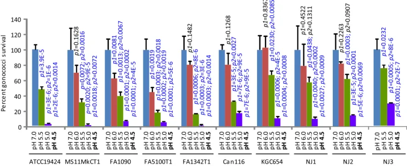
Lactic acid kills GC in a low pH-dependent manner
decreased killing was observed when pH was raised from 5.5 to 6.0 although thep-values only trended toward significance in triplicate experiments (Fig 1).
Lactic acid treatment causes propidium iodide (PI) to enter GC
We used the DNA-binding fluorochrome PI to determine the mechanism underlying GC kill-ing by lactic acid uskill-ing two bacterial strains, ATCC19424 and NJ2. As expected, the membrane impermeable molecule is largely excluded from the cells of both strains treated with GCL-LA adjusted to pH 7.0 (Fig 2). However, apparent staining with the fluorochrome was observed in bacteria treated with GCL-LA starting at pH 5.5, which became increasingly prominent as the pH decreased to 5.0 and 4.5 (Fig 2). These findings suggest that lactic acid-mediated low pH kills GC by compromising the integrity of the bacterial cell wall/membrane.
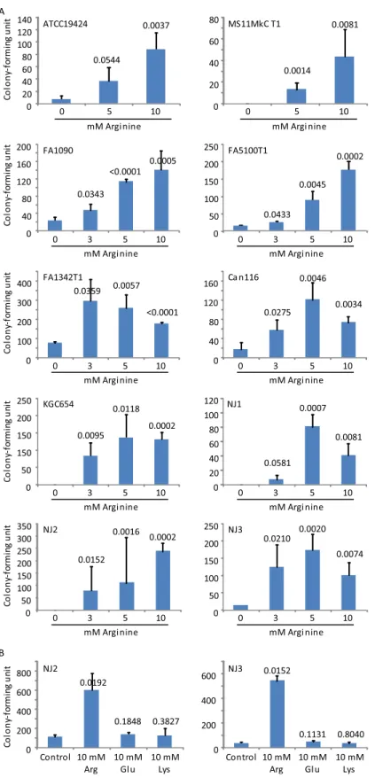
Arginine decreases lactic acid-mediated GC killing
Arginine, glutamic acid and lysine increase the resistance ofE.coliand other bacteria to the microbicidal activity of low pH generated by HCl [27,28]. To determine whether similar acid resistance mechanisms exist in GC in response to lactic acid, we substituted the rich GCL medium with NEG, an amino acid-free medium, for acid killing experiments. As expected, lac-tic acid also demonstrated low pH-dependent GC-killing activity in NEG. Interestingly, 9 of the 10 GC strains showed highly significantly improved survival in the presence of arginine at both 10 mM and 5 mM, whereas thepvalue for ATCC19424 treated with 10 mM arginine was also highly significant (0.0037), and with 5 mM arginine trended toward significance at 0.0544 (Fig 3A). Eight strains were also tested with 3 mM arginine, and all demonstrated increased
Fig 1. Lactic acid-mediated, pH-dependent GC killing.The GCL medium was supplemented with 100 mM lactic acid and adjusted to indicated pH using NaOH. The resulting GCL-LA and control lactic acid-free medium (pH 7.0) were used to treat GC. Surviving bacteria were quantified by plating the treatments onto GC Agar plates. Number of input bacteria per assay were 10,000–70,000 colony-forming units (CFU), as determined by treatment with control GCL. Note that treatment with pH 4.5 (shown in boldface) resulted in complete killing in all GC strains. Data represent averages±standard deviations of triplicate experiments.p-values for difference in numbers of colony-forming units between samples treated with GCL-LA and control GCL (pH 7.0) were calculated usingttests (two-tailed between control and pH 6.0, and one-tailed for all other comparisons).p1is thep-values for difference between samples treated with GCL-LA and control GCL (pH 7.0), whereasp2is the difference between samples treated with GCL-LA at the pH that thep-value appears above, and samples treated with GCL-LA that has a 0.5 unit higher pH.p-values of<0.05, which indicate statistically significant differences, are shown in blue font, while those of>0.05 are shown in black font.
doi:10.1371/journal.pone.0147637.g001
Fig 2. Lactic acid-mediated, pH-dependent entry of propidium iodide (PI) into GC cells.The membrane-impermeable fluorescent molecule PI was added to GC after the bacteria were treated with GCL-LA adjusted to indicated pH. After a wash to remove the free fluorochrome, smears were prepared and images were acquired. A scale bar is at the bottom of the figure.
Fig 3. Arginine-induced acid resistance in GC.Bacteria were treated with the defined medium NEG supplemented with 75 mM lactic acid (NEG-LA) with or without further supplementation with arginine at the indicated concentration (A), or with arginine, glutamic acid or lysine at 10 mM (B). All media were adjusted to pH 5.0. Numbers of surviving bacteria were determined by plating the treatments onto GC Agar plates. Data represent averages±standard deviations of triplicate experiments. Numbers above the bars werep-values
survival although thepvalue for NJ1 trended toward significance at 0.0581 (Fig 3A). In con-trast to 10 mM arginine, neither 10 mM glutamic acid nor 10 mM lysine showed any effect on bacterial survival when tested concurrently with arginine against strains NJ2 and NJ3 (Fig 3B). Taken together, our data suggest that arginine but not glutamic acid or lysine is effective in inducing acid resistance in GC.
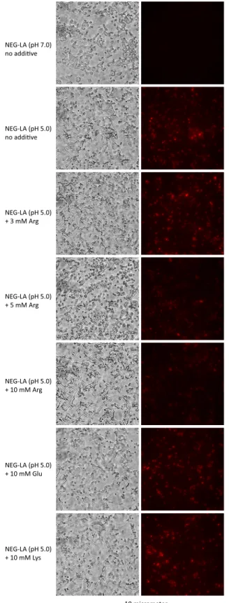
Arginine reduces lactic acid-mediated PI entry
Arginine causes acid resistance through a hydrogen ion-consuming reaction catalyzed by the arginine decarboxylase in the bacterial cytoplasm [27,28]. Thus, the demonstration of argi-nine-dependent lactic acid resistance in GC (Fig 3) suggests that hydrogen ions also cause inac-tivation of intracellular molecules, in addition to disruption of cell wall/membrane as
demonstrated inFig 2. Surprisingly, compared to GC treated with control NEG-LA (pH 5.0), bacteria treated with the same medium supplemented with either 10 or 5 mM arginine dis-played noticeably decreased PI staining although the effect of 3 mM was not obvious (Fig 4). In comparison, neither 10 mM glutamic acid nor 10 mM lysine had an apparent effect on PI entry. These results suggest that arginine not only serves to decrease the intracellular hydrogen ion concentration, but also can ameliorate lactic acid-induced cell wall/membrane disruption.
Agmatine lessens lactic acid-induced GC killing and PI entry
Agmatine is a triamine product of arginine decarboxylase. Interestingly, agmatine also demon-strated strong protective effects in all 7 GC strains treated with NEG-LA (pH 5.0) (Fig 5A). In all 5 strains for which both arginine and agmatine were tested, inclusion of 10 mM agmatine in the medium resulted more bacteria survival than 10 mM arginine; the difference was statisti-cally (highly) significant for 4 of the 5 strains, and trended towards significance for the remain-ing 1 strain (FA1342T1). Consistent with its protective function in lactic acid-induced GC killing, agmatine also strongly reduced the entry of PI into GC (Fig 5B), with 10 mM agmatine displaying a stronger effect than 10 mM arginine in decreasing PI staining. These data suggest that agmatine is responsible for the ability of arginine to stabilize the cell wall/membrane in GC in the presence of lactic acid.
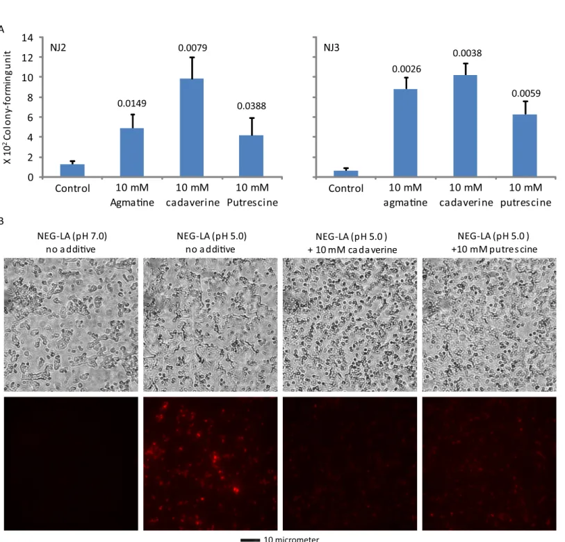
Cadaverine and putrescine decrease lactic acid-mediated GC killing and
PI entry
In light of the induction of acid resistance by agmatine, we also tested the effects of two addi-tional polyamines, cadaverine and putrescine, on lactic acid-mediated GC killing. Similar to agmatine, both cadaverine and putrescine significantly increased the survival (Fig 6A) and decreased PI entry (Fig 6B). These findings suggest that polyamines in general can decrease lac-tic acid-mediated GC killing by stabilizing the bacterial cell wall/membrane.
Discussion
Lactic acid-producing bacteria create an acidic vaginal environment in most, but not all, repro-ductive-age women. Although previous studies have indirectly linked STI to increased vaginal pH, resulting from decreases or absence of lactobacilli [7,15,16], the role of lactic acid in the
(one-tailedttests) for difference in surviving bacteria between samples treated with the control NEG-LA (pH 5.0) and those treated with NEG-LA supplemented with an indicated concentration of arginine (A), glutamate or lysine (B). Number of input bacteria per assay were 10,000–40,000 CFU, as determined by treatment with control NEG-LA (pH 7.0).
Fig 4. Arginine-, but not glutamate- or lysine-mediated inhibition of lactic acid-induced wall/ membrane permeation.NJ2 were first treated with NEG-LA (pH 5.0) or NEG-LA containing indicated supplements as described in the legend toFig 3legend, then exposed to PI, and finally imaged with a fluorescent microscope (Fig 2). Matching phase contrast images are presented on the left. A scale bar is at the bottom.
doi:10.1371/journal.pone.0147637.g004
Fig 5. Tramine agmatine-induced acid resistance to LA-mediated killing (A) and wall/membrane permeation (B).A. Killing assays were performed as described inFig 3legend. B. PI staining was performed as described inFig 3legend. Data represent averages±standard deviations of triplicate
staining of NJ2 were performed as described in the legend toFig 4. Matching phase contrast images (upper) and fluorescent images (lower) are presented. A scale bar is at the bottom.
doi:10.1371/journal.pone.0147637.g005
Fig 6. Diamines cadaverine and putrescine-induced acid resistance to lactic acid-mediated killing (A) and wall/membrane permeation (B).A. Killing assays were performed as described inFig 3legend. B. PI staining was performed as described inFig 3legend. Data represent averages±standard
deviations of triplicate experiments. Numbers above the above the bars werep-values (one-tailedttests) for difference in surviving bacteria between samples treated with the control NEG-LA (pH 5.0) and those treated with NEG-LA supplemented with 10 mM cadaverine or 10 mM putrescine. Number of input bacteria per assay for NJ2 and NJ3 were 20,000 and 60,000 colony-forming units, respectively. B. Treatment and PI staining of NJ2 were performed as described in the legend toFig 4. Matching phase contrast images (upper) and fluorescent images (lower) are presented. A scale bar is at the bottom.
doi:10.1371/journal.pone.0147637.g006
gonorrheal pathogenesis has not been experimentally examined to the best of our knowledge. In this report, we have demonstrated acidity-dependent GC killing by lactic acid. Thus, even though all GCL-LA contained 100μM lactic acid, only GCL-LA at pH 4.5 or pH 4.0 were fully
bactericidal, whereas the killing efficiency decreased progressively as the pH rose to 5.0 and above (Fig 1). These results are consistent with observations that neutralization of lactic acid completely abolishes the microbicidal activities towardsChlamydia trachomatisand HIV [23,
24]. They support the notion that the hydrogen ion but not the anion of lactic acid is responsi-ble for killing microbes.
Many bacteria are armed with acid resistance mechanisms. InE.coli, arginine, lysine, and particularly glutamic acid can induce acid resistance. Inside the bacterial cells, these amino acids undergo decarboxylation, which consumes hydrogen ions. The decarboxylation products exit the bacterial cell in exchange for additional substrate amino acids [27,28]. Interestingly, only arginine but not glutamic acid or lysine induced acid resistance in GC (Fig 3). Apparently, there is a correlation between the amino acid that induces resistance and the corresponding amino acid decarboxylase in GC, because only arginine decarboxylase but not glutamate decar-boxylase or lysine decardecar-boxylase is encoded by the GC genome.
Our data indicate two mechanisms are involved in GC killing by lactic acid-dissociated hydrogen ions. First, staining of acid-treated GC with the otherwise membrane-impermeable PI indicates that lactic acid causes loss of bacterial cell wall/membrane integrity. Acid-induced cell wall/membrane disruption has been demonstrated for other bacteria [29,30]. We have not examined how lactic acid compromises the gonococcal cell wall/membrane integrity. However, previous studies have shown membrane protein expression profile changes in GC cultured in medium acidified with HCl [22,31]. How those protein expression profile changes affect the wall/membrane integrity and bacterial survival have yet to be determined.
The ability of arginine decarboxylase, an enzyme located in the bacterial cytoplasm, to res-cue GC from lactic acid-generated low pH suggests hydrogen ion-mediated inactivation of intracellular molecules as the second mechanism for lactic acid-induced GC killing. It is gener-ally believed that arginine decarboxylase confers acid resistance by consuming hydrogen ions while removing the carboxyl group from arginine to produce agmatine and H2O [27,32–36].
Interestingly, we noticed that at concentrations above 5 mM, arginine also reverses acidity-induced PI entry into GC, whereas 3 mM arginine does not appear to have such an effect (Fig 4). These observations indicate that, whereas at low concentrations, arginine induces acid resis-tance mainly by consuming hydrogen ions, at higher concentrations, arginine induces acid resistance by both decreasing intracellular hydrogen ions and lessening acidity-induced cell wall/membrane disruption.
In addition to arginine, its decarboxylated derivative agmatine also induces acid resistance (Figs5and6). The gonococcal genome encodes an arginine-agmatine antiporter. Since the antiporter couples the exit of agmatine with the entry of arginine, agmatine may cooperate with arginine in mediating acid resistance. However, our demonstration of agmatine-induced acid resistance was made in defined medium without exogenous arginine. PI staining data indi-cate that inhibition of gonococcal wall/membrane disruption is the underlying mechanism for agmatine-induced, arginine-independent acid resistance. We further suggest that agmatine is partly responsible for mediating the protective effect of high arginine concentrations on the gonococcal wall/membrane integrity.
Thus, it is highly probable that the level of arginine in semen is high enough to promote GC survival and colonization in the vagina.
We have shown that agmatine, cadaverine and putrescine all protect GC from acid assault by stabilizing the bacterial cell wall/membrane (Figs5Band6B). Interestingly, previous studies have shown that polyamines including cadaverine and putrescine protect GC against human antimicrobial peptides that target lipid membranes [39]. Therefore, it is likely that other poly-amines exert protective effects on acid-mediated GC killing, similar to the three polypoly-amines that we have examined here.
Like arginine-induced acid resistance, polyamine-induced resistance also has practical implications for the pathogenesis of gonorrhea. First, there are various levels of polyamines in semen [40–42]. Thus, similar to seminal arginine, seminal polyamines may protect GC in the vagina. Second, cervicovaginal fluids also contain polyamines; bacterial vaginosis-associated microbes produce particularly high levels of diamines including cadaverine and putrescine [43–47]. Therefore, microbe-derived polyamines may increase the risks of gonorrhea by pro-tecting the pathogen in women with mild bacterial vaginosis that still retain a relatively low vaginal pH, and by conferring resistance to antimicrobial peptides [39].
In summary, we have demonstrated lactic acid-mediated, low pH-dependent GC killing, and arginine- and polyamine-induced acid resistance. These observations are consistent with previous findings of increased risks of gonorrhea in women with bacterial vaginosis character-ized by increased vaginal pH and high abundance of polyamines including cadaverine and putrescine. The revelation of arginine-induced acid resistance suggests that semen, with a basic pH and rich in arginine, protects GC through both neutralization-dependent and independent mechanisms. Likewise, the demonstration of polyamine-induced acid resistance indicates that polyamines generated by bacterial vaginosis-associated organisms can increase gonorrheal risks before the vaginal pH is raised significantly. Creation and maintenance of a sustainable acidic pH and means to decrease arginine and polyamine levels should aid in protection against GC in women and their male sexual partners in turn.
Acknowledgments
We thank Dr. Ann E. Jerse for the kind supply of FA1090, Dr. Loren Runnels for giving us access to his fluorescence microscope equipped with an oil immersion objective, Drs. Theresa L. Chang, Gloria Bachmann and Lingli Tang for discussions and Mr. Malhar Desai for assis-tance in counting bacterial colonies. This work was supported by a National Institutes of Health grant (grant number AI071954) and a New Jersey Health Foundation Grant (grant number PC7-13) to HF. XW was a recipient of an oversea research scholarship awarded by the Hunan University of Traditional Chinese Medicine-affiliated Ningxiang People’s Hospital.
Author Contributions
Conceived and designed the experiments: HF. Performed the experiments: ZG MMT XW. Analyzed the data: ZG MMT XW NP DG GAJ HF. Contributed reagents/materials/analysis tools: DG GAJ. Wrote the paper: ZG DG GAJ HF.
References
1. Center for Disease Control and Prevention. Notifiable diseases and mortality tables. Morb Mortal Wkly Rep. 2013; 62(31):424–37.
2. Center for Disease Control and Prevention. CDC Fact Sheet. STD trends in the United States: 2012 national data for chlamydia, gonorrhea, and syphilis. 2014.
3. Center for Disease Control and Prevention. Sexually transmitted disease surveillance 2014. Atlanta: US Department of Health and Human Services. 2015.
4. Miettinen A, Heinonen PK, Teisala K, Hakkarainen K, Punnonen R. Serologic evidence for the role of
Chlamydia trachomatis,Neisseria gonorrhoeae, andMycoplasma hominisin the etiology of tubal factor infertility and ectopic pregnancy. Sex Transm Dis. 1990; 17(1):10–4. PMID:2106171.
5. Stamm WE, Handsfield HH, Rompalo AM, Ashley RL, Roberts PL, Corey L. The association between genital ulcer disease and acquisition of HIV infection in homosexual men. JAMA. 1988; 260(10):1429–
33. PMID:3404600.
6. Weir SS, Feldblum PJ, Roddy RE, Zekeng L. Gonorrhea as a risk factor for HIV acquisition. AIDS. 1994; 8(11):1605–8. Epub 1994/11/01. PMID:7848598.
7. Martin HL, Richardson BA, Nyange PM, Lavreys L, Hillier SL, Chohan B, et al. Vaginal lactobacilli, microbial flora, and risk of human immunodeficiency virus type 1 and sexually transmitted disease acquisition. J Infect Dis. 1999; 180(6):1863–8. PMID:10558942.
8. Center for Disease Control and Prevention. Update to CDC's Sexually transmitted diseases treatment guidelines, 2010: oral cephalosporins no longer a recommended treatment for gonococcal infections. Morb Mortal Wkly Rep. 2012; 61(31):590–4. PMID:22874837.
9. Ravel J, Gajer P, Abdo Z, Schneider GM, Koenig SSK, McCulle SL, et al. Vaginal microbiome of repro-ductive-age women. Proc Natl Acad Sci USA. 2011; 108 (Supplement 1):4680–7. doi:10.1073/pnas. 1002611107
10. Antonio MA, Hawes SE, Hillier SL. The identification of vaginal Lactobacillus species and the demo-graphic and microbiologic characteristics of women colonized by these species. J Infect Dis. 1999; 180 (6):1950–6. PMID:10558952.
11. O'Hanlon DE, Moench TR, Cone RA. Vaginal pH and microbicidal lactic acid when lactobacilli dominate the microbiota. PLoS One. 2013; 8(11):e80074. Epub 2013/11/14. doi:10.1371/journal.pone.0080074
PONE-D-13-31594 [pii]. PMID:24223212; PubMed Central PMCID: PMC3819307.
12. Schwebke JR, Muzny CA, Josey WE. Role ofGardnerella vaginalisin the pathogenesis of bacterial vag-inosis: a conceptual model. J Infect Dis. 2014; 210(3):338–43. doi:10.1093/infdis/jiu089PMID:
24511102
13. Tamrakar R, Yamada T, Furuta I, Cho K, Morikawa M, Yamada H, et al. Association between Lactoba-cillusspecies and bacterial vaginosis-related bacteria, and bacterial vaginosis scores in pregnant Japa-nese women. BMC Infect Dis. 2007; 7:128. doi:10.1186/1471-2334-7-128PMID:17986357; PubMed Central PMCID: PMC2212641.
14. Ronnqvist PD, Forsgren-Brusk UB, Grahn-Hakansson EE. Lactobacilli in the female genital tract in relation to other genital microbes and vaginal pH. Acta Obstet Gynecol Scand. 2006; 85(6):726–35. PMID:16752267.
15. Wiesenfeld HC, Hillier SL, Krohn MA, Landers DV, Sweet RL. Bacterial vaginosis is a strong predictor ofNeisseria gonorrhoeaeandChlamydia trachomatisinfection. Clin Infect Dis. 2003; 36(5):663–8. Epub 2003/02/21. CID21159 [pii] doi:10.1086/367658PMID:12594649.
16. Martius J, Krohn MA, Hillier SL, Stamm WE, Holmes KK, Eschenbach DA. Relationships of vaginal
Lactobacillusspecies, cervicalChlamydia trachomatis, and bacterial vaginosis to preterm birth. Obstet Gynecol. 1988; 71(1):89–95. Epub 1988/01/01. PMID:3336545.
17. Brotman RM, Klebanoff MA, Nansel TR, Yu KF, Andrews WW, Zhang J, et al. Bacterial vaginosis assessed by gram stain and diminished colonization resistance to incident gonococcal, chlamydial, and trichomonal genital infection. J Infect Dis. 2010; 202(12):1907–15. Epub 2010/11/12. doi:10.1086/ 657320PMID:21067371; PubMed Central PMCID: PMC3053135.
18. Hillier SL, Critchlow CW, Stevens CE, Roberts MC, Wolner-Hanssen P, Eschenbach DA, et al. Microbi-ological, epidemiological and clinical correlates of vaginal colonisation byMobiluncusspecies. Geni-tourin Med. 1991; 67(1):26–31. Epub 1991/02/01. PMID:1916772; PubMed Central PMCID: PMC1194609.
19. Gallo MF, Macaluso M, Warner L, Fleenor ME, Hook EW 3rd, Brill I, et al. Bacterial vaginosis, gonor-rhea, and chlamydial infection among women attending a sexually transmitted disease clinic: a longitu-dinal analysis of possible causal links. Ann Epidemiol. 2012; 22(3):213–20. doi:10.1016/j.annepidem. 2011.11.005PMID:22192490.
20. Sewankambo N, Gray RH, Wawer MJ, Paxton L, McNaim D, Wabwire-Mangen F, et al. HIV-1 infection associated with abnormal vaginal flora morphology and bacterial vaginosis. Lancet. 1997; 350 (9077):546–50. Epub 1997/08/23. S0140673697010635 [pii]. PMID:9284776.
21. Pettit RK, McAllister SC, Hamer TA. Response of gonococcal clinical isolates to acidic conditions. J Med Microbiol. 1999; 48(2):149–56. doi:10.1099/00222615-48-2-149PMID:9989642.
23. Gong Z, Luna Y, Yu P, Fan H. Lactobacilli inactivateChlamydia trachomatisthrough lactic acid but not H2O2. PLoS One. 2014; 9(9):e107758. doi:10.1371/journal.pone.0107758PMID:25215504.
24. Aldunate M, Tyssen D, Johnson A, Zakir T, Sonza S, Moench T, et al. Vaginal concentrations of lactic acid potently inactivate HIV. J Antimicrob Chemother. 2013; 68(9):2015–25. doi:10.1093/jac/dkt156
PMID:23657804
25. Zini A, O'Bryan MK, Schlegel PN. Nitric oxide synthase activity in human seminal plasma. Urology. 2001; 58(1):85–9. PMID:11445485.
26. Jerse AE, Cohen MS, Drown PM, Whicker LG, Isbey SF, Seifert HS, et al. Multiple gonococcal opacity proteins are expressed during experimental urethral infection in the male. J Exp Med. 1994; 179 (3):911–20. PMID:8113683.
27. Kanjee U, Houry WA. Mechanisms of acid resistance inEscherichia coli. Annu Rev Microbiol. 2013; 67:65–81. doi:10.1146/annurev-micro-092412-155708PMID:23701194.
28. Richard HT, Foster JW. Acid resistance inEscherichia coli. Adv Appl Microbiol. 2003; 52:167–86. Epub 2003/09/11. PMID:12964244.
29. Alakomi H-L, Skyttä E, Saarela M, Mattila-Sandholm T, Latva-Kala K, Helander IM. Lactic acid permea-bilizes gram-negative bacteria by disrupting the outer membrane. Appl Environ Microbiol. 2000; 66 (5):2001–5. doi:10.1128/aem.66.5.2001-2005.2000PMID:10788373
30. Coconnier-Polter M-H, Liévin-Le Moal V, Servin AL. ALactobacillus acidophilus strain of human gas-trointestinal microbiota origin elicits killing of enterovirulentSalmonella entericaserovar Typhimurium by triggering lethal bacterial membrane damage. Appl Environ Microbiol. 2005; 71(10):6115–20. doi:
10.1128/aem.71.10.6115-6120.2005PMID:16204528
31. Pettit RK, Whelan TM, Woo KS. Acid stress upregulated outer membrane proteins in clinical isolates of
Neisseria gonorrhoeae, but not most commensalNeisseria. Can J Microbiol. 2001; 47(9):871–6. PMID:
11683469.
32. Fang Y, Kolmakova-Partensky L, Miller C. A bacterial arginine-agmatine exchange transporter involved in extreme acid resistance. J Biol Chem. 2007; 282(1):176–82. doi:10.1074/jbc.M610075200PMID:
17099215.
33. Iyer R, Williams C, Miller C. Arginine-agmatine antiporter in extreme acid resistance inEscherichia coli. J Bacteriol. 2003; 185(22):6556–61. PMID:14594828; PubMed Central PMCID: PMC262112. 34. Kieboom J, Abee T. Arginine-dependent acid resistance inSalmonella entericaserovar Typhimurium. J
Bacteriol. 2006; 188(15):5650–3. doi:10.1128/JB.00323-06PMID:16855258; PubMed Central PMCID: PMC1540025.
35. Richard H, Foster JW.Escherichia coliglutamate- and arginine-dependent acid resistance systems increase internal pH and reverse transmembrane potential. J Bacteriol. 2004; 186(18):6032–41. Epub 2004/09/03. doi:10.1128/JB.186.18.6032-6041.2004186/18/6032 [pii]. PMID:15342572; PubMed Central PMCID: PMC515135.
36. Tsai MF, Miller C. Substrate selectivity in arginine-dependent acid resistance in enteric bacteria. Proc Natl Acad Sci USA. 2013; 110(15):5893–7. doi:10.1073/pnas.1301442110PMID:23530220; PubMed Central PMCID: PMC3625328.
37. Owen DH, Katz DF. A review of the physical and chemical properties of human semen and the formula-tion of a semen simulant. J Androl. 2005; 26(4):459–69. doi:10.2164/jandrol.04104PMID:15955884. 38. Mitchell C, Paul K, Agnew K, Gaussman R, Coombs RW, Hitti J. Estimating volume of cervicovaginal
secretions in cervicovaginal lavage fluid collected for measurement of genital HIV-1 RNA levels in women. J Clin Microbiol. 2011; 49(2):735–6. doi:10.1128/JCM.00991-10PMID:21106793; PubMed Central PMCID: PMC3043490.
39. Goytia M, Shafer WM. Polyamines can increase resistance ofNeisseria gonorrhoeaeto mediators of the innate human host defense. Infect Immun. 2010; 78(7):3187–95. doi:10.1128/IAI.01301-09PMID:
20439477; PubMed Central PMCID: PMC2897401.
40. Fair WR, Clark RB, Wehner N. A correlation of seminal polyamine levels and semen analysis in the human. Fertil Steril. 1972; 23(1):38–42. PMID:5008948.
41. Lefevre PL, Palin MF, Murphy BD. Polyamines on the reproductive landscape. Endocrine reviews. 2011; 32(5):694–712. doi:10.1210/er.2011-0012PMID:21791568.
42. Williamson JD. Semen polyamines in AIDS pathogenesis. Nature. 1984; 310(5973):103. PMID:
6738707.
43. Chvapil M, Eskelson C, Jacobs S, Chvapil T, Russell DH. Studies on vaginal malodor. I. Study in humans. Obstet Gynecol. 1978; 52(1):88–93. PMID:28501.
44. Srinivasan S, Morgan MT, Fiedler TL, Djukovic D, Hoffman NG, Raftery D, et al. Metabolic signatures of bacterial vaginosis. MBio. 2015; 6(2). doi:10.1128/mBio.00204-15PMID:25873373; PubMed Cen-tral PMCID: PMC4453549.
45. Chen KC, Amsel R, Eschenbach DA, Holmes KK. Biochemical diagnosis of vaginitis: determination of diamines in vaginal fluid. J Infect Dis. 1982; 145(3):337–45. PMID:7061879.
46. Mendonca K, Costa C, Ricci V, Pozzi G. Enzymatic assay to test diamines produced by vaginal bacte-ria. New Microbiol. 2015; 38(2):267–70. PMID:25938752.