Protein-RNA linkage and posttranslational
modifications of feline calicivirus and
murine norovirus VPg proteins
Allan Olspert1, Myra Hosmillo2, Yasmin Chaudhry2, Lauri Peil3, Erkki Truve1and Ian Goodfellow2
1Faculty of Science, Department of Gene Technology, Tallinn University of Technology, Tallinn, Estonia
2Division of Virology, Department of Pathology, University of Cambridge, Cambridge, United Kingdom
3Faculty of Science and Technology, Institute of Technology, University of Tartu, Tartu, Estonia
ABSTRACT
Members of the Caliciviridae family of positive sense RNA viruses cause a wide range of diseases in both humans and animals. The detailed characterization of the calicivirus life cycle had been hampered due to the lack of robust cell culture systems and experimental tools for many of the members of the family. However, a number of caliciviruses replicate efficiently in cell culture and have robust reverse genetics systems available, most notably feline calicivirus (FCV) and murine norovirus (MNV). These are therefore widely used as representative members with which to examine the mechanistic details of calicivirus genome translation and replication. The replication of the calicivirus RNA genome occurs via a double-stranded RNA intermediate that is then used as a template for the production of new positive sense viral RNA, which is covalently linked to the virus-encoded protein VPg. The covalent linkage to VPg occurs during genome replication via the nucleotidylylation activity of the viral RNA-dependent RNA polymerase. Using FCV and MNV, we used mass spectrometry-based approach to identify the specific amino acid linked to the 5′end of the viral nucleic acid. We observed that both VPg proteins are covalently linked to guanosine diphosphate (GDP) moieties via tyrosine positions 24 and 26 for FCV and MNV respectively. These data fit with previous observations indicating that mutations introduced into these specific amino acids are deleterious for viral replication and fail to produce infectious virus. In addition, we also detected serine phosphorylation sites within the FCV VPg protein with positions 80 and 107 found consistently phosphorylated on VPg-linked viral RNA isolated from infected cells. This work provides the first direct experimental characterization of the linkage of infectious calicivirus viral RNA to the VPg protein and highlights that post-translational modifications of VPg may also occur during the viral life cycle.
Subjects Molecular Biology, Virology
Keywords Protein-RNA linkage, Posttranslational modifications, Calicivirus, Feline calicivirus, Murine norovirus, VPg
Submitted12 April 2016
Accepted24 May 2016
Published28 June 2016
Corresponding author
Ian Goodfellow, ig299@cam.ac.uk
Academic editor
Ana Grande-Pe´rez
Additional Information and Declarations can be found on page 11
DOI10.7717/peerj.2134
Copyright
2016 Olspert et al.
Distributed under
INTRODUCTION
The RNA genomes of several positive sense RNA viruses are covalently linked to virus-encoded protein, referred to as VPg. Vertebrate RNA viruses that encode a VPg protein include picornaviruses, astroviruses and caliciviruses (Goodfellow, 2011). In picornaviruses, VPg is a 22 amino acid peptide that is linked to the 5′end of the RNA via a phosphodiester bond between the hydroxyl group of a tyrosine residue and the 5′phosphate group of the viral genomic RNA, which invariably starts with a pUpU sequence (Ambros & Baltimore, 1978;Rothberg et al., 1978). The linkage of VPg to picornavirus RNA occurs during viral genome replication in a process whereby the viral RNA-dependent RNA polymerase uses an RNA structure as a template for the addition of a pUpU moiety to a highly conserved tyrosine residue within the VPg peptide (Hewlett & Florkiewicz, 1980;Goodfellow et al., 2000;
Paul et al., 2000). The VPg protein of caliciviruses and astroviruses is typically 13–15 kDa in size and is essential for the infectivity of viral RNA (Goodfellow et al., 2005;Chaudhry et al., 2006;Fuentes et al., 2012;Hosmillo et al., 2014). In the case of feline calicivirus (FCV) and murine norovirus (MNV), VPg plays an essential role in viral protein synthesis that has been linked to the ability of VPg to interact with cellular translation initiation factors, eIF4E in the case of FCV and eIF4G in the case of MNV (Goodfellow et al., 2005;Chaudhry et al., 2006;Chung et al., 2014). In plants, members ofSecoviridae, Potyviridae, Luteoviridae families andSobemovirusgenus possess VPg proteins of 2–24 kDa in size, covalently linked to the 5′-terminal nucleotide of the viral RNA via tyrosine, serine or threonine residues within VPg (Jiang & Laliberte´, 2011;Olspert et al., 2011a;Olspert et al., 2011b). Whilst VPg of plant viruses play multiple roles in the viral life cycle, the best characterized potyvirus VPg interacts with the canonical factor eIF4F also confirming a role in virus translation and the regulation of host gene expression (Jiang & Laliberte´, 2011).
The covalent linkage of VPg to the 5′end of calicivirus RNAs has previously been examined by iodination of purified viral RNA, confirming that VPg is linked to both genomic and subgenomic viral RNAs (Herbert, Brierley & Brown, 1997). Reverse genetics has also been used to identify the key amino acids in VPg required for viral infectivity of both FCV and MNV (Mitra, Sosnovtsev & Green, 2004;Subba-Reddy, Goodfellow & Kao, 2011;Leen et al., 2013); however, the multifunctional role of VPg has complicated the direct identification of amino acids involved in linkage to viral RNA as well as the precise nature of the nucleotide that is linked. Therefore, direct experimental confirmation of the amino acid-RNA linkage to infectious calicivirus RNA is still lacking.
Here we have used mass-spectrometry based characterization to identify the amino acids involved in VPg-linkage to viral RNA in both FCV and MNV, demonstrating a direct linkage to pGp moieties. In addition, we identified the possible posttranslational modifications that may contribute to the regulation of VPg function during the calicivirus life cycle.
MATERIALS AND METHODS
Virus culture and isolation of viral VPg-linked RNA
respectively. For each preparation, at least five 170 cm2flasks were infected with a multiplicity of infection of 0.2 TCID50/cell. Infected cells were harvested at∼15 h post infection. Cells were resuspended directly in lysis buffer and the total RNA was isolated using the GenElute mammalian total RNA miniprep kit as per the manufacturer’s instructions. Eluted samples were combined, further concentrated by ethanol
precipitation and resuspended in nuclease free water. Where required, preparations of VPg-linked RNA were treated with RNase cocktail (Ambion) at 37C for 1 h prior to the
addition of SDS-PAGE sample buffer and separation by 15% SDS-PAGE.
Recombinant protein expression and purification
Untagged derivatives of FCV and MNV VPg proteins were expressed and purified inE. coli as previously described (Goodfellow et al., 2005;Chaudhry et al., 2006).
Mass-spectrometric analysis of FCV and MNV VPg-linked RNA
VPg, covalently bound to the RNA, was trypsin digested and the RNA was subsequently hydrolyzed in 10% trifluoroacetic acid for 48 h at room temperature. 10mg of total RNA, measured by an absorbance-based quantitation (NanoDrop), was used per analysis. The samples were then dried under vacuum, purified with StageTips (Rappsilber, Mann & Ishihama, 2007) and analyzed by LC–MS/MS using an Agilent 1,200 series nanoflow system (Agilent Technologies) connected to a LTQ Orbitrap mass-spectrometer (Thermo Electron) equipped with a nanoelectrospray ion source (Proxeon), as described previously (Olspert et al., 2011a). LTQ Orbitrap was operated in the data dependent mode with a full scan in the Orbitrap (mass range m/z 300–1,900, resolution 60,000 at m/z 400, target value 1 Aˆ 106 ions) followed by up to five MS/MS scans in the LTQ part of the instrument (normalized collision energy 35%, wideband activation enabled, target value 5,000 ions). Fragment MS/MS spectra from raw files were extracted as MSM files and then merged to peak lists using Raw2MSM version 1.11, selecting top eight peaks for each 100 Da (Olsen et al., 2005). MSM files were searched with the Mascot 2.3 search engine (Matrix Science) against the protein sequence database composed of VPg sequences and common contaminant proteins such as trypsin, keratins etc. Search parameters were as follows: 5 ppm precursor mass tolerance and 0.6 Da MS/MS mass tolerance, three missed trypsin cleavages plus a number of variable modifications such as oxidation (M), oxidation (HW), ethyl (DE), phospho (ST), phospho (Y), pAp (STY), pGp (STY), pCp (STY) and pUp (STY). For both viruses at least two independent biological samples were analyzed. For publication the spectra were auto-annotated with xiSPEC (http://spectrumviewer.org) and images were prepared using Inkscape (http://www.inkscape.org). The levels of viral RNA present in the samples used for mass spec were not routinely quantified, however yields from identical preparations were typically in the range of∼107genome equivalents permg of total RNA.
RESULTS AND DISCUSSION
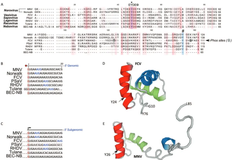
Conservation of the calicivirus 5′ end and VPg sequences
VPg sequences vary in length from 65 amino acids for bovine nebovirus to 138 amino acids for Norwalk virus. The recent solution of the structures of the VPg proteins from MNV, FCV, and porcine sapovirus (PSaV) highlight the presence of conserved helical bundles at the core of VPg, tightly bound in hydrophobic interactions (Leen et al., 2013;
either in vitro biochemical assays (Machı´n, Martı´n Alonso & Parra, 2001;Belliot et al., 2008;Han et al., 2010) or a novel cell-based assay (Subba-Reddy, Goodfellow & Kao, 2011). However, discordant data was obtained for MNV where in vitro biochemical assays identified tyrosine at position 117 as the site for nucleotidylylation (Han et al., 2010). Importantly, mutational analysis of this amino acid in the context of the MNV infectious clone confirmed that Y117 was not required for viral infectivity whereas Y24 was essential (Subba-Reddy, Goodfellow & Kao, 2011). This highlights that, at least for MNV, in vitro biochemical approaches using recombinant purified proteins, are confounded by an apparent lack of specificity of the viral RdRp.
Alignment of the 5′end sequences of representative calicivirus genomes has demonstrated that almost all genomic and subgenomic RNAs start with a GU
dinucleotide (Figs. 1Band1C). Tulane virus is an exception to this, as published data would indicate that the genome begins with a GGG sequence (Farkas et al., 2008). It is worth noting that there is only a single full-length genome sequence available for Tulane virus, therefore confirmation of the 5′end may require additional viral sequences. Based entirely on sequence conservation, we would expect calicivirus VPg proteins to be guanylylated on the conserved tyrosine within the EYDEsequence equivalent to positions 24 and 26 for FCV and MNV respectively. These amino acids are predicted to lie within the structured region of the FCV and MNV proteins as highlighted inFigs. 1Dand1E, respectively.
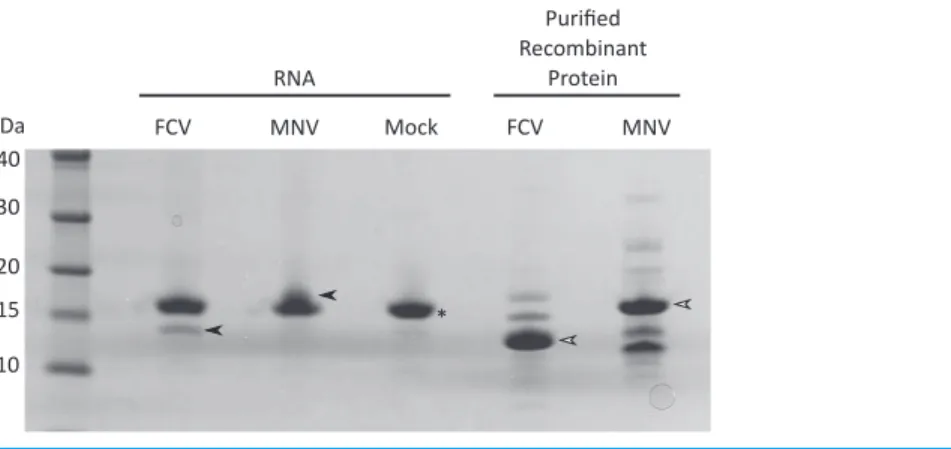
Purification of viral VPg-linked RNA
and subsequent mass spectrometry (see below) was used to confirm the isolation of the MNV VPg protein.
Detection of FCV and MNV VPg peptides and post-translational modifications
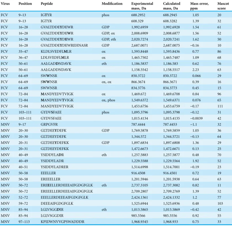
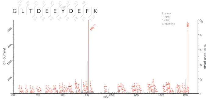
Initial attempts were made to analyse the RNase treated and trypsin-digested VPg-linked RNA preparations as a method to identify the nucleotide and amino acids involved in the covalent linkage; however, this approach failed to produce spectra that allowed for the detection of the nucleotide-linked VPg peptides. As an alternative approach, we used tryptic digestion of VPg-linked RNA preparations followed by acid hydrolysis of the RNA-linked peptides as described previously (Olspert et al., 2011a;Olspert et al., 2011b). The RNA-linked amino-acid residue modification after RNA hydrolysis using this method is known to be a 5′,3′-diphosphate nucleotide, pNp (N denoting adenosine, cytidine, guanosine or uridine), and the possible phosphodiester bond acceptor residues are serine, tyrosine and threonine. FCV and MNV VPg-linked RNA preparations were prepared and analyzed by Orbitrap MS. The sequence coverage obtained for the FCV and MNV VPg proteins were 70 and 63%, respectively. The identified peptides are shown in Table 1,
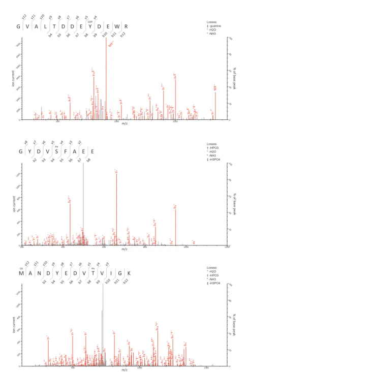
Figs. 3and4. The regions not detected were most likely absent due to trypsin digestion producing peptides too short for detection or confident identification.
Using this approach we determined that the FCV VPg is linked to RNA through the tyrosine residue at position 24 (Y24) and the corresponding modification was pGp, as assigned by the modification delta mass and the corresponding fragmentation spectrum (Fig. 3B). This is in agreement with the high degree of conservation of a 5′G nucleotide in all calicivirus genomes (Figs. 1B and1C). The spectra were searched against all possible nucleotides (pGp, pUp, pCp and pAp) but no other matches were detected indicating that all the detected VPg peptides were derived from linkage to the positive strand of viral RNA. The corresponding FCV VPg peptide was never detected without pGp modification.
In FCV VPg we also identified two potential phosphorylation sites; threonine at position 80 (Fig. 3C) and serine at position 107 (Fig. 3D) were consistently detected as
phosphorylated but the same peptides were also detected without the modification. This might indicate loss of modification during sample handling and/or transient Table 1 Examples of detected peptides identified by fragmentation spectra.The post-translational modifications are described and the modified position is in bold in the peptide.
Virus Position Peptide Modification Experimental
mass, Da
Calculated mass, Da
Mass error, ppm
Mascot score
FCV 9–13 IGTYR phos 688.2952 688.2945 1.05 20
FCV 9–13 IGTYR 608.329 608.3282 1.39 32
FCV 16–28 GVALTDDEYDEWR GDP 1,992.6959 1,992.6928 1.54 61
FCV 16–28 GVALTDDEYDEWR GDP, ox 2,008.6909 2,008.6877 1.56 52
FCV 16–28 GVALTDDEYDEWR GDP, eth 2,020.7274 2,020.7241 1.62 50
FCV 16–28 GVALTDDEYDEWREHNASR GDP 2,687.0071 2,687.0075 -0.16 10
FCV 35–47 KLDLSVEDFLMLR ox 1,593.8448 1,593.8436 0.77 86
FCV 36–47 LDLSVEDFLMLR ox 1,465.7502 1,465.7487 1.09 68
FCV 50–61 AALGADDNDAVK eth 1,186.5837 1,186.583 0.62 76
FCV 50–61 AALGADDNDAVK 1,158.5542 1,158.5517 2.23 63
FCV 64–69 SWWNSR ox 850.3722 850.3722 0.066 29
FCV 64–69 SWWNSR ox, ox 866.3674 866.3671 0.39 16
FCV 64–69 SWWNSR 834.3776 834.3773 0.45 15
FCV 72–84 MANDYEDVTVIGK ox 1,469.672 1,469.6708 0.84 96
FCV 72–84 MANDYEDVTVIGK ox, phos 1,549.6372 1,549.6371 0.076 65
FCV 72–84 MANDYEDVTVIGK 1,453.6756 1,453.6759 -0.17 111
FCV 103–111 GYDVSFAEE phos 1,095.3796 1,095.3798 -0.12 31
FCV 103–111 GYDVSFAEE 1,015.4134 1,015.4135 -0.0039 42
MNV 9–17 GRPGVFR 787.4444 787.4453 -1.1 32
MNV 20–30 GLTDEEYDEFK GDP 1,769.5878 1,769.5859 1.05 36
MNV 20–30 GLTDEEYDEFK 1,344.572 1,344.5721 -0.13 64
MNV 20–31 GLTDEEYDEFKK GDP 1,897.6834 1,897.6808 1.36 29
MNV 20–31 GLTDEEYDEFKK 1,472.6673 1,472.6671 0.13 25
MNV 40–49 YSIDDYLADR eth 1,257.5883 1,257.5877 0.48 70
MNV 40–49 YSIDDYLADR 1,229.5588 1,229.5564 1.92 52
MNV 40–51 YSIDDYLADRER 1,514.6998 1,514.7001 -0.19 23
MNV 50–58 EEELLER 916.4508 916.4501 0.72 19
MNV 50–58 EREEELLER 1,201.5946 1,201.5938 0.64 63
MNV 50–72 EREEELLERDEEEAIFGDGFGLK eth 2,737.3105 2,737.3082 0.82 11
MNV 50–72 EREEELLERDEEEAIFGDGFGLK 2,709.2807 2,709.2769 1.39 52
MNV 52–72 EEELLERDEEEAIFGDGFGLK 2,424.1361 2,424.1332 1.2 77
MNV 59–72 DEEEAIFGDGFGLK 1,525.6944 1,525.6936 0.48 103
MNV 85–94 LGLVSGGDIR eth 1,013.5865 1,013.5869 -0.42 42
MNV 85–94 LGLVSGGDIR 985.5566 985.5556 0.92 55
MNV 97–113 KPIDWNVVGPSWADDDR 1,968.9345 1,968.933 0.75 33
Note:
nature of the modification. Threonine at position 11 was also detected in a
phosphorylated form (Table 1), however this was identified in only one of the two FCV samples. Unfortunately, due to peptide’s short length and low amount of matched fragmentation ions, this is a low confidence observation. Threonine at position 11 is not conserved between FCV isolates, with all other isolated possessing a proline at this position, while T80 and S107 are 100% conserved across all FCV isolates. Amino acids T80 and S107 are in disordered region of the FCV VPg protein (Fig. 1D). It is unclear as to whether the phosphorylation would have resulted in a change in the mobility of VPg on SDS-PAGE.
The significance of VPg phosphorylation in the FCV life cycle clearly warrants further study using reverse genetics and mutagenesis. VPg is a multifunctional protein; it functions in viral RNA synthesis via an interaction with NS7, in viral protein synthesis via an interaction with translation initiation factors, and possibly in viral encapsidation via an interaction with VP1 (Goodfellow, 2011). Therefore, phosphorylation may provide a mechanism with which to regulate these roles in the virus life cycle.
The MNV VPg protein was identified as also linked to RNA through a tyrosine residue at position 26 and the modification was also pGp (Fig. 4B). Surprisingly for MNV, the corresponding peptide was also detected without the RNA modification, suggesting that RNA modification was lost during sample preparation. In addition, we detected random aspartate and glutamate ethylation, methionine and tryptophan oxidations (Table 1), which are known to be generated in vitro during sample preparation (Stadtman & Levine, 2003;Xing et al., 2008;Olspert et al., 2011a) and were therefore not considered of biological relevance. We did not observe evidence of MNV VPg phosphorylation. This difference between FCV and MNV may be due to inherent biological variation or may be a simple reflection of the lower abundance of MNV VPg in the samples analyzed and the sensitivity of the assay. Further studies are required to specifically address whether MNV VPg is post-translationally modified.
Previous studies on caliciviruses VPg nucleotidylylation have examined the ability of the viral RdRp to uridylylate VPg by leading to the formation of a VPg-pUpU(OH) moiety (Rohayem et al., 2006;Belliot et al., 2008). As all calicivirus genomic and
subgenomic RNAs begin with a single G, uridylylated VPg, if formed, would be expected to prime only negative sense RNA synthesis on the 3′poly A tail present on the viral genomic RNA. The end result would be VPg-pUpU at the 5′end of negative sense viral RNA. Whether VPg is uridylylated during calicivirus replication remains to be
determined, however our data would indicate that only guanosine diphosphate (GDP) was found linked to either the FCV or the MNV VPg proteins isolated from infected cells. Given the asymmetry of calicivirus genome replication, the number of negative sense RNA molecules present within an infected cell can be > 1,000 fold lower than the corresponding positive sense RNA molecule (Vashist, Urena & Goodfellow, 2012), which even if it were linked to uridylylated VPg, may be present in such low quantities to make detection challenging. We have previously demonstrated that the norovirus RdRp possesses the ability to initiate RNA synthesis de novo and that this activity is regulated by binding to the viral capsid protein VP1 (Subba-Reddy, Goodfellow & Kao, 2011). This has allowed us to propose a model whereby negative sense RNA synthesis occurs via a primer
independent de novo mechanism of RNA synthesis but positive sense RNA synthesis occurs via a VPg-primed mechanism (Reviewed inThorne & Goodfellow (2014)). In this model, only guanylylated VPg would be produced, fitting with our current experimental observations.
In conclusion, this work has provided experimental confirmation that at least for FCV and MNV, the covalent linkage of the VPg proteins to the 5′end of the viral genome occurs specifically via a highly conserved tyrosine residue to the 5′G nucleotide. The identification of potential phosphorylated sites in the FCV VPg protein may provide a mechanism by which the function of VPg is temporally regulated during the viral life cycle.
ADDITIONAL INFORMATION AND DECLARATIONS
Funding
Funding is provided by Wellcome Trust (Ref: 097997/Z/11/Z) and by the institutional research funding IUT 19-3 of the Estonian Ministry of Education and Research. The funders had no role in study design, data collection and analysis, decision to publish, or preparation of the manuscript.
Competing Interests
The authors declare that they have no competing interests.
Author Contributions
Allan Olspert conceived and designed the experiments, performed the experiments,
analyzed the data, contributed reagents/materials/analysis tools, wrote the paper, prepared figures and/or tables, reviewed drafts of the paper.
Myra Hosmillo conceived and designed the experiments, analyzed the data, contributed
reagents/materials/analysis tools, wrote the paper, prepared figures and/or tables, reviewed drafts of the paper.
Yasmin Chaudhry conceived and designed the experiments, performed the experiments,
analyzed the data, contributed reagents/materials/analysis tools, wrote the paper, prepared figures and/or tables, reviewed drafts of the paper.
Lauri Peil conceived and designed the experiments, performed the experiments,
analyzed the data, contributed reagents/materials/analysis tools, wrote the paper, prepared figures and/or tables, reviewed drafts of the paper.
Erkki Truve conceived and designed the experiments, analyzed the data, contributed
reagents/materials/analysis tools, wrote the paper, prepared figures and/or tables, reviewed drafts of the paper.
Ian Goodfellow conceived and designed the experiments, performed the experiments,
analyzed the data, contributed reagents/materials/analysis tools, wrote the paper, prepared figures and/or tables, reviewed drafts of the paper.
Data Deposition
The following information was supplied regarding data availability: All raw data is included in the manuscript.
REFERENCES
Belliot G, Sosnovtsev SV, Chang K-O, McPhie P, Green KY. 2008.Nucleotidylylation of the VPg protein of a human norovirus by its proteinase-polymerase precursor protein.Virology 374(1):33–49DOI 10.1016/j.virol.2007.12.028.
Chaudhry Y, Nayak A, Bordeleau M-E, Tanaka J, Pelletier J, Belsham GJ, Roberts LO, Goodfellow IG. 2006.Caliciviruses differ in their functional requirements for eIF4F components.Journal of Biological Chemistry281(35):25315–25325DOI 10.1074/jbc.M602230200.
Chung L, Bailey D, Leen EN, Emmott EP, Chaudhry Y, Roberts LO, Curry S, Locker N, Goodfellow IG. 2014.Norovirus translation requires an interaction between the C terminus of the genome-linked viral protein VPg and eukaryotic translation initiation factor 4G.Journal of Biological Chemistry289(31):21738–21750DOI 10.1074/jbc.M114.550657.
Farkas T, Sestak K, Wei C, Jiang X. 2008.Characterization of a rhesus monkey calicivirus representing a new genus of Caliciviridae.Journal of Virology82(11):5408–5416
DOI 10.1128/JVI.00070-08.
Fuentes C, Bosch A, Pinto RM, Guix S. 2012.Identification of human astrovirus genome-linked protein (VPg) essential for virus infectivity.Journal of Virology86(18):10070–10078
DOI 10.1128/JVI.00797-12.
Goodfellow I, Chaudhry Y, Richardson A, Meredith J, Almond JW, Barclay W, Evans DJ. 2000. Identification of a cis-acting replication element within the poliovirus coding region.Journal of Virology74(10):4590–4600DOI 10.1128/JVI.74.10.4590-4600.2000.
Goodfellow I, Chaudhry Y, Gioldasi I, Gerondopoulos A, Natoni A, Labrie L, Laliberte´ J-F, Roberts L. 2005.Calicivirus translation initiation requires an interaction between VPg and eIF4E.EMBO Reports6(10):968–972DOI 10.1038/sj.embor.7400510.
Goodfellow I. 2011.The genome-linked protein VPg of vertebrate viruses—a multifaceted protein.Current Opinion in Virology1(5):355–362DOI 10.1016/j.coviro.2011.09.003. Han KR, Choi Y, Min BS, Jeong H, Cheon D, Kim J, Jee Y, Shin S, Yang JM. 2010.Murine
norovirus-1 3Dpol exhibits RNA-dependent RNA polymerase activity and nucleotidylylates on Tyr of the VPg.Journal of General Virology91(7):1713–1722DOI 10.1099/vir.0.020461-0. Herbert TP, Brierley I, Brown TD. 1997.Identification of a protein linked to the genomic and
subgenomic mRNAs of feline calicivirus and its role in translation.Journal of General Virology 78(5):1033–1040DOI 10.1099/0022-1317-78-5-1033.
Hewlett MJ, Florkiewicz RZ. 1980.Sequence of picornavirus RNAs containing a radioiodinated 5′-linked peptide reveals a conserved 5′sequence.Proceedings of the National Academy of Sciences of the United States of America77(1):303–307DOI 10.1073/pnas.77.1.303.
Hosmillo M, Chaudhry Y, Kim D-S, Goodfellow I, Cho K-O. 2014.Sapovirus translation requires an interaction between VPg and the cap binding protein eIF4E.Journal of Virology
88(21):12213–12221DOI 10.1128/JVI.01650-14.
Hwang H-J, Min HJ, Yun H, Pelton JG, Wemmer DE, Cho K-O, Kim J-S, Lee CW. 2015.Solution structure of the porcine sapovirus VPg core reveals a stable three-helical bundle with a conserved surface patch.Biochemical and Biophysical Research Communications459(4):610–616
DOI 10.1016/j.bbrc.2015.02.156.
Jiang J, Laliberte´ J-F. 2011.The genome-linked protein VPg of plant viruses—a protein with many partners.Current Opinion in Virology1(5):347–354DOI 10.1016/j.coviro.2011.09.010. Langereis MA, Feng Q, Nelissen FHT, Virgen-Slane R, van der Heden van Noort GJ,
Maciejewski S, Filippov DV, Semler BL, van Delft FL, van Kuppeveld FJM. 2014.
Leen EN, Kwok KYR, Birtley JR, Simpson PJ, Subba-Reddy CV, Chaudhry Y, Sosnovtsev SV, Green KY, Prater SN, Tong M, Young JC, Chung LMW, Marchant J, Roberts LO, Kao CC, Matthews S, Goodfellow IG, Curry S. 2013.Structures of the compact helical core domains of feline calicivirus and murine norovirus VPg proteins.Journal of Virology87(10):5318–5330
DOI 10.1128/JVI.03151-12.
Machı´n A, Martı´n Alonso JM, Parra F. 2001.Identification of the amino acid residue involved in rabbit hemorrhagic disease virus VPg uridylylation.Journal of Biological Chemistry276:27787– 27792DOI 10.1074/jbc.M100707200.
Maciejewski S, Nguyen JHC, Go´mez-Herreros F, Corte´s-Ledesma F, Caldecott KW, Semler BL. 2016.Divergent requirement for a DNA repair enzyme during enterovirus infections.mBio 7(1):e1931-15DOI 10.1128/mBio.01931-15.
Mitra T, Sosnovtsev SV, Green KY. 2004.Mutagenesis of tyrosine 24 in the VPg protein is lethal for feline calicivirus.Journal of Virology78(9):4931–4935
DOI 10.1128/JVI.78.9.4931-4935.2004.
Olsen JV, de Godoy LM, Li G, Macek B, Mortensen P, Pesch R, Makarov A, Lange O, Horning S, Mann M. 2005.Parts per million mass accuracy on an Orbitrap mass spectrometer via lock mass injection into a C-trap.Molecular and Cellular Proteonomics4(12):2010–2021
DOI 10.1074/mcp.T500030-MCP200.
Olspert A, Arike L, Peil L, Truve E. 2011a.Sobemovirus RNA linked to VPg over a threonine residue.FEBS Letters585(19):2979–2985DOI 10.1016/j.febslet.2011.08.009.
Olspert A, Peil L, He´brard E, Fargette D, Truve E. 2011b.Protein-RNA linkage and post-translational modifications of two sobemovirus VPgs.Journal of General Virology 92(2):445–452DOI 10.1099/vir.0.026476-0.
Paul AV, Rieder E, Kim DW, van Boom JH, Wimmer E. 2000.Identification of an RNA hairpin in poliovirus RNA that serves as the primary template in the in vitro uridylylation of VPg.Journal of Virology74(22):10359–10370DOI 10.1128/JVI.74.22.10359-10370.2000.
Rappsilber J, Mann M, Ishihama Y. 2007.Protocol for micro-purification, enrichment, pre-fractionation and storage of peptides for proteomics using StageTips.Nature Protocols 2(8):1896–1906DOI 10.1038/nprot.2007.261.
Rohayem J, Robel I, Ja¨ger K, Scheffler U, Rudolph W. 2006.Protein-primed and de novo initiation of RNA synthesis by norovirus 3Dpol.Journal of Virology80(14):7060–7069
DOI 10.1128/JVI.02195-05.
Rothberg PG, Harris TJ, Nomoto A, Wimmer E. 1978.O4-(5′-uridylyl)tyrosine is the bond between the genome-linked protein and the RNA of poliovirus.Proceedings of the National Academy of Sciences of the United States of America75(10):4868–4872
DOI 10.1073/pnas.75.10.4868.
Stadtman ER, Levine RL. 2003.Free radical-mediated oxidation of free amino acids and amino acid residues in proteins.Amino Acids25(3–4):207–218DOI 10.1007/s00726-003-0011-2. Subba-Reddy CV, Goodfellow I, Kao CC. 2011.VPg-primed RNA synthesis of norovirus
RNA-dependent RNA polymerases by using a novel cell-based assay.Journal of Virology 85(24):13027–13037DOI 10.1128/JVI.06191-11.
Thorne LG, Goodfellow IG. 2014.Norovirus gene expression and replication.Journal of General Virology95(Pt 2):278–291DOI 10.1099/vir.0.059634-0.
Virgen-Slane R, Rozovics JM, Fitzgerald KD, Ngo T, Chou W, van der Heden van Noort GJ, Filippov DV, Gershon PD, Semler BL. 2012.An RNA virus hijacks an incognito function of a DNA repair enzyme.Proceedings of the National Academy of Sciences of the United States of America109(36):14634–14639DOI 10.1073/pnas.1208096109.