Capping Enzyme
Brittney R. Henderson1, Bejan J. Saeedi1, Grace Campagnola2, Brian J. Geiss1,2*
1Department of Microbiology, Immunology, and Pathology, Colorado State University, Fort Collins, Colorado, United States of America,2Department of Biochemistry and Molecular Biology, Colorado State University, Fort Collins, Colorado, United States of America
Abstract
Flaviviruses are small, capped positive sense RNA viruses that replicate in the cytoplasm of infected cells. Dengue virus and other related flaviviruses have evolved RNA capping enzymes to form the viral RNA cap structure that protects the viral genome and directs efficient viral polyprotein translation. The N-terminal domain of NS5 possesses the methyltransferase and guanylyltransferase activities necessary for forming mature RNA cap structures. The mechanism for flavivirus guanylyltransferase activity is currently unknown, and how the capping enzyme binds its diphosphorylated RNA substrate is important for deciphering how the flavivirus guanylyltransferase functions. In this report we examine how flavivirus NS5 N-terminal capping enzymes bind to the 59end of the viral RNA using a fluorescence polarization-based RNA binding assay. We observed that the KDfor RNA binding is approximately 200 nM Dengue, Yellow Fever, and West Nile virus capping enzymes. Removal of one or both of the 59phosphates reduces binding affinity, indicating that the terminal phosphates contribute significantly to binding. RNA binding affinity is negatively affected by the presence of GTP or ATP and positively affected by S-adensyl methoninine (SAM). Structural superpositioning of the dengue virus capping enzyme with the Vaccinia virus VP39 protein bound to RNA suggests how the flavivirus capping enzyme may bind RNA, and mutagenesis analysis of residues in the putative RNA binding site demonstrate that several basic residues are critical for RNA binding. Several mutants show differential binding to 59di-, mono-, and un-phosphorylated RNAs. The mode of RNA binding appears similar to that found with other methyltransferase enzymes, and a discussion of diphosphorylated RNA binding is presented.
Citation:Henderson BR, Saeedi BJ, Campagnola G, Geiss BJ (2011) Analysis of RNA Binding by the Dengue Virus NS5 RNA Capping Enzyme. PLoS ONE 6(10): e25795. doi:10.1371/journal.pone.0025795
Editor:K.T. Jeang, National Institute of Health, United States of America
ReceivedJuly 28, 2011;AcceptedSeptember 11, 2011;PublishedOctober 12, 2011
Copyright:ß2011 Henderson et al. This is an open-access article distributed under the terms of the Creative Commons Attribution License, which permits unrestricted use, distribution, and reproduction in any medium, provided the original author and source are credited.
Funding:This project was supported by a grant from the Rocky Mountain Regional Center for Excellence 10 (U54 AI-065357) to BJG. The funders had no role in study design, data collection and analysis, decision to publish, or preparation of the manuscript.
Competing Interests:The authors have declared that no competing interests exist.
* E-mail: [email protected]
Introduction
Dengue viruses are members of the Flaviviridae family (genus Flavivirus), which are small RNA viruses of 10–11 Kb in length
with capped non-polyadenylated positive strand genomes. Dengue virus proteins are produced from a single open reading frame via translation of the genomic viral RNA as a single polyprotein that is co-translationally processed into 3 structural proteins (Capsid, prM, and Envelope) and 8 non-structural proteins (NS1, NS2A, NS2B, NS3, NS4A, 2K, NS4B, and NS5). The non-structural proteins are responsible for directing viral genomic RNA replication, including synthesizing negative- and positive-strand RNAs and forming the viral RNA cap structure.
The flavivirus RNA cap is critical for viral polyprotein translation and RNA replication. The RNA cap allows the viral RNA to be efficiently translated by the cellular translational machinery and provides protection for the genome from cellular exonucleases. Flavivirus genomic RNA replication occurs on rough endoplasmic reticulum membranes in membranous com-partments away from the cellular capping machinery, requiring the viruses to develop a mechanism for generating an RNA cap structure. Dengue and other flaviviruses have evolved a complete RNA capping machinery to form an RNA cap on the 59end of the positive-strand genomic RNA. Cellular RNA cap structures are
formed via the action of an RNA triphosphatase (RTPase), guanylyltransferase (GTase), N7-methyltransferase (N7-MTase), and 29-O methyltransferase (29O-MTase) [1]. Flavivirus genomic RNA is modified at the 59 end of positive strand genomic RNA with a cap 1 structure (me7-GpppA-me2) generated by the virus encoded RTPase (NS3), GTase (NS5), 29-OMTase (NS5), and Guanine-N7-MTase (NS5) [2,3,4,5,6,7,8,9]. X-ray crystal struc-tures for each of these viral enzymes have been solved [3,8,10,11] providing a wealth of information about how these enzymes may function. The RTPase resides within the helicase domain of NS3 and appears to utilize the helicase ATP hydrolysis site to remove thec-phosphate from the 59end of the RNA [12]. The NS5 N-terminal capping enzyme domain (dengue virus NS5 AA 1–265) possesses the 29-O-MTase, Guanine-N7-MTase, and GTase activities and the NS5 C-terminal domain possesses the RNA dependent RNA polymerase [7,8,13,14,15,16,17].
how the protein binds the uncapped diphosphorylated RNA substrate for the GTase reaction. The current location of the RNA binding region has been suggested based on the presence of basic residues andin silicomolecular dynamics docking of an RNA into
the crystal structure of the dengue capping enzyme [18]. A recent structure of the dengue virus type 3 capping enzyme in complex with an octomeric capped RNA demonstrated interactions between the guanosine cap structure and the capping enzyme showed no interactions between the RNA and the capping enzyme putative RNA binding region [19]. This structure may represent the post-capping product, but does not shed light onto how the capping enzyme may bind diphosphorylated RNA during capping. The flavivirus NS5 capping enzyme does not encode a canonical Kx[D/N]G motif or any other known GTase motifs [20,21,22,23]. Since the flavivirus capping enzyme is able to form a guanylated intermediate (a GMP linked to the protein via a phosphoamide bond) and transfer GMP to a diphosphorylated RNA [7], it stands to reason that the capping enzyme must have a non-canonical GTase motif. Understanding how the capping enzyme binds its diphosphorylated RNA substrate is critical for deciphering how this non-canonical GTase functions, but at this point how it binds diphosphorylated RNA is unclear.
In this manuscript we examine the binding of the viral 59
diphosphorylated RNA substrate to the dengue virus capping enzyme. We developed a fluorescence polarization-based RNA binding assay to monitor the association of a short dipho-sphorylated RNA corresponding to the conserved 59 end of the flavivirus genome and determined the RNA binding affinity to the capping enzyme. We assessed the effects of the various ligands used by the capping enzyme on RNA binding affinity, and determined that binding is negatively affected by GTP and ATP and positively affected by SAM. We also performed a structure-directed mutational analysis of the dengue 2 capping enzyme to determine which amino acids may be involved with RNA binding based on the structural similarity of the dengue virus capping enzyme with the Vaccinia virus VP39 methyltransferase protein bound to RNA. We identified several residues that are critical for binding to RNA and report their relative contribution to binding. We have also explored the contribution of the 59 phosphates to RNA binding and found that the 59 b- and a- phosphates are critical for diphosphorylated RNA binding to the capping enzyme.
Materials and Methods
Expression and purification of flavivirus capping enzyme proteins
Recombinant dengue virus type 2, yellow fever virus, and West Nile virus capping enzymes were previously described [7,11]. Dengue capping enzyme was produced in BL21 (DE3) pLysSE. colicells (Novagen). Cultures (750 ml) were induced with 400mM IPTG overnight at 22uC, and the bacterial pellets were collected and stored at280uC in low imidizole lysis buffer. Frozen pellets were thawed and lysed with a M-110-L Pneumatic microfluidizer (Microfluidics Inc.), and the lysate was clarified by centrifugation at 18 K RPM in a SS-24 rotor and filtered through a 0.22mM syringe filter. The histidine-tagged protein was purified from clarified lysates using a Hi-Trap Nickel column (GE Healthcare) on an AKTA Purifier FPLC system. The eluted proteins were concentrated using 10 K Amicon Ultra concentrators (Millipore), and buffer exchanged into 400 mM NaCl, 20 mM Tris-Base pH 7.5, 0.02% sodium azide, 20% glycerol, and 5 mM Tris(2-Carboxyethyl) phosphine hydrochloride (TCEP-HCl) on a Super-dex 200 gel filtration column (Amersham). Purified proteins were concentrated using 10 K Amicon Ultra concentrators to 100mM
and the concentrations were determined by the absorbance at 280 nm on a NanoDrop 2000 spectrophotometer (Nanodrop, Inc.) using extinction coefficients obtained from the ExPASy web site. Isolated proteins were.98% pure as estimated from SDS-PAGE and Coomassie Blue staining.
Fluorescence Polarization RNA binding assay
Fluorescence polarization (FP) RNA binding assays were performed with purified wild-type and mutant dengue 2 capping enzymes and 39-fluoroscein (FAM) labeled 5-base RNAs (59
ppAGUAA-FAM, 59 pAGUAA-FAM, 59 AGUAA-FAM). The RNAs were chemically synthesized (Biosynthesis, Inc), HPLC purified, and verified by mass spectrometry. SAM and SAH were purchased from New England Biolabs. GTP, ATP, GDP, and GMP were purchased from Sigma-Aldrich. All FP experiments were performed in 50ml volumes in black 384-well microtiter
plates. Binding reactions were carried out in final concentrations of 50 mM Tris pH 7.5, 0.1% NP-40, 2 mM DTT, 50 mM NaCl, and 50 nM RNA. Wild-type and mutant dengue capping enzymes were serially diluted in 400 mM NaCl, 50 mM Tris pH 7.5, and 2 mM DTT and added to the RNA mix. The binding reactions were incubated at 28uC for 1 hr, then FP and total fluorescence signals were detected using a Victor 3 V multimode platereader set to 28uC (Perkin Elmer). All FP experiments were performed in triplicate with free RNA and total bound RNA controls. KDvalues
for RNA binding were determined using Kalidagraph (Synergy Software) using an equation based on [24]. In cases where the curves did not reach saturation, we used the average milli-polarization (mP) values for fully bound control samples and force-fitted the curves to those values to estimate KD. ppAGUAA-FAM
and pAGUAA-FAM had average free and bound mP values of 125 mP and 325 mP, respectively, whereas AGUAA-FAM had average free and bound values of 155 mP and 425 mP.
Structural Alignment and visualization
Structural alignment of the dengue virus capping enzyme (PDB code: 2P1D) with the Vaccinia VP39 methyltransferase domain (PDB code: 1AV6) was performed with the TopMatch Alignment Server (http://topmatch.services.came.sbg.ac.at) [25,26]. Struc-tural figures were generated with the PyMOL Molecular Graphics System [27].
Results
Development of the fluorescence polarization RNA binding assay
We established a fluorescence polarization-based RNA binding assay to monitor binding of the capping enzyme to the 59end of the genomic RNA. The 59 end of the dengue, yellow fever, and West Nile virus genomic RNA is conserved as an ‘‘59-AG(U/A)’’ sequence, and we observed that a 59ppAG terminated RNA not related to a flavivirus 59 UTR can be capped by the capping enzyme [7], indicating that only the very 59base sequences of the RNA are necessary for the GTase reaction and that RNA structures important for MTase activity are not necessary for the RNA guanyltransfer step [16,17]. We have used short 5 base AGUAA RNAs with different 59ends (59ppAGUAA, 59pAGUAA, or 59AGUAA) and 39 6-carboxyfluorescein (FAM) as tools to monitor binding in solution. These RNAs show a polarization of 115–130 mP units in solution when unbound to the capping enzyme, but upon binding polarization signal increases to,330
We first determined the KD for ppAGUAA RNA binding to
dengue 2, yellow fever, and West Nile virus capping enzymes. We obtained similar KDvalues for ppAGUAA bound to the dengue
and West Nile virus proteins (KD= 18766 nM, and 13668 nM
respectively; Figure 1) and slightly weaker to the yellow fever virus capping enzyme (KD= 420620 nM, Figure 1). The RNA binds
similarly between the three viruses, so we chose to focus on the dengue capping enzyme for the remainder of the project due to its ease of purification and the availability of mutants from our previous work [7,11].
Effects of 59phosphates on RNA binding
The presence of two additional phosphates at the 59end of the viral RNA likely contribute to the overall binding affinity between the RNA and the capping enzyme. To determine what roles thea -and b-phosphates at the 59end of the RNA play in binding, we determined the affinities of ppAGUAA, pAGUAA, and AGUAA RNAs for binding wild-type dengue capping enzyme (18766 nM, 967688 nM, and 3.860.2mM respectively) (Figure 2). The Hill slopes of the ppAGUAA and pAGUAA curves are 1.37 and 1.46 as compared to 1.1 for AGUAA, which may indicate very weak positive cooperativity in binding for the 59phosphorylated species. However, the Hill slopes for West Nile and yellow fever capping enzymes in Figure 1 were both,1.1, so the increased Hill slopes
observed in Figure 2 may be dengue specific. These data indicate that the 59 terminal phosphates contribute significantly to RNA binding, and thea- andb-phosphates both contribute to binding
affinity, although b-phosphate appears to contribute more significantly to binding affinity.
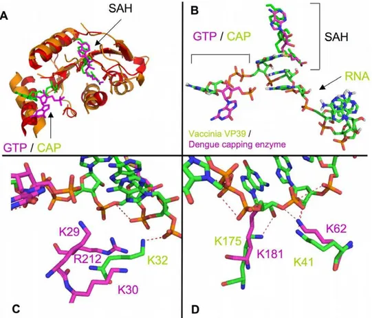
Structural superposition of the Vaccinia Virus VP39 with the yellow fever capping enzyme
The flavivirus capping enzyme does not have structural homology to known GTases but does have significant structural homology to methyltransferase enzymes. We performed a TopMatch structural alignment with the Vaccinia virus VP39 methyltransferase protein (PDB code: 1AV6) [28] which was crystallized with a bound capped RNA, and the dengue virus capping enzyme in complex with GTP (PDB code: 2P1D) (Figure 3). The alignment showed strong structural homology in the methyltransferase section of the capping enzyme (Figure 3A), but more interestingly the bound cap/GTP and SAH ligands were in very similar positions in both structures, and the RNA bound in 1AV6 is in close proximity to the basic patch of residues that has been postulated to be the RNA binding site (Figure 3B). Residues K32, K41, and K175 in the VP39 structure (1AV6) appear to interact with the phosphates of the RNA. Dengue virus capping enzyme residues K62, K181, and R212/K29/K30 appear to structurally overlap with VP39 residues K41, K175, and K32, respectively. The R212/K29/R30 cluster is slightly farther away from the RNA than VP39 K32 (Figure 3C), but would strongly interact with the RNA if it bent towards the residues. K181 (dengue) appears to structurally clash with the RNA ribose group in the 1AV6 structure, whereas K175 (VP39) interacts with the
Figure 1. Comparison of dengue, yellow fever, and West Nile virus capping enzyme KDvalues for ppAGUAA-FAM RNA.50 nM
ribose (Figure 3D). This may indicate that the RNA would need to be pushed away from its position in the VP39 structure to accommodate binding to K181.
Mutational Analysis of dengue virus capping enzyme RNA binding
Based on the structural alignment of VP39 and the dengue virus capping enzyme, we performed a mutagenesis analysis of the dengue capping enzyme to evaluate individual residue contribu-tions to RNA binding. Protein:RNA interaccontribu-tions commonly occur between basic residues (Arg and Lys) interacting with phosphates within and at the end of the RNA. The region between the GTP and SAM binding site on the capping enzyme is rich with basic residues, and has been hypothesized to be the RNA binding site. To examine the contribution of residues to RNA binding, we individually mutated conserved and semi-conserved (eg. K/R) residues on the GTP/SAM binding face of the capping enzyme and determined how each mutation affected binding affinity di-, mono-, and unphosphorylated RNAs (Table 1).
We observed that mutation of residues F25, K30, R57, K181, and R212 reduced the binding affinity to the greatest extent of all residues tested (#5-fold reduction of KD(Table 2)). The remaining
mutations had little effect on RNA binding affinity. K62A in the proposed RNA bind site did not affect RNA binding with any RNA species, indicating that it is not involved in binding RNA. K30A appeared to have a strong effect on binding with ppAGUAA and AGUAA, indicating that it interacts with a phosphate present in both RNA species, most likely the phosphate between the A and G bases of the AGUAA RNA. F25A was
initially added as a control because it interacts with the guanosine cap that is not present on the RNAs in this study. However, we observed that the mutant had significantly reduced binding to ppAGUAA RNA but no significant effect on pAGUAA and AGUAA binding. The phenylalanine group likely does not interact directly with the RNA, but a possible explanation is that mutation to alanine alters the position of helix A2 and moves K30 out of the optimal position to keep the 59diphosphate in line with other binding residues. R57A appeared to have its greatest effect when binding to ppAGUAA but less effect with pAGUAA and AGUAA, suggesting that R57 may interact predominately with theb-phosphate of the diphosphorylated RNA. K181A severely reduced ppAGUAA binding but had greatly reduced effects on pAGUAA and AGUAA binding, suggesting that K181 strongly interacts with theb-phosphate. R212A showed significant effects with binding to ppAGUAA and mild effects on pAGUAA binding (2.8 fold), suggesting that R212 interacts primarily with the b -phosphate but may weakly interact with the a-phosphate. In summary, based on these experiments R57, K181, and R212 likely interact with theb-phosphate, R212 may interact with thea -or b-phosphate, and K30 may interact with the phosphate between the A and G bases.
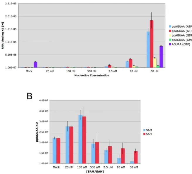
Effects of GTP and SAM on RNA binding affinity
Diphosphorylated RNA is one of three ligands involved in the guanyltransferase and methyltransferase reactions, the other two being GTP and SAM. We assessed the effects of GTP and SAM on RNA binding to wild-type dengue capping enzyme to determine if either could positively or negatively affected binding affinity. We
Figure 2. Effects of 59RNA phosphates on dengue capping enzyme binding affinity.50 nM of AGUAA-FAM, pAGUAA-FAM, and ppAGUAA-FAM was incubated with increasing concentrations of wild-type dengue capping enzyme for 1 hr, then fluorescence polarization signal was detected. n = 3.
first determined the KDof ppAGUAA in presence of increasing
amounts of GTP, GDP, GMP, and ATP (Figure 4A). We observed that high concentrations of GTP and ATP (50mM) significantly
weakened ppAGUAA binding (KD=,18mM), whereas GDP had
a moderate affect on ppAGUAA binding binding (KD=,3mM)
and GMP had a very minor effect on ppAGUAA binding
Figure 3. Structural superposition of the Vaccinia virus VP39 with the dengue virus capping enzyme. A) Global overlap of 1AV6 (VP39) and 2P1D (dengue virus capping enzyme). Superposition was performed using the TopMatch webserver, and figures were generated in PyMol. Red/ orange indicate structural overlap between 1AV6 and 2P1D. Non-overlapping regions are not shown. Bound GTP/Cap and SAH are shown. RNA has been removed for clarity.B) Overlay of bound ligands from 1AV6 (green) and 2P1D (magenta).C) Overlap of 1AV6 residue K32 (interacting with RNA phosphate#6) to 2P1D residues K29, K30, and R212.D) Overlap of 1AV6 residues K41 (interacting with RNA phosphate#4) and K175 (interacting with RNA ribose#1 hydroxyl) to 2P1D K62 and K181, respectively.
doi:10.1371/journal.pone.0025795.g003
Table 1.Binding affinities of mutant dengue capping enzyme proteins for different diphosphorylated RNA species and GTP.
AGUAA pAGUAA ppAGUAA GTP
Mutant Average KD SD Average KD SD Average KD SD Average KD SD
WT 3.7mM 217 nM 967 nM 88 nM 187 nM 6 nM 77 nM 4 nM
K22A 3.9mM 470 nM 862 nM 201 nM 689 nM 450 nM 120 nM 24 nM
F25A 6.0mM 687 nM 1.8mM 440 nM 1.4mM 501 nM 4.6mM 495 nM
K29A 4.1mM 1.0mM 1.4mM 75 nM 652 nM 59 nM 609 nM 154 nM
K30A 18.5mM 4.7mM 1.8mM 243 nM 4.0mM 778 nM 133 nM 41 nM
E35A 3.4mM 813 nM 524 nM 69 nM 247 nM 30 nM 67 nM 15 nM
R57A 2.1mM 462 nM 735 nM 137 nM 2.66mM 775 nM 78 nM 17 nM
G58A 2.9mM 337 nM 687 nM 71 nM 641 nM 54 nM 85 nM 36 nM
K62A 5.5mM 299 nM 1.3mM 649 nM 305 nM 122 nM 271 nM 59 nM
K181A 8.5mM 2.0mM 2.75mM 742 nM 4.6mM 1.9mM 359 nM 32 nM
R212A 7.9mM 845 nM 1.6mM 79 nM 1.0mM 222 nM 193 nM 31 nM
50 nM of the indicated AGUAA-FAM, pAGUAA-FAM, ppAGUAA-FAM, or 10 nM GTP-Bodipy were incubated with increasing concentrations of the indicated protein for 1 hr at 28uC then fluorescence polarization signal was detected. KDand standard deviation values are reported for each. n = 3.
(KD=,800 nM). The effect of GTP and ATP on ppAGUAA RNA
binding (an 80 fold reduction in binding) as compared to GDP and GMP levels suggest that thec- andb-phosphates on the nucleotides compete with the diphosphate on the RNA for binding. To further examine if the GTP b- and c- phosphates compete with diphosphorylated RNA binding, we determined the affinity of AGUAA RNA in the presence of 50mM GTP. We observed that AGUAA binding was weakened 2.4 fold in the presence of 50mM
GTP (KD(Mock) = 3.5mM, KD(50mM GTP) = 8.4mM),
indicat-ing that GTP had only minor effects on AGUAA bindindicat-ing. These results suggest that the GTP phosphates interfere with the diphosphate on the ppAGUAA RNA during binding.
To examine the effect of SAM and the post-methylation product SAH, we determined the KDof ppAGUAA binding to the
dengue capping enzyme in the presence of increasing amounts of SAM or SAH (Figure 4B). At low concentrations of SAM and SAH we observed no effect of either SAM or SAH on ppAGUAA binding affinity (KD=,200 nM), but we did observe a slight
increasing in ppAGUAA RNA affinity in the presence of increasing concentrations of both SAM and SAH. A slightly stronger effect on RNA binding affinity was evident with SAM than SAH, suggesting that SAM was able to stabilize RNA binding slightly better than SAH.
Mapping diphosphorylated RNA binding to the flavivirus capping enzyme
Based on our biochemical data, we mapped residues that significantly interacted with RNAs on RNA binding residues on the dengue virus capping enzyme in complex with GTP (PDB code: 2P1D). Figure 5 shows residues that were tested in this manuscript, and color codes the effects of each residue on RNA binding. Residues K22, K29, E35, and R62 had minimal effects on RNA binding (magenta), whereas residues F25, K30, R57, K181, and R212 (green) had significant effects on binding affinities (reduction greater than 5-fold). A clear clustering around the base of helix A2 is apparent, and suggests that the diphosphorylated RNA may enter the capping enzyme to be capped through the groove region between helices A2 and A3.
Discussion
In this study we performed a detailed characterization of the RNA binding characteristics of the dengue 2 capping enzyme. We
present data demonstrating that dengue, yellow fever, and West Nile virus capping enzyme proteins bind 59diphosphorylated end of the viral RNA with similar affinity, although yellow fever virus capping enzyme bound RNA with 2-fold weaker affinity than dengue or West Nile virus capping enzymes. The yellow fever virus capping enzyme has an arginine at position 30 whereas the dengue and West Nile virus capping enzymes have a lysine at position 30, which may explain the small difference in affinity between the viruses (Figure 1). Based on our biochemical and mutagenesis data, we present a preliminary model for capping enzyme binding to the 59 diphosphorylated end of the viral genomic RNA strand (Figure 5). The mechanism of action for the guanylyltransferase and methyltransferase activities of the flavivi-rus NS5 capping enzyme have been the focus of intensive study since the dengue capping enzyme structure was solved in 2002 [8,13,16,17]. Most of the effort has been focused on understanding the methyltransferase function. Because how the GTase activity within the capping enzyme functions is unknown, understanding how the capping enzyme binds one of its GTase substrates will help clarify how the enzyme works.
The overlap of the Vaccinia VP39 protein with the dengue capping enzyme is very strong in the methyltransferase region of the capping enzyme as had been previously noted, and superposition and alignment of residues involved in RNA cap binding have been described [8]. The location of the SAH in both structures is almost identical, and the GTP in the dengue virus capping enzyme is in close proximity to the cap structure in VP39. An obvious difference between the two structures is that the GTP/ Cap structures are flipped in respect to each other, indicating differing modes of guanine recognition. The VP39 cap and the dengue virus capping enzyme GTP are shifted approximately 5 A˚ from each other, indicating that a capped RNA bound to the dengue virus capping enzyme would also be shifted,5 A˚ from
where the RNA in the VP39 structure is situated.
Mutation of K62 in the dengue capping enzyme did not affect RNA binding, whereas the homologous lysine residue in the Vaccinia virus VP39 did interact with a phosphate (Figure 3D). K181 mutation strongly affected binding, suggesting that the superposition was partially correct. We observed a steric clash between K181 and the superimposed VP39 RNA, suggesting that the mode of K181 (dengue) interaction with RNA may be somewhat different than K175 (VP39) binding to the ribose of the first RNA nucleotide. The dramatic effect of mutating K30 on
Table 2.Comparison of RNA and GTP binding affinities.
Mutant Ratio Mutant/WT AGUAA KD Ratio Mutant/WT pAGUAA KD Ratio Mutant/WT ppAGUAA KD Ratio Mutant/WT GTP KD
WT 1.0 1.0 1.0 1.0
K22A 1.1 0.9 3.7 1.6
F25A 1.6 1.9 7.3 60.3
K29A 1.1 1.4 3.5 7.9
K30A 5.0 1.9 21.2 1.7
E35A 0.9 0.5 1.3 0.9
R57A 0.6 0.8 14.2 1.0
G58A 0.8 0.7 3.4 1.1
K62A 1.5 1.3 1.6 3.5
K181A 2.3 2.8 24.5 4.7
R212A 2.1 1.7 5.5 2.5
Figure 4. Effects of capping enzyme ligands on RNA binding. A) Effect of purine nucleotides on ppAGUAA-FAM and AGUAA-FAM RNA binding. KDvalues for ppAGUAA binding to wild-type dengue capping enzyme were determined in the presence of increasing concentrations of the indicated nucleotide. AGUAA binding was determined only in the presence of 50mM GTP or Mock.B) Effect of SAM and SAH on ppAGUAA-FAM RNA
binding affinity. n = 3.
doi:10.1371/journal.pone.0025795.g004
Figure 5. RNA binding residues on the dengue capping enzyme.All residues that were tested in this study were mapped on the dengue virus capping enzyme structure (2P1D) bound to GTP [11].A) Residues that showed greater than 5-fold reduction in RNA binding affinity against AGUAA, pAGUAA, or ppAGUAA are colored in green. Residues that showed less than 5-fold reduction in binding affinity against AGUAA, pAGUAA, or ppAGUAA are colored in magenta. Bound GTP and SAH are shown.B) Surface representation of 2P1D with RNA binding residues colored green and non-binding residues colored magenta.
RNA binding suggests that the K30 strongly interacts with the RNA. VP39 K32, which partially overlaps with the K29/K30/ R212 cluster, interacts with a phosphate, but K30 and R212 are situated about 5 A˚ away from the superimposed RNA. The lack of effect by mutating K62 and the strong effect by mutating K30 and R212 (to a lesser extent) suggest that the RNA is pulled back toward K30/R212 and away from K62 in the dengue capping enzyme structure. Supporting these observations is the effect seen on diphosphorylated RNA binding to the F25A mutant. This mutation severely affects GTP binding, but also appears to significantly affect RNA binding. Removing a hydrophobic phenylalanine likely perturbs the stability of the helix A2 (K30 is at the base of helix A2 (Figure 5)) and potentially reduces K30 binding the RNA by reducing its stability or moving it out of optimal binding position. Combining the differences in the locations of the cap and GTP with the effects on binding seen with K30 and R212 but not K62 suggests that the capped RNA would wrap tightly around helix A2 in the capping enzyme and have little to no interaction with the SAM binding face of the capping enzyme. This does not preclude the RNA ribose hydroxyl groups from being in an appropriate position to undergo 29-O methylation, but additional structural studies would need to be performed to determine their positions.
Addition of GTP and ATP to the ppAGUAA-FAM binding experiments showed that the phosphates on the nucleotides are able to compete with diphosphorylated RNA binding, and that reducing the number of phosphates on the nucleoside reduces the observed competitive effect. We also observed that high concen-trations of GTP had only minor effects on the binding of an unphosphorylated AGUAA RNA, suggesting that GTP predom-inately interferes with diphosphate binding. The position of GTP binding to the capping enzyme is well known [8,11], and GTP phosphates interact with several residues that are also involved in RNA interaction based on this study. R212 and K181 bind to GTP phosphates as well as to RNA (Table 1 and [11]), suggesting that the residues could interact with GTP or RNA at different times during capping. Since GTP was able to displace dipho-sphorylated RNA from the capping enzyme, it is likely that the capping enzyme would first bind GTP and form a guanylated protein intermediate prior to interacting with the diphosphory-lated RNA to form the cap. This could inform the order of substrate binding during the GTase reaction, which would help with the development of a catalytic mechanism for the GTase. The small increase in ppAGUAA-FAM affinity that was observed in the presence of SAM suggests either that SAM stabilizes the interaction between the RNA and the capping enzyme or that SAM transfers a methyl group to the RNA that increases the
affinity of the RNA for the capping enzyme. The effect observed with SAM may have a component of each situation, as SAH (which cannot methylate the RNA) show a weak stabilizing effect on RNA binding. We are currently testing if the diphosphorylated RNA is methylated in the presence of SAM by mass spectrometry. If we determine that the diphosphorylated RNA substrate is methylated in the presence of SAM, it would suggest that the genomic RNA may be methylated at the 29-O position prior to RNA capping, providing important information about the order of cap formation.
These studies provide a solid foundation for further exploration into how the flavivirus capping enzymes bind to the 59
diphosphorylated end of the viral genomic RNA during RNA capping. It provides a starting point for further investigations into to determinants of RNA binding, such as further clarifying which sidechains interact with which phosphate, determining the role of ribose hydroxyls in binding, and the specificity for guanine versus adenine in the first nucleotide position during capping [7]. Additional studies will help clarify these questions. Testing ppAGUAA-FAM RNAs with specific methylphosphanate substi-tutions in combination with capping enzyme mutants would help to map out specific amino acid:phosphate interactions [29]. Substituting ribose groups with 29deoxy ribose at specific positions in the RNA would allow for an examination of hydroxyl interactions with specific amino acids. Substitution of each base in the AGUAA RNA with analogs (eg. ppIGUAA-FAM, where I = inosine) would be critical for understanding the correlates of adenine vs. guanine specificity for capping the RNA [7]. With these data, molecular docking experiments could be performed that take into account the various distance restraints to build a biochemically-derived model of RNA binding. Our increased understanding of how the flavivirus capping enzyme binds to the substrates it uses to cap the genome will aid in our understanding of how this non-canonical capping enzyme functions, and will be valuable for the development of rationally designed capping enzyme specific drugs.
Acknowledgments
We would like to thank Dr. Olve Peersen, members of the Peersen lab, Dr. Martin Bisaillon, and Dr. Susan Keenan for helpful discussions about this project.
Author Contributions
Conceived and designed the experiments: BJG BRH. Performed the experiments: BRH BJS GC BJG. Analyzed the data: BJG BRH. Wrote the paper: BJG.
References
1. Bisaillon M, Lemay G (1997) Viral and cellular enzymes involved in synthesis of mRNA cap structure. Virology 236: 1–7.
2. Yon C, Teramoto T, Mueller N, Phelan J, Ganesh VK, et al. (2005) Modulation of the nucleoside triphosphatase/RNA helicase and 59-RNA triphosphatase activities of Dengue virus type 2 nonstructural protein 3 (NS3) by interaction with NS5, the RNA-dependent RNA polymerase. J Biol Chem 280: 27412–27419.
3. Xu T, Sampath A, Chao A, Wen D, Nanao M, et al. (2005) Structure of the Dengue virus helicase/nucleoside triphosphatase catalytic domain at a resolution of 2.4 A. J Virol 79: 10278–10288.
4. Warrener P, Tamura JK, Collett MS (1993) RNA-stimulated NTPase activity associated with yellow fever virus NS3 protein expressed in bacteria. J Virol 67: 989–996.
5. Ray D, Shah A, Tilgner M, Guo Y, Zhao Y, et al. (2006) West nile virus 59-cap structure is formed by sequential Guanine N-7 and ribose 29-o methylations by nonstructural protein 5. J Virol 80: 8362–8370.
6. Kuo MD, Chin C, Hsu SL, Shiao JY, Wang TM, et al. (1996) Characterization of the NTPase activity of Japanese encephalitis virus NS3 protein. J Gen Virol 77(Pt 9): 2077–2084.
7. Issur* M, Geiss* BJ, Bougie I, Picard-Jean F, Despins S, et al. (2009) The flavivirus NS5 protein is a true RNA guanylyltransferase that catalyzes a two-step reaction to form the RNA cap structure. RNA 15: 2340–2350. * = Equal Contributors.
8. Egloff MP, Benarroch D, Selisko B, Romette JL, Canard B (2002) An RNA cap (nucleoside-29-O-)-methyltransferase in the flavivirus RNA polymerase NS5: crystal structure and functional characterization. EMBO J 21: 2757–2768. 9. Benarroch D, Selisko B, Locatelli GA, Maga G, Romette JL, et al. (2004) The
RNA helicase, nucleotide 59-triphosphatase, and RNA 59-triphosphatase activities of Dengue virus protein NS3 are Mg2+-dependent and require a functional Walker B motif in the helicase catalytic core. Virology 328: 208–218. 10. Wu J, Bera AK, Kuhn RJ, Smith JL (2005) Structure of the Flavivirus helicase: implications for catalytic activity, protein interactions, and proteolytic process-ing. J Virol 79: 10268–10277.
11. Geiss BJ, Thompson AA, Andrews AJ, Sons RL, Gari HH, et al. (2009) Analysis of flavivirus NS5 methyltransferase cap binding. J Mol Biol 385: 1643–1654. 12. Bartelma G, Padmanabhan R (2002) Expression, purification, and
13. Zhou Y, Ray D, Zhao Y, Dong H, Ren S, et al. (2007) Structure and function of flavivirus NS5 methyltransferase. J Virol 81: 3891–3903.
14. Suaya JA, Shepard DS, Siqueira JB, Martelli CT, Lum LC, et al. (2009) Cost of dengue cases in eight countries in the Americas and Asia: a prospective study. Am J Trop Med Hyg 80: 846–855.
15. Koonin EV (1993) Computer-assisted identification of a putative methyltrans-ferase domain in NS5 protein of flaviviruses and lambda 2 protein of reovirus. J Gen Virol 74(Pt 4): 733–740.
16. Dong H, Ren S, Zhang B, Zhou Y, Puig-Basagoiti F, et al. (2008) West Nile virus methyltransferase catalyzes two methylations of the viral RNA cap through a substrate-repositioning mechanism. J Virol 82: 4295–4307.
17. Dong H, Ray D, Ren S, Zhang B, Puig-Basagoiti F, et al. (2007) Distinct RNA elements confer specificity to flavivirus RNA cap methylation events. J Virol 81: 4412–4421.
18. Milani M, Mastrangelo E, Bollati M, Selisko B, Decroly E, et al. (2009) Flaviviral methyltransferase/RNA interaction: structural basis for enzyme inhibition. Antiviral Res 83: 28–34.
19. Yap LJ, Luo D, Chung KY, Lim SP, Bodenreider C, et al. (2010) Crystal structure of the dengue virus methyltransferase bound to a 59-capped octameric RNA. PLoS One 5.
20. Cong P, Shuman S (1995) Mutational analysis of mRNA capping enzyme identifies amino acids involved in GTP binding, enzyme-guanylate formation, and GMP transfer to RNA. Mol Cell Biol 15: 6222–6231.
21. Qiu T, Luongo CL (2003) Identification of two histidines necessary for reovirus mRNA guanylyltransferase activity. Virology 316: 313–324.
22. Ogino T, Banerjee AK (2008) Formation of guanosine(59)tetraphospho(59 )ade-nosine cap structure by an unconventional mRNA capping enzyme of vesicular stomatitis virus. J Virol 82: 7729–7734.
23. Li J, Rahmeh A, Morelli M, Whelan SP (2008) A conserved motif in region v of the large polymerase proteins of nonsegmented negative-sense RNA viruses that is essential for mRNA capping. J Virol 82: 775–784.
24. Martin RL, Renosto F, Segel IH (1991) A simple method for calculating the dissociation constant of a receptor (or enzyme).unlabeled ligand complex from radioligand displacement measurements. Arch Biochem Biophys 284: 26–29. 25. Sippl MJ, Suhrer SJ, Gruber M, Wiederstein M (2008) A discrete view on fold
space. Bioinformatics 24: 870–871.
26. Sippl MJ, Wiederstein M (2008) A note on difficult structure alignment problems. Bioinformatics 24: 426–427.
27. PyMol website. Available: WWW.PyMol.Org. Accessed 2011 Sept 14. 28. Hodel AE, Gershon PD, Quiocho FA (1998) Structural basis for
sequence-nonspecific recognition of 59-capped mRNA by a cap-modifying enzyme. Mol Cell 1: 443–447.