J. Evid. Based Med. Healthc., pISSN- 2349-2562, eISSN- 2349-2570/ Vol. 3/Issue 81/Oct. 10, 2016 Page 4398
HYSTEROSCOPIC EVALUATION OF WOMEN IN REPRODUCTIVE AGE GROUP WITH
ABNORMAL UTERINE BLEEDING
E. Vanaja Reddy1, M. Nagalakshmi2, V. Sridevi3
1Assistant Professor, Department of Obstetrics and Gynaecology, MGMH, Petlaburj, Hyderabad.
2Assistant Professor, Department of Obstetrics and Gynaecology, MGMH, Petlaburj, Hyderabad.
3Postgraduate, Department of Obstetrics and Gynaecology, MGMH, Petlaburj, Hyderabad.
ABSTRACT
BACKGROUND
Abnormal uterine bleeding is the most common complaint in gynaecology and an important source of morbidity. This study evaluates the usefulness of hysteroscopy in the diagnosis of abnormal uterine bleeding in comparison to dilatation and curettage in reproductive age group.
MATERIALS AND METHODS
Between September 2011 to July 2013, women with AUB attending Gynaec OP were subjected to hysteroscopy and subsequent dilatation and curettage. Data was collected and analysed.
RESULTS
AUB was more common in 30-34 yrs. The most common presenting complaint was menorrhagia. Normal hysteroscopic view was seen in 50% cases. Abnormalities seen were endometrial hyperplasia, polyps, submucous myoma synechiae and rue. Both hysteroscopy and curettage gave specificity of 70%, but the ability to diagnose focal lesion (sensitivity) was more with hysteroscopy in comparison to curettage 70 vis. 36. 43 patients had the same tissue diagnosis in both hysteroscopy and curettage. Hysteroscopy revealed more information than curettage in 42% and curettage had more information in 15% cases, 100% accuracy was seen in case of myoma, IUCD, adhesions and polyps with hysteroscopy.
CONCLUSION
This study confirms the conclusion of many others that hysteroscopy is superior to dilatation and curettage in evaluating patients with abnormal uterine bleeding.
KEYWORDS
Uterine Bleeding, Hysteroscopy, Curettage.
HOW TO CITE THIS ARTICLE: Reddy EV, Nagalakshmi M, Sridevi V.Hysteroscopic evaluation of women in reproductive age
group with abnormal uterine bleeding.J. Evid. Based Med. Healthc. 2016; 3(81), 4398-4402. DOI: 10.18410/jebmh/2016/936
BACKGROUND: Although, uterine bleeding is a normal physiologic episodic occurrence to most women, its characteristics nevertheless vary considerably. The broad range of normal variation causes difficulty in identifying abnormal patterns. The problem is that uterine bleeding has a wide range of diagnostic possibilities and confusion is generated when review and reports fail to outline the diagnostic evaluation of the patient who presents with abnormal uterine bleeding patterns. Goals of clinical management are primarily dependent upon attaining a correct aetiological diagnosis. The history, physical and pelvic examination attempt to determine the site of the bleeding and its source. Information gathered from this will suggest what direction the investigation would take. Traditionally, Dilatation and Curettage and Ultrasonography
were the most common investigations employed in the evaluation of the causes of abnormal uterine bleeding. Dilatation and Curettage is a blind procedure and the endometrium has to be sent to the pathologist to study histological patterns and for the report. Ultrasonography clearly depicts the uterine contour and the status of the ovary, but fails to provide adequate information regarding the endometrium. Hysteroscopy has ushered a new era in the evaluation of abnormal uterine bleeding. By direct visualisation of the uterine cavity, it is able to pinpoint the abnormal focal area for biopsy. Abnormal uterine bleeding is one of the most common complaints with which a patient presents to a gynaecologist. D and C has long been the diagnostic gold standard for abnormal uterine bleeding. However, only 70%-80% of the endometrium can be curetted, polyps and submucous fibroids are frequently undetected by curettage alone. The judicious use of hysteroscopy to manage this medical entity adds a new dimension in handling this often perplexing problem. This study has been taken up to analyse the usefulness of hysteroscopy in the evaluation of Abnormal Uterine Bleeding in terms of accuracy of hysteroscopic findings and the contribution of the procedure to clinical diagnosis. It also Financial or Other, Competing Interest: None.
Submission 16-09-2016, Peer Review 24-09-2016, Acceptance 30-09-2016, Published 10-10-2016. Corresponding Author:
Dr. M. Nagalakshmi,
#502, Suchitra Residency, BAGH Amberpet, Hyderabad-13. E-mail: [email protected]
J. Evid. Based Med. Healthc., pISSN- 2349-2562, eISSN- 2349-2570/ Vol. 3/Issue 81/Oct. 10, 2016 Page 4399
aims to correlate hysteroscopic findings with
histopathological results.
MATERIALS AND METHODS: This study was conducted in Modern Government Maternity Hospital, Petlaburj, under Osmania Medical College, Hyderabad, which is a tertiary referral centre. The period of study was between September 2011 to July 2013, i.e. 23 months. Women of reproductive age group (15 to 44 years) attending Gynaecology OP at MGMH. All the patients in this study underwent Hysteroscopy followed by Dilatation and Curettage and the curettings were sent for Histopathology analysis. The results of Hysteroscopy and Endometrial Histopathology were studied and analysed. The analysed data was compared with other series in literature and discussed. A master chart dealing with all aspects has been designed and presented. All patients were well informed about the study in all aspects and informed consent was obtained. Ethical clearance obtained.
Inclusion Criteria: Patients with age between 15-44 years with abnormal uterine bleeding. Both parous and nulliparous women. Patients who do not require any emergency management.
Exclusion Criteria: Pregnancy/Abortions/Ectopic pregnancy, Uterine and cervical infections and PID, STD's and vaginitis, Lower genital tract malignancies, Medical contraindications to any invasive procedures, Thyroid disease bleeding disorder, Adnexal mass.
Cases were selected by diagnosis on history, general physical examination, abdomen and pelvic examination and basic investigations. Proforma specially made for the study was used. Patients were advised to have a light dinner before lOPM on the night prior to hysteroscopy. The patients were prepared as for any other surgical procedure.
Laboratory Investigation: Complete blood picture, Complete urine examination, urine culture and sensitivity, Blood grouping and Rh typing, BT, CT, HIV, HbsAg, Blood urea, Serum creatinine, random blood sugar, chest x-ray, ECG, ultrasonography.
In this study, hysteroscopy was performed under N sedation.
Procedure: The patient is put in lithotomy position, the Pubis and Perineum are washed with Savlon. The perineum is draped. Cervix and vagina washed with Betadine. Under anaesthesia, after catheterising the bladder, a bimanual pelvic examination was done. After introducing Sims speculum, the anterior lip of the cervix was held with vulsellum. After measuring the length of the uterine cavity, the internal os was dilated with Hegar's dilator (whenever necessary). Up to 8 Hegar's dilator was needed in some patients. The Hysteroscope was introduced into the cervical canal under vision. The uterine cavity was distended with 0.9% normal saline and examined. The pressure is applied up to 150 mm of Hg telescope connected to light source.
The following points were noted like the nature of surface and colour of endometrium, the glandular openings, the vascular pattern and the tubal ostia and any other abnormalities. Patients with normal uterine cavities without any questionable areas were labeled as "Normal Hysteroscopic View" when the following 3 criteria were met: Good visualisation of entire uterine cavity, No structural abnormalities in the cavity, A uniformly thin, homogenous-appearing endometrium without variation and thickness.
Dilatation and Curettage: Under the same anaesthesia, endometrial curettage was done with a sharp curette and the curettings were sent for histopathological examination. Postoperatively, patient was put on a broad-spectrum antibiotics and were observed for any complications. Most of the patients were discharged on the next day.
RESULTS: In the present study, hysteroscopy was performed using hysteroscope in 100 patients who presented with Abnormal Uterine Bleeding followed by Dilatation and Curettage. The curetted endometrium was sent for histopathological analysis.
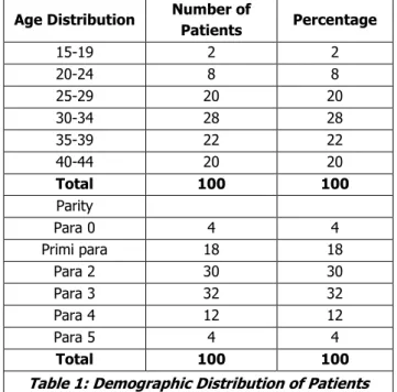
Age Distribution Number of
Patients Percentage
15-19 2 2
20-24 8 8
25-29 20 20
30-34 28 28
35-39 22 22
40-44 20 20
Total 100 100
Parity
Para 0 4 4
Primi para 18 18
Para 2 30 30
Para 3 32 32
Para 4 12 12
Para 5 4 4
Total 100 100
Table 1: Demographic Distribution of Patients
Mean age is 29.5 years, in the present study, maximum age incidence was between 30-34 years, 28 patients. Mean parity is 2.5. 4% of patients were nulliparous, 18% were primiparous and 32% were para 3.
Presentation Number of
Patients Percentage
Menorrhagia 28 28
Metrorrhagia 15 15
Menometrorrhagia 14 14
Polymenorrhea 10 10
Oligomenorrhea 13 13
Polymenorrhagia 10 10
Hypomenorrhoea 10 10
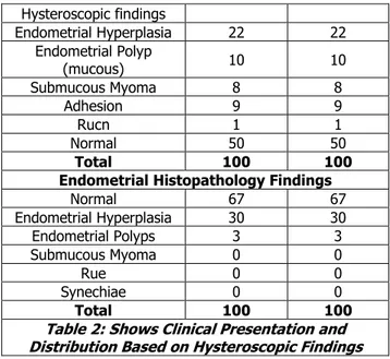
J. Evid. Based Med. Healthc., pISSN- 2349-2562, eISSN- 2349-2570/ Vol. 3/Issue 81/Oct. 10, 2016 Page 4400 Hysteroscopic findings
Endometrial Hyperplasia 22 22
Endometrial Polyp
(mucous) 10 10
Submucous Myoma 8 8
Adhesion 9 9
Rucn 1 1
Normal 50 50
Total 100 100
Endometrial Histopathology Findings
Normal 67 67
Endometrial Hyperplasia 30 30
Endometrial Polyps 3 3
Submucous Myoma 0 0
Rue 0 0
Synechiae 0 0
Total 100 100
Table 2: Shows Clinical Presentation and Distribution Based on Hysteroscopic Findings
Majority of the patients, 28% presented with
menorrhagia, the second commonest group had
metrorrhagia 15% and followed by menometrorrhagia 14%. Abnormal findings were seen in 50 patients (50%), while in the remaining 50 patients, no abnormality was detected (negative hysteroscopic view). The most common abnormality was endometrial hyperplasia (22 cases, 22%) followed by Endometrial polyps (10 cases, 10%). There were also 8 cases (8%) of submucous myoma, 9 cases (9%) of adhesions, 1 case of rue. In the 50 cases (50%) of negative hysteroscopic view, 15 cases abnormal findings were detected on Histopathology showed hyperplasia, 15 cases shown as hyperplasia on Hysteroscopy were normal on Histopathology. One of the most consistent findings in this study has been the detection of intrauterine pathology. Endometrial polyp (10 cases, 10%) and submucous myoma (8 cases, 8%) with adhesions 9 cases, rue 1 case with 100% accuracy with Hysteroscopy.
The diagnosis of 8 cases of endometrial polyps and 8 cases of submucous myoma, 9 cases of adhesions, 1 case of rue was missed by endometrial histopathology by Dilatation and Curettage.
Variables Hysteroscopy in %
Dilatation and Curettage in %
Sensitivity 70% 36%
Specificity 70% 70%
PPV 70% 54.5%
NPV 70% 52.2%
Accuracy 70% 53%
Table 3: Shows Validity of Hysteroscopy and Dilatation and Curettage
Validity of Hysteroscopy:
Sensitivity: a/a+c x 100 = 35/50 x 100 = 70%, Specificity: d/b+d x 100 = 35/50 x 100 = 70%,
Positive Predictive value: a/a+b x 100 = 35/50x100 = 70%, Negative Predictive value: d/c+d x 100 = 35/50x100 = 70%, False Positive Rate: b/ b+d x 100 = 15/50 x 100 = 30%, False Negative Rate: c/a+c x 100 = 15/50 x 100 = 30%,
Concordance (Accuracy): a+d/a + b + c + d x 100 = 70/100 x 100 = 70%.
Validity of Dilatation and Curettage:
Sensitivity: a/a+c x 100 = 18/50 x 100 = 36%, Specificity: d/b+d x 100 = 35/50 x 100 = 70%,
Positive Predictive value: a/a+b x 100 = 18/33 x 100 = 54.5%,
Negative Predictive value: d/c+d x 100 = 35/67 x 100 = 52.2%,
False Positive Rate: b/b+d x 100 = 15/50 x 100 = 30%, False Negative Rate: c/a+c x 100 = 32/50 x 100 = 64%, Concordance (Accuracy): a+d/a + b + c + d x 100 = 53/100 x 100 = 53%.
Both hysteroscopy and curettage were accurate giving a specificity of 70% for both. The ability to diagnose a lesion (Sensitivity) was more with Hysteroscopy in comparison to Curettage (70% vis. 36%), while a negative diagnosis was less wrongly made with Hysteroscopy (false negative ratio: 30% vis. 64%).
DISCUSSION: In the present study, "Hysteroscopic evaluation of women in reproductive age group with abnormal uterine bleeding" diagnostic hysteroscopy was performed in 100 consecutive cases of AUB and its correlation with histopathological findings were sought. The age group in this study was between 15-44 years and maximum incidence was between 30-34 yrs. Panda found that maximum age incidence was between 35-45 yrs. in
range between 25-70 yrs. In Gianninoto's1 series, age range
was 38-80 yrs. and commonest incidence was between
30-45 yrs. Trotsenburg1 reported maximum age incidence
between 41-50 yrs. The commonest presenting complaint in this study was menorrhagia (28%) followed by metrorrhagia
(15%) and menometrorrhagia (14%). Panda's2 series had
60% cases of menorrhagia followed by Polymenorrhagia and Metrorrhagia. In this study, abnormal findings on hysteroscopy were found in 50 patients (46%) while in remaining 50 patients (54%), no abnormality was detected. Of the 50 cases with abnormal findings on hysteroscopy, commonest seen was endometrial hyperplasia 22 cases (22%), followed by endometrial polyps 10 cases (10%) and submucous myoma 8 cases (8%), Synechiae 9 cases (9%), rue 1%, Panda found endometrial hyperplasia is 28.3%. Wamsteker found endometrial polyp is 19%, endometrial hyperplasia is 12.2% and submucous myoma is 7.8%.
Trotsenburg3 observed myomas and polyps is 14% and
deLewit4 reported myomas is 21% and polyps is 14.4%. In
Wamsteker series, number of cases were 199, normal findings at hysteroscopy was 41.5% and abnormal findings
at hysteroscopy was 58.5%. In Gimpelson RJ, Rappold HO5
series, number of cases were 276, normal findings at hysteroscopy was 60% and abnormal findings at
hysteroscopy was 40%. In Loffer6 series, number of cases
were 91, normal findings at hysteroscopy was 48.66% and
abnormal findings at hysteroscopy was 51.44%. In Sheth7
J. Evid. Based Med. Healthc., pISSN- 2349-2562, eISSN- 2349-2570/ Vol. 3/Issue 81/Oct. 10, 2016 Page 4401
hysteroscopy was 56%. In Parasnis8 series, number of cases
were 96, normal findings at hysteroscopy was 73.95% and abnormal findings at hysteroscopy was 26.05%. In
Neumann9 series, number of cases were 85, normal findings
at hysteroscopy was 55.2% and abnormal findings at
hysteroscopy was 44.8%. In Panda3 series, number of cases
were 66, normal findings at hysteroscopy was 46.6% and abnormal findings at hysteroscopy was 53.4%. In
Trotsenburg3 series, number of cases were 819, normal
findings at hysteroscopy was 66% and abnormal findings at
hysteroscopy was 34%. In Garuti10 series, number of cases
were 1500, normal findings at hysteroscopy was 61.8% and abnormal findings at hysteroscopy was 38.2%. In
Gianninoto2 series, number of cases were 512, normal
findings at hysteroscopy was 25% and abnormal findings at
hysteroscopy was 75%. In de Wit AC4 series, number of
cases were 1045, normal findings at hysteroscopy was 54.2% and abnormal findings at hysteroscopy was 45.8%.
Hysteroscopy diagnosed all cases of endometrial
hyperplasia, polyps and myomas with a specificity of 100%.
Sheth7 reported 81.8% accuracy in diagnosis of polyps and
myomas, while Garuti10 reported 95.4% specificity in
diagnosis of polyps. In the present study, hysteroscopy made a false positive diagnosis of hyperplasia in 15 cases, which were normal in histology. The accuracy of hysteroscopy in this study was 70% and that of endometrial histopathology was 53%.
Author Accuracy Misinterpretation
Baggish11 87.5 12.5
Barbot12 84 16
Sheth 82 18
Parasnis 92 8
Panda 92.69 7.31
Present Series 70 30
Comparison of Validity Factors-7 Hysteroscopy
Author Sensitivity Specificity
Levvero13 98 95
Garuti 94.2 88.8
Loffer 98 100
Parasnis 92 100
Panda 92.5 78.78
Present series 70 70
Comparison of Validity Factors-7 Dilatation and Curettages
Levvero 79.2 95
Garuti 78 94
Loffer 65 100
Parasnis 76 100
Present series 36 70
Table 4: Shows Comparison of Accuracy of Hysteroscopy Findings, Comparison of Validity Factors-7 Hysteroscopy, Comparison of 7 Dilatation
and Curettages
For hysteroscopy findings, F test P=1>0.05 NS. For validity factors-7 hysteroscopy, F test P=0.2688, >0.05, for validity factors-7 dilatation and curettages, F test P=0.9962, >0.05. A statistical analysis of the accuracy obtained by various authors and of the present study shows that there is no significant difference between the values. There is no
difference between sensitivity and specificity obtained in this study and that obtained by various authors. This confirms the validity of hysteroscopy done in the present study. A comparison of sensitivity and specificity of D and C obtained in the present study with those obtained by other authors shows no significant difference between the obtained values.
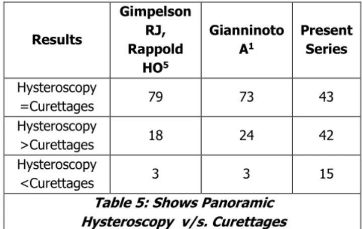
Results
Gimpelson RJ, Rappold
HO5
Gianninoto A1
Present Series
Hysteroscopy
=Curettages 79 73 43
Hysteroscopy
>Curettages 18 24 42
Hysteroscopy
<Curettages 3 3 15
Table 5: Shows Panoramic Hysteroscopy v/s. Curettages
F test P=1>0.05, in the present study, the results of hysteroscopy and dilatation and curettage were in agreement in 43% patients. Hysteroscopy revealed more information than curettage in 42% patients and curettage revealed more information than hysteroscopy in 15% patients. This is comparable to other similar studies, which shows that Hysteroscopy is better than Curettage in the evaluation of abnormal uterine bleeding.
CONCLUSION: This study confirms that hysteroscopy IS superior to curettage III evaluating patients with abnormal uterine bleeding. Hysteroscopy is a safe, reliable and quick procedure in the diagnosis of cases with abnormal uterine bleeding with high sensitivity, specificity and negative predictive value.
REFERENCES
1. Gianninoto A, Morana C, Campione C. Diagnostic
hysteroscopy in abnormal uterine bleeding.
Five-year’s experience. Minerva Ginecol
2003;55(1):57-61.
2. Panda A, Parulekar SV, Gupta A. Diagnostic
hysteroscopy in abnormal uterine bleeding and its histopathological correlation. J Obstet Gynaecol India 1999;49:74-76.
3. Van Trotsenburg M, Wieser F, Naegle F. Diagnostic
hysteroscopy for the investigation of abnormal uterine bleeding in premenopausal patients. Contrib Gynecol Obstet 2000;20:21-26.
4. de Wit AC, Vleugels MP, de Kruif JH. Diagnostic
J. Evid. Based Med. Healthc., pISSN- 2349-2562, eISSN- 2349-2570/ Vol. 3/Issue 81/Oct. 10, 2016 Page 4402
5. Gimpelson RJ, Rappold HO. A comparative study
between panoramic hysteroscopy with directed biopsies and dilatation and curettage: A review of 276 cases. Am J Obstet Gynecol 1988;158(3 Pt 1):489-492.
6. Loffer FD. Hysteroscopy with selective endometrial
sampling compared with dilatation and curettage for abnormal uterine bleeding: the value of negative hysteroscopic view. Obstet Gynecol 1989;73(1):16-20.
7. Sheth SS, Nerurkar NM, Mangeshkar PS.
Hysteroscopy in abnormal uterine bleeding. J Obstet Gynaecol India 1990;40(3):451-454.
8. Parasnis HB, Parulekar SV. Significance of negative
hysteroscopic view in abnormal uterine bleeding. J Postgrad Med 1992;38(2):62-64.
9. Neumann T, Astudillo J. Hysteroscopic study in
patients with abnormal uterine bleeding. Rev Chil Obstet Gynecol 1994;59(5):349-352.
10. Garuti G, Sambruni I
,
Colonnelli M, et al. Accuracy ofhysteroscopy in predicting histopathology of
endometrium in 1500 women. J Am Assoc Gynecol Laparosc 2001;8(2):207-213.
11. Baggish MS. Operative hysteroscopy. In: Rock JA.
Jones HW, eds. TeLinde's operative gynecology. 9th
edn. Philadelphia: Lippincott Williams & Wilkins 2003:379-411.
12. Barbot J, Parent B, Dubuisson JB. Contact
hysteroscopy: another method of endoscopic examination of the uterine cavity. Am J Obstet Gynecol 1980;136(6):721-726.
13. Loverro G, Bettocchi S, Cormio G, et al. Diagnostic