J. Evid. Based Med. Healthc., pISSN- 2349-2562, eISSN- 2349-2570/ Vol. 3/Issue 47/June 13, 2016 Page 2312
TRANSVAGINAL SONOGRAPHY COMBINED WITH SALINE CONTRAST
SONOHYSTEROGRAPHY IN EVALUATING THE UTERINE CAVITY IN PREMENOPAUSAL
PATIENTS WITH ABNORMAL UTERINE BLEEDING
K. G. M. Premleela1, Shivaprasad V. R2, Nimisha Etayangara Katankot3, Vijaykanth Reddy4
1Professor, Department of Obstetrics & Gynaecology, Sri Venkateshwaraa Medical College, Hospital and Research Centre. 2Associate Professor, Department of Radiodiagnosis, Sri Venkateshwaraa Medical College, Hospital and Research Centre. 32nd Year Post Graduate, Department of Radiodiagnosis, Sri Venkateshwaraa Medical College, Hospital and Research Centre. 42nd Year Resident Doctor, Department of Radiodiagnosis, Sri Venkateshwaraa Medical College, Hospital and Research Centre.
ABSTRACT
OBJECTIVES
To evaluate whether saline contrast sonohysterography (SCSH) improved the diagnostic accuracy of transvaginal sonography (TVS) for predicting endometrial abnormality in premenopausal patients with abnormal uterine bleeding.
PATIENTS AND METHODS
The uterine cavity was evaluated with TVS and SCSH in 60 premenopausal patients with abnormal uterine bleeding. All 58 patients underwent operative hysteroscopy or hysterectomy within 4 months which provided a detailed description of the uterine cavity and was used as the true value for exclusion of polyps and submucous myomas.
RESULT
Out of 60 patients, 45 had uterine abnormalities on TVS and SCSH and rest of the patients who appeared normal but had other abnormalities such as ovarian haemorrhagic cyst. Out of the 45 patients, 9 patients had submucous myomas and 9 were diagnosed as endometrial polyp. The findings were confirmed using hysterectomy/hysteroscopy/endometrial sampling.
CONCLUSION
The use of TVS without saline contrast left nine submucosal fibroids and five in nine of the polyps undiagnosed in referred patients with complaints of abnormal bleeding. It also helps in reducing the rate of more invasive procedures such as hysteroscopy. However, studies carried out for longer duration and large study population are required to validate our findings.
KEYWORDS
Transvaginal Sonography, Saline Contrast Sonohysterography, Abnormal Uterine Bleeding, Hysteroscopy, Hysterectomy.
HOW TO CITE THIS ARTICLE: Premleela KGM, Shivaprasad VR, Katankot NE, et al. Transvaginal sonography combined with saline contrast sonohysterography in evaluating the uterine cavity in premenopausal patients with abnormal uterine bleeding. J. Evid. Based Med. Healthc. 2016; 3(47), 2312-2317. DOI: 10.18410/jebmh/2016/511
INTRODUCTION: Abnormal uterine bleeding (AUB) results from a wide variety of causes such as anovulation, uterine
pathology and coagulopathies.1 The main diagnostic
methods for the evaluation of abnormal uterine bleeding are transvaginal ultrasonography and diagnostic hysteroscopy.2
Hysteroscopy with biopsy is the gold standard for evaluation of the uterine cavity and is a reliable and safe method in routine outpatient settings.3 Transvaginal sonography (TVS)
is a non-invasive method that has been routinely used for the evaluation of endometrium and uterine cavity in postmenopausal patients.
Being an operator dependent procedure, TVS cannot be relied on when it is performed by unexperienced consultants. Moreover, recent studies have found that about 21.8% patients with abnormal uterine bleeding with normal TVS findings were found to have some abnormality on
hysteroscopy and recommended that even after a normal TVS, a second-step office hysteroscopy should be considered.4
Saline contrast hysterosonography can be used as a diagnostic procedure in the evaluation of the uterine cavity in pre- and postmenopausal women suffering from abnormal uterine bleeding.5,6 The diagnostic accuracy of saline
contrast sonohysterography is found to be equal to that of hysteroscopy in detecting focally growing lesions in the uterine cavity in women with postmenopausal bleeding7,8.
But there are certain limitations to the use of saline contrast sonohysterography as they are contraindicated in patients with a pelvic infection or in case of unexplained pelvic tenderness.9
In the present study, the diagnostic potential of TVS, and that of TVS combined with saline contrast, were compared with the findings at operative hysteroscopy or hysterectomy in a group of premenopausal patients who presented to the obstetrics department with complaints of abnormal uterine bleeding. Initially, we evaluated whether the presence of a straight regular endometrial lining at TVS or SCSH, excluded the presence of polyps and myomas. Then, a straight regular endometrial lining was combined Financial or Other, Competing Interest: None.
Submission 27-04-2016, Peer Review 16-05-2016, Acceptance 23-05-2016, Published 10-06-2016. Corresponding Author:
Dr. K. G. M. Premleela,
Sri Venkateshwaraa Medical College, Ariyur, Pondicherry.
J. Evid. Based Med. Healthc., pISSN- 2349-2562, eISSN- 2349-2570/ Vol. 3/Issue 47/June 13, 2016 Page 2313 with an endometrial thickness of <12 mm for exclusion of all
abnormalities including hyperplasia.
Moreover, findings of polyps or myomas at TVS were compared to findings at the subsequent SCSH in the selected group of premenopausal patients who were referred for abnormal uterine bleeding.
METHODS: Patients were recruited based on their clinical complaints i.e. presence of abnormal uterine bleeding (Menorrhagia, Metrorrhagia, and Menometrorrhagia), and menstrual history preferably premenopausal (defined as being within 1 year of arrest of bleeding) and were below the age of 55 years. Patients on hormone replacement therapy (HRT) and who had an indefinite menopausal status were included when the duration of HRT was less than 3 years.10 By these selections, we tried to exclude cases with
endometrial cancers, as SCSH has a theoretical but not proven risk of spreading an endometrial cancer by transport of endometrial cells through fallopian tubes.11
In each patient, the gynaecological history was taken, the procedures were explained and consent was obtained. Bimanual palpation of the pelvis was performed with the patient in the dorsal lithotomy position and findings were recorded. Transvaginal sonography was then done in these patients and their findings were recorded. Then, a catheter was introduced into the uterus by a well-experienced gynaecologist assisted by radiologist. Saline contrast sonohysterography was then performed and findings recorded respectively. As per the literature, Transvaginal ultrasound was the procedure best accepted by the patients, followed by SCSH, hysteroscopy and endometrial sampling.
Moreover, patients would prefer SCSH over
hysteroscopy as an initial diagnostic approach in the evaluation of abnormal uterine bleeding.12 it is also found
that SCSH was more sensitive and specific in diagnosing polyp, myoma and adenomyosis with high positive and negative predictive value. 13
Procedure of Transvaginal sonography (TVS):
Transvaginal sonography was performed using a 5–7.5-MHz
transvaginal transducer (Siemens Acuson X300). The contours of the endometrial cavity were studied from the internal os to the fundus in the longitudinal and transverse planes. The midline echo was considered to be normal when a straight regular endometrial lining, with well-defined margins and without echodense foci was found. When the midline echo was disturbed, polyps were defined as echogenic masses with a fairly homogeneous texture without disruption of the myometrial–endometrial interface, while submucous myomas had an inhomogeneous texture with possible continuity with the myometrium. Myomas disturbing the midline echo or exceeding a diameter of 15 mm in the myometrium were counted.
Procedure of Saline Contrast Sonohysterography (SCSH): A sterile speculum was passed and the cervix was visualised and disinfected with Betadine solution. A flexible Foley catheter number 8 with inflatable balloon was inserted through the cervical canal into the uterine cavity. After confirmation of the position of the catheter, 10 mL of 0.9% sterile saline solution was injected into the uterine cavity slowly and continued to obtain optimal views of endometrial cavity.
By using concomitant transvaginal sonography, the uterine cavity was evaluated for detecting any abnormality or pathological condition. The uterine cavity was evaluated in sagittal and coronal views and pictures were taken for documentation. Findings at SCSH were noted according to a standard form. A normal finding implied the presence of a straight regular endometrial lining without echodense foci and projections from the myometrium. This procedure was performed by a two well-experienced investigators without the use of local anaesthesia. Most of the patients had diagnostic operative hysteroscopy under a general anaesthesia and few underwent hysterectomy.
Hysteroscopy was performed using cervix dilatation, 2 Misoprostol tablets (6 hours before operation) and prophylactic antibiotic. The hysteroscopies were done by the expert operator. Endometrial biopsy was carried out directly after hysteroscopy.
Submucous Fibroid in a 52-year-old patient on SCSH
Analysis: We analysed findings at TVS and SCSH.
Normal endometrial morphology alone (straight
regular endometrial lining, with well-defined margins and with no echogenic foci, was seen irrespective of the endometrial thickness).
Endometrial and myometrial morphology.
RESULTS: All 60 patients were selected based on the inclusion criteria. Twenty two patients did not complain of pain during the examination, while acceptable discomfort was reported in 38 patients. Some difficulties were faced while performing the procedure as the patients in premenopausal state with heavy bleeding had complaints of additional pain and discomfort. But all patients tolerated well. Patients complaining of severe pain were managed using analgesics and were kept under observation. All sixty patients met the inclusion criteria during the study period. Saline contrast sonohysterography could not be performed in 4 cases, leaving findings from 60 patients for analysis. In one case SCSH was inconclusive.
J. Evid. Based Med. Healthc., pISSN- 2349-2562, eISSN- 2349-2570/ Vol. 3/Issue 47/June 13, 2016 Page 2314 We had come across some additional findings such as
chronic cervicitis, cervical hyperplasia, cervical intraepthelial
neoplasia, ovarian haemorrhagic cyst, endometrial
hyperplasia.
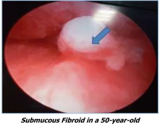
Endometrial Polyp in a 50-year-old Patient on TVS
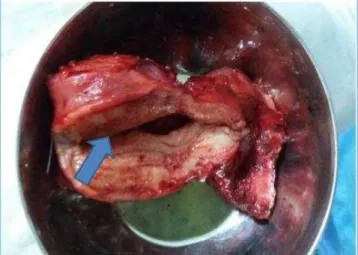
Hysterectomy Specimen of a 50-year-old Patient who was diagnosed as Submucous Fibroid
TVS
SCSH
Findings Abnormal Normal Total
Abnormality
present 31 14 45
Normal 0 15 15
Total 31 29 60
Table 1: Numbers of Patients with Various Abnormalities on SCSH Compared with Hysteroscopy/Hysterectomy are Listed
The Numbers of Abnormalities Diagnosed in the Different Group are listed in Table 2.
TVS SSG
Fibroids 18 27
Adenomyosis 6 6
Polyps 4 9
Normal 29 15
Endometrial hyperplasia 2 1
Combined (adeno + polyp) 0 1
Combined (adeno + fibroid) 1 1
Total 60 60
Table 2
Diagnostic Potential of Transvaginal Sonography for Diagnosis of Submucous Myomas and Polyps Compared with Hysteroscopy/Hysterectomy are Listed in Table 3 and 4
MYOMA TVS Hysteroscopy/Hysterectomy
Submucous 0 8
Intramural 16 15
Subserous 2 2
Total 18 25
Table 3
MYOMA SCSH Hysteroscopy/Hysterectomy
Submucous 9 8
Intramural 16 15
Subserous 2 2
Total 27 25
Table 4
TVS SCSH Hysteroscopy/
Hysterectomy
POLYPS 4 9 9
Total 9
Table 5
DISCUSSION: In this prospective study, TVS and TVS combined with SCSH were used to examine the uterine cavity in a population of patients with premenopausal bleeding disorders. At the end of both procedures, the findings were confirmed using operative hysteroscopy or hysterectomy which is taken as the gold standard.
TVS SCSH
Sensitivity 66.6% 100%
Specificity 100% 100%
Table 6: Comparing the Sensitivity and Specificity of TVS and SCSH in Detecting Submucosal Fibroid. (n=58)
TVS SCSH
Sensitivity 44.4% 100%
Specificity 100% 100%
Table 7: Comparing the Sensitivity and Specificity of TVS and SCSH in Detecting Endometrial Polyp. (n=58)
While TVS was the least invasive of the two procedures, SCSH combined with TVS displayed a higher diagnostic potential compared to TVS alone. Moreover, the midline echo seen on TVS could not be described as definitely abnormal or normal in few patients. In these cases, the uterine cavity had to undergo further evaluation such as curettage and HPE. Thus TVS alone displayed a positive predictive value (PPV – 90%), and in patients scheduled for surgery, routine use of TVS alone without further investigations might leave a significant number of abnormalities undiagnosed.
TVS SCSH
Negative predictive
value (NPV) 100% 100%
Positive predictive
value (PPV) 77.5% 100%
J. Evid. Based Med. Healthc., pISSN- 2349-2562, eISSN- 2349-2570/ Vol. 3/Issue 47/June 13, 2016 Page 2315
TVS SCSH
Negative predictive value (NPV) 100% 100%
Positive predictive value (PPV) 90% 100%
Table 9: Comparing the Predictive Values of TVS and SCSH in Detecting Polyps. (n=58)
Saline contrast sonohysterography when combined with TVS displayed a high sensitivity (100%) for diagnosis of abnormalities in the uterine cavity and revealed a high negative predictive value (100%). Thus, routine use of this method even in non-expert hands implies a low number of undiagnosed lesions. A normal sonogram combined with an endometrial thickness of <12 mm excluded even abnormalities such as hyperplasia, but the small number of patients with hyperplasia made the benefits and evidence limited for exclusion of this abnormality.
Submucous Fibroid in a 50-year-old Patient on SCSH
The main disadvantage of SCSH was that small irregularities caused by blood clots or endometrial protrusions were frequently interpreted as polyps. In addition, slight projections of myomas in the intramural portion of the uterus were seen at SCSH, but in some cases these projections might disappear after marked distension during operative hysteroscopy. These factors may have contributed to the relatively low specificity for SCSH. Moreover, detailed mapping of more than one lesion was difficult. In addition, a large study population is required to validate our finding. The same examiner performed TVS and SCSH for each one of the patients. This study design eliminated the difference within patients caused by differences in skill between observers.
The use of examiners who were not blinded at SCSH and at operative hysteroscopy may have biased the results slightly, but operative hysteroscopy could not be performed blinded to the ultrasound diagnosis for ethical reasons, and the results appear more reliable as the true values compared to the possible alternative of diagnostic hysteroscopy. As per earlier studies such as, Kelekci et al study, for detecting endometrial polyp using saline infusion sonography; sensitivity, specificity, PPV and NPV were 70%, 100%, 100% and 90.9% retrospectively whereas all of these parameters were 100% in detecting submucous myoma.14 Validating our
study, Soares et al indicated that sonohysterography had 100% sensitivity, 100% PPV and 100% diagnostic accuracy
for endometrial polyps, fibroids and endometrial
hyperplasia.15
In addition, Nanda et al reported that there is no missing in diagnosis of endometrial polyp using sonohysterography.16 In one study, 135 patients with AUB
and subfertility were evaluated and the result showed that SCHS is a very accurate method for detecting focal
endometrial pathology, compared to diagnostic
hysteroscopy.17 and in another study claimed that
hysteroscopy can be replaced by Saline-infusion sonography in more than half of AUB cases.18 and also there is very good
agreement between sonohysterography and hysteroscopy for diagnosis endometrial abnormalities in postmenopausal women.19
Submucous Fibroid in a 50-year-old Patient who Underwent Hysteroscopy
Transvaginal sonography was compared to SCSH in all patients referred for abnormal bleeding. In accordance with other studies, polyps or myomas were diagnosed in 31 cases. At least 77.5% false-negative findings occurred at subsequent SCSH, when the midline echo was found to be normal at TVS. Nevertheless, SCSH is an invasive method; it may thus have resulted in unnecessary procedures in 2% of the performed hysteroscopies. Moreover, only a few submucous myomas, but several polyps were missed. Thus, the higher diagnostic accuracy of SCSH at the expense of a few unnecessary hysteroscopies and slight patient discomfort may motivate the combined use of TVS with SCSH in a population of patients referred with abnormal bleeding.20
J. Evid. Based Med. Healthc., pISSN- 2349-2562, eISSN- 2349-2570/ Vol. 3/Issue 47/June 13, 2016 Page 2316
HPE Illustration of Endometrial Polyp in a 42-year-old Patient
In conclusion, especially when operative treatment is planned, SCSH is superior to TVS. Normal findings at TVS reduce intervention rates but fail to eliminate particularly polyps, while submucous myomas may be identified. The use of TVS without SCSH would leave 5 out of 9 polyps undiagnosed in patients referred for abnormal uterine bleeding. Thus, SCSH was found to be a very sensitive tool for prediction of the abnormal uterine cavity, and routine use of this method as an alternative to diagnostic hysteroscopy in the primary investigation of patients with bleeding disorders would potentially lead to two in three hysteroscopies being avoided.21
CONCLUSION: Saline contrast sonohysterosalpingography appears to be more sensitive (100%) and specific (100%), compared to transvaginal sonography alone. Endometrial cavity appears well distended on saline contrast sonohysterosalpingography, hence endometrial lesions such as submucous myomas and polyps could be accurately diagnosed. On TVS, endometrial cavity lesions appear to be inconclusive and hence has low sensitivity (66.6% and 44.4%) and poor positive predictive value (77.5% and 90%). Being a very sensitive tool in predicting abnormalities in endometrial cavities, routine use of SCSH aids in detecting lesions which might have been left undiagnosed and helps in reducing the rate of more invasive procedures such as hysteroscopy. Moreover, SCSH has no contrast related adverse reactions, no ionising radiation and is cost effective and easy to perform.
REFERENCES
1. Fritz MA, Speroff L, eds. Clinical gynecologic endocrinology and infertility. Philadelphia: LWW 2011;8th edn:591-606.
2. Farquhar C, Ekeroma A, Furness S, et al. A systematic
review of transvaginal sonography,
sonohysterography and hysteroscopy for the investigation of abnormal uterine bleeding in premenopausal women. Acta Obstet Gynecol Scand 2003;82(6):493-504.
3. Epstein E, Ramirez A, Skoog L, et al. Dilatation and curettage fails to detect most focal lesions in the uterine cavity in women with postmenopausal
bleeding. Acta Obstet Gynecol Scand
2001;80(12):1131–1136.
4. Barati M, Masihi S, Moramezi F, et al. Office hysteroscopy in patients with abnormal uterine bleeding and normal transvaginal sonography. Int J Fertil Steril 2008;1(4):175-179.
5. De Kroon CD, de Bock GH, Dieben SW, et al. Saline
contrast hysterosonography in abnormal uterine bleeding: a systematic review and meta-analysis.
BJOG 2003;110(10):938–947.
6. Dijkhuizen FP, Mol BW, Bongers MY, et al. Cost-effectiveness of transvaginal sonography and saline infused sonography in the evaluation of menorrhagia. Int J Gynaecol Obstet 2003;83(1):45-52.
7. Epstein E, Ramirez A, Skoog L, et al. Transvaginal sonography, saline contrast sonohysterography and hysteroscopy for the investigation of women with postmenopausal bleeding and endometrium >5mm. Ultrasound Obstet Gynecol 2001;18(2):157–162. 8. Leone FP, Lanzani C, Ferrazzi E. Use of strict
sonohysterographic methods for preoperative
assessment of submucous myomas. Fertil Steril 2003;79(4):998–1002.
9. Breitkopf D, Goldstein SR, Seeds JW. ACOG
technology assessment in obstetrics and gynecology saline infusion sonohysterography. Obstet Gynecol 2003;102(3):659–662.
10. Ferrazzi E, Leone FP. Investigating abnormal bleeding on HRT or tamoxifen: the role of ultrasonography. Best Pract Res Clin Obstet Gynaecol 2004;18(1):145– 156.
11. Alcazar JL, Errasti T, Zornoza A. Saline infusion
sonohysterography in endometrial cancer:
assessment of malignant cells dissemination risk. Acta Obstet Gynecol Scand 2000;79(4):321–322.
12. Van den Bosch T, Verguts J, Daemen A, et al. Pain experienced during transvaginal ultrasound, saline contrast sonohysterography, hysteroscopy and office sampling: comparative study. Ultrasound Obstet Gynecol 2008;31(3):346-351.
13. Alborzi S, Parsanezhad ME, Mahmoodian N, et al. Sonohysterography versus transvaginal sonography for screening of patients with abnormal uterine bleeding. Int J Gynaecol Obstet 2007;96(1):20–23. 14. Kelekci S, Kaya E, Alan M, et al. Comparison of
transvaginal sonography, saline infusion sonography, and office hysteroscopy in reproductive-aged women with or without abnormal uterine bleeding. Fertil Steril 2005;84(3):682-686.
15. Soares SR, Barbosa dos Reis MM, Camargos AF.
Diagnostic accuracy of sonohysterography,
transvaginal sonography, and hysterosalpingography in patients with uterine cavity diseases. Fertil Steril 2000;73(2):406-411.
16. Kamel HS, Darwish AM, Mohamed SA. Comparison of
transvaginal ultrasonography and vaginal
J. Evid. Based Med. Healthc., pISSN- 2349-2562, eISSN- 2349-2570/ Vol. 3/Issue 47/June 13, 2016 Page 2317 17. Nanda S, Chadha N, Sen J, et al. Transvaginal
sonography and saline infusion sonohysterography in the evaluation of abnormal uterine bleeding. Aust N Z J Obstet Gynaecol 2002;42(5):530-534.
18. Milingos S, Kallipolitis G, Stefanidis K, et al. Saline contrast hysterosonography in infertile patients and in women with abnormal uterine bleeding. Eur J Gynaecol Oncol 2005;26(5):564-567.
19. Brölmann HA, Bongers MY, Moret E, et al.
Transvaginal contrast sonography of the uterus in the diagnosis of abnormal uterine blood loss: less hysteroscopies needed. Ned Tijdschr Geneeskd 2003;147(11):502-506.
20. Gumus II, Keskin EA, Kiliç E, et al. Diagnostic value
of hysteroscopy and hysterosonography in
endometrial abnormalities in asymptomatic
postmenopausal women. Arch Gynecol Obstet 2008;278(3):241-244.