Fitness and Phenotypic Characterization of
Miltefosine-Resistant
Leishmania major
Kimbra G. Turner, Paola Vacchina, Maricela Robles-Murguia, Mariha Wadsworth, Mary Ann McDowell, Miguel A. Morales*
Eck Institute for Global Health, Department of Biological Sciences, University of Notre Dame, Notre Dame, Indiana, United States of America
*miguel.morales@nd.edu
Abstract
Trypanosomatid parasites of the genusLeishmaniaare the causative agents of leishmania-sis, a neglected tropical disease with several clinical manifestations.Leishmania majoris the causative agent of cutaneous leishmaniasis (CL), which is largely characterized by ulcerative lesions appearing on the skin. Current treatments of leishmaniasis include penta-valent antimonials and amphotericin B, however, the toxic side effects of these drugs and difficulty with distribution makes these options less than ideal. Miltefosine (MIL) is the first oral treatment available for leishmaniasis. Originally developed for cancer chemotherapy, the mechanism of action of MIL inLeishmaniaspp. is largely unknown. While treatment with MIL has proven effective, higher tolerance to the drug has been observed, and resistance is easily developed in anin vitroenvironment. Utilizing stepwise selection we generated MIL-resistant cultures ofL.majorand characterized the fitness of MIL-resistantL.major. Resistant parasites proliferate at a comparable rate to the wild-type (WT) and exhibit similar apoptotic responses. As expected, MIL-resistant parasites demonstrate decreased suscep-tibility to MIL, which reduces after the drug is withdrawn from culture. Our data demonstrate metacyclogenesis is elevated in MIL-resistantL.major, albeit these parasites display atten-uatedin vitroandin vivovirulence and standard survival rates in the natural sandfly vector, indicating that development of experimental resistance to miltefosine does not lead to an increased competitive fitness inL.major.
Author Summary
Cutaneous Leishmaniasis (CL) is characterized by the appearance of ulcerative lesions on the skin, and results from infection with trypanosomatid parasites such asLeishmania major. Current treatments for CL are expensive and have a wide range of toxic side effects of variable severity. Miltefosine, a recently introduced treatment option, is the first oral drug for leishmaniasis treatment. Although widespread clinical resistance has not yet been established, miltefosine-resistant parasite populations are easily created in a laboratory environment. Through step-wise selection, we have created populations ofL.major resis-tant to miltefosine. These resisresis-tant parasites grow at a similar rate to miltefosine-sensitive
OPEN ACCESS
Citation:Turner KG, Vacchina P, Robles-Murguia M, Wadsworth M, McDowell MA, Morales MA (2015) Fitness and Phenotypic Characterization of Miltefosine-ResistantLeishmania major. PLoS Negl Trop Dis 9(7): e0003948. doi:10.1371/journal. pntd.0003948
Editor:Louis Maes, University of Antwerp, BELGIUM
Received:April 21, 2015
Accepted:July 3, 2015
Published:July 31, 2015
Copyright:© 2015 Turner et al. This is an open access article distributed under the terms of the
Creative Commons Attribution License, which permits unrestricted use, distribution, and reproduction in any medium, provided the original author and source are credited.
Data Availability Statement:All relevant data are within the paper and its Supporting Information files.
Funding:This work was supported by the Eck Institute for Global Health and capitalization funds from the University of Notre Dame. The funders had no role in study design, data collection and analysis, decision to publish, or preparation of the manuscript.
parasites and exhibit similar stress responses. Accordingly, miltefosine-resistant parasites display a decrease in tolerance when selective pressure of MIL is withdrawn from the pop-ulation. There is no conferred resistance to treatment with other antileishmanial agents, though increased sensitivity to alternative treatments is observed in some instances. Leish-maniaundergoes a complex life cycle including the differentiation to highly infective forms, in a process termed metacyclogenesis. Experimental resistance to miltefosine increases metacyclogenesis inL.major, however resistant parasites display a lower fitness than their sensitive counterparts, as judged by their attenuated virulencein vitroandin vivo.
Introduction
Leishmaniasis is caused by protozoan parasites of the genusLeishmania, and presents as a vari-ety of clinical manifestations ranging from lesions on the skin to disseminated visceral infec-tions [1]. Cutaneous leishmaniasis (CL) often results in self-resolving lesions, whereas visceral leishmaniasis (VL) is habitually fatal when left untreated. With an annual incidence of 2 mil-lion cases and a prevalence of more than 12 milmil-lion, leishmaniasis is responsible for 70,000 deaths annually [2]. 88 countries have reported infection, resulting in 350 million individuals at risk for infection and an estimated 2.4 million disability-adjusted life years (DALYs) [2]. These statistics are grossly underestimated due to misdiagnosis and insufficient disease surveil-lance systems.
Leishmaniaspecies have a digenetic life cycle including both extracellular promastigote and obligate intracellular amastigote forms. Extracellular flagellated promastigotes reside in the midgut of the phlebotomine sandfly vector. Following infection in the mammalian host, pro-mastigotes are engulfed by macrophages where they differentiate into non-motile apro-mastigotes in the phagolysosome. This differentiation is triggered by environmental cues, mainly pH and temperature [3]. Current antileishmanial drugs include pentavalent antimony, amphotericin B, paromomycin, pentamidine, and miltefosine; most are toxic and expensive. To date, no suc-cessful vaccine exists, and the few antileishmanial drugs mentioned either risk becoming inef-fective due to emerging resistance, or are limited in their use due to cost and parental
identified in MIL-resistantL.donovaniinclude W210 (LdMT) and M1 (LdRos3) [14]. Sequencing of the entire miltefosine transporter was performed in bothL.majorandL. infan-tum, and all identified sequence mutations differed from those previously detailed inL. dono-vani(L856P, T420N, W210, and M1) [15]. In the same study, no mutations were observed in theβ-subunit Ros3 in any of the MIL-resistant populations. Widespread clinical resistance has not yet been demonstrated, nonetheless twoL.infantumisolates from HIV co-infected patients have been reported to exhibit MIL resistance [16,17]. The analysis of clinical isolates from patients infected withL.donovanithat had relapsed to standard MIL therapeutic regimes dem-onstrated that the recovered parasites were significantly more tolerant to MIL [14]. None of the resistance markers i.e. point mutations aforementioned were found in the isolates. In the absence of a definitive mechanism of miltefosine resistance, the concept of fitness or“ profi-ciency”of drug resistant pathogens is becoming more relevant and how the acquisition of resis-tance may impact the life cycle of the parasite, particularly its capacity to survive both in the insect and mammalian hosts and thus its ability to compete with wild type (sensitive) parasites [18–20]. Most of these studies are focused on antimony resistance inL.donovaniand more recently, drug combinations [21]. Here we present the characterization and fitness of clonal lines ofL.majorthat have experimentally acquired resistance to miltefosine, with relevance to survival in the mammalian host and phlebotomine vector.
Materials and Methods
Ethics statement
All studies using vertebrate animals were conducted in accordance with the U. S. Public Health Service Policy on Humane Care and Use of Laboratory Animals and followed the standards as described in theGuide for the Care and Use of Laboratory Animals. Per these standards, all ver-tebrate animal studies were conducted following review by the University of Notre Dame Insti-tutional Animal Care and Use Committee under protocol #15–047 (approved October 16, 2012). The University of Notre Dame is credited through the Animal Welfare Assurance #A3093-01.
Cell culture conditions
Leishmania majorstrain Friedlin V1 (MHOM/JL/80/Friedlin) promastigotes were cultured at 27°C in M199 medium (medium 199 (CellGro) supplemented with 10% heat-inactivated fetal bovine serum (FBS), 20 mM HEPES, 10 mM adenine, penicillin/streptomycin, hemin, biotin, L-glutamine, and 7.5% NaHCO3) and passaged every 3–4 days. Macrophages (RAW264.7 cell
line) were cultured at 37°C with 5% CO2in RPMI supplemented with 10% heat-inactivated
FBS, penicillin/streptomycin, and L-glutamine, and passaged every 2–3 days.
Generation of MIL-resistant populations
MIL-resistant cultures ofL.majorwere generated using step-wise selection. Cultures were pas-saged every 3–4 days at an initial concentration of 5x10⁵promastigotes/mL. Increasing concen-trations of MIL (Sigma) were introduced to the cultures beginning with 2.5μM MIL and
successively to 5, 8, 10, 15, 20, 30, and 40μM MIL. Cultures were exposed to an increased
Growth rates were measured for each set of resistant populations and compared with the WT strain. Parasites were counted at an initial concentration of 5x10⁵parasites/mL and growth was measured daily using a Neubauer chamber until the population reached stationary phase.
To further assess stability and fitness, two fluorescent FACS-based apoptotic markers were used to evaluate MIL-selection. Membrane permeability was assessed using the kit YO-PRO1 (Invitrogen) according to manufacturer’s recommendations. Briefly, samples were pelleted and washed in 1X M199 complete media. Following the wash, samples were resuspended in 1X M199 complete media and YO-PRO (Invitrogen) and Propidium Iodide (Invitrogen) were added and incubated for 20 minutes. Exposure of phosphatidylserine (PS) residues was investi-gated with Annexin-V-FITC (Miltenyi Biotec) following manufacturer’s instructions. Analyses were performed in a Beckman Coulter FC500 Flow Cytometer.
Assessment of drug resistance
In order to assess the MIL-resistance achieved, the half-maximal effective concentration, EC50,
was performed using the resazurin-based CellTiter-Blue (Promega) method as previously described [23]. Cultures were counted using a Neubauer chamber. 1x10⁶parasites/mL were incubated for 48 hours at 27°C in M199 medium (CellGro) and appropriate concentrations of MIL (Sigma), pentamidine isethionate (Sigma), amphotericin B (Sigma), potassium antimony (III) tartrate hydrate (Sigma) and paromomycin sulfate salt (Sigma), were used in order to accurately evaluate the resistance. Solvent (DMSO) controls were used where appropriate. HundredμL from each well were incubated at 37°C at 5% CO2for 4 hours with 20μL Cell
Titer Blue (Promega). FiftyμL of 10% SDS were added to each well, and fluorescence was
mea-sured (555 nmλexc/580 nmλem) using a Typhoon FLA-9500 laser scanner (GE Healthcare) and analyzed with ImageQuant TL software (GE Healthcare). EC50values were calculated by
non-linear regression analysis using SigmaPlot (v 11.0). All experiments were done in triplicate with appropriate controls in each case.
Partial sequencing of LmMT and LmRos3
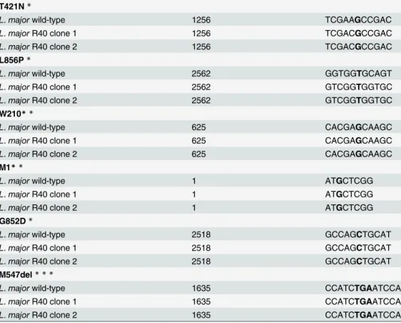
Both WT and MIL-resistant cultures were sequenced for previously described point mutations in theL.donovaniMT (T421N, L856P, W210) and Ros3 subunit (M1) [14] and inL.major (G852D, M547del) [15]. DNA was amplified with primers outlined inS1 Table. PCR product sizes ranging from 149–277 bp were purified using the GeneJET Gel Extraction Kit (Thermo) and sent to the Genomics Core Facility at the University of Notre Dame for sequencing. Sequences were analyzed using ClustalX [24].
RNA extraction and real-time PCR analysis
Metacyclogenesis
Two different methods were utilized to assess metacyclogenesis as described previously [26]. Briefly, a Ficoll (Sigma) gradient was set-up using 4 mL of 20% Ficoll overlaid with 4 mL 10% Ficoll in M199 medium without FBS and 4 mL of 5-day stationary-phase culture in M199 medium laid on top. The step gradients were centrifuged at room temperature for 10 min at 1300 x g without braking or acceleration to separate out the layers. The top two layers of the gradient were recovered and the percentage of metacyclic parasites was determined by count-ing in a Neubauer chamber before and after the enrichment procedure. For agglutination anal-ysis, 5-day stationary-phase cultures were pelleted and resuspended in 1 mL M199 medium (CellGro) and 10μL peanut agglutinin (50μg/mL) (Sigma) was added. After 30 minutes of
room temperature incubation, samples were centrifuged at 200g for 10 minutes. The superna-tant was recovered and the percentage of metacyclic parasites was determined by counting in a Neubauer chamber before and after the enrichment procedure. All experiments were done in triplicate.
Macrophage infections
RAW264.7 murine macrophage cells were counted using Trypan Blue (Amresco) and plated at 5x10⁵cells/well in 12-well plates. Infections were performed with metacyclic parasites isolated as described above. Infections were carried out at a multiplicity of infection (MOI) of 10 para-sites per macrophage. Free parapara-sites were removed by one wash with RPMI without FCS 6 h post-infection and samples collected at 6, 12, 24 and 48 h post-infection by DiffQuick staining of cytospin whole-cell preparations and visualized with light microscopy. All infections were done in triplicate and at least two independent experiments were performed.
Sandfly infections
Phlebotomus papatasi(Origin: Turkey, PPTK) was reared in the Department of Biological Sci-ences, University of Notre Dame, according to conditions previously described [27]. For the experiment, three-to-five day old female sandflies were used. Two groups, one experimental and one control, each containing 50 female and 10 male sandflies were placed in a 500 mL plastic container (ø = 6.3 cm, height = 6.5 cm) (Thermo-Nalgene) covered with a piece of nylon mesh (0.5mm). Blood feeding was performed through a young chicken skin membrane attached to a feeding device. Prior to sandfly feeding, fresh mouse blood was heat inactivated for 30 min at 56°C. Infection of sandflies withL.majorFVI strain promastigotes was done by addition of 1×10⁷logarithmic parasites/mL into the blood meal. Sixteen to twenty four hours after blood feeding, the presence or absence of blood in the sandfly digestive tract was verified by anesthetizing flies with CO2and observing the midgut distension under a stereomicroscope
(Carl Zeiss). One week post-blood meal, midguts of blood-fed sandflies were individually dis-sected and thoroughly homogenized in 30μl PBS buffer (pH 7.4) using a hand held tissue
homogenizer and pestle. Parasites were counted in a Neubauer chamber.
Mouse strains and infections
Statistics
Significance was determined by p-values calculated from a two-tailed student’s T-test in GraphPad Prism 6.0 unless otherwise stated.
Accession numbers
L.donovaniMT: GenBank accession number AY321397.1;L.donovaniRos3 GenBank accession number DQ205096.1; SHERP: GenBank accession number XM_001683391; GAPDH: GenBank accession number XP_001684904, and SOD: GenBank accession number XP_001685502.
Results and Discussion
Selection of MIL-resistant populations of
L
.
major
L.majorFVI MIL-resistant parasites were generated using step-wise selection up to 40μM
MIL. Parasites were unable to proliferate in higher MIL concentrations, likely due to reaching the critical micellar concentration of MIL leading to degradation of the membrane due to the detergent effects of MIL [29]. FVI WT promastigotes were plated in solid M199 media and two random clones were used for MIL selection in flasks. In order to assess the degree of MIL-resis-tance in our lab populations ofL.majorwe measured EC50values using the resazurin-based
CellTiter-Blue (Promega) assay. MIL-resistant cultures exposed to the highest concentrations of MIL (30μM, 40μM), and labeled R30 and R40 herein, have accordingly higher EC50values
than R10 and R20 (Fig 1). MIL-resistant cultures growing in the absence of MIL exhibited lower EC50values than their counterparts under constant MIL-selection. However, it is
impor-tant to note that this decreased EC50value of MIL-resistantL.majoris still higher than the
EC50of WTL.majorcultures (Fig 1, dotted line) after at least 95 passages (2 passages per week,
ca11 months). This suggests that once any degree of resistance is accrued MIL-resistant cul-tures do not revert back to WT phenotype, despite the removal of MIL selective pressure (Fig 1). It is worth noting that a different resistant phenotype may be obtained if drug selection is performed in axenic promastigotes or intracellular amastigotes, as shown for paromomycin selection in antimony-resistantL.donovani[17,30].
Phenotypic characterization
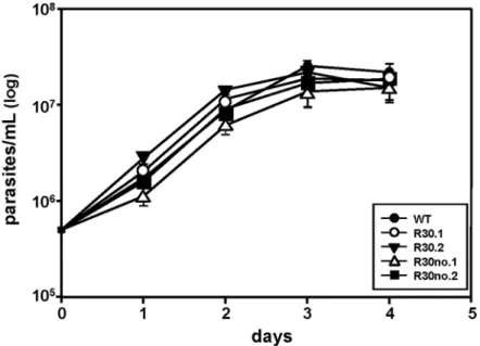
We next determined any difference in growth patterns between the sensitive (WT), resistant (R30) and resistant grown in the absence of MIL (R30no)L.majorpopulations. Growth curves showed that MIL-resistantL.majorproliferation is similar toL.majorWT and cured lines (Fig 2), indicating that increased MIL exposure has no effect on proliferation inL.major. We used a FACS-based approach to detect two different apoptotic markers i) membrane permeability and ii) PS exposure to determine the response of parasite to stress after MIL selection.L.majorR30 cell lines exhibit minimal stress and are comparable to WT populations judging the histogram levels corresponding to Annexin V and YO-PRO as analyzed by flow cytometry (S1 Fig).
possibility of other unidentified genetic mutations having a role in MIL-resistance inL.major, it is interesting to observe that even at higher concentrations (R40) and after long-term expo-sure to MIL (at least 75 passages) none of the reported mutations were found.
Fig 1. Susceptibility of MIL-resistantL.majorFVI populations generated by step-wise selection and determined by EC50analysis.1×106Log-phase parasites were incubated in the presence of a range of
drug concentrations for 48 hours at 27°C, and the surviving cells were quantified with Cell Titer Blue proliferation assay using a Typhoon FLA-9500 laser scanner. Populations of parasites were grown in increasing concentrations of MIL ranging from 10μM (R10) to 40μM (R40), showing increased resistance to
MIL. Horizontal dashed line represents WT threshold for MIL resistance.“Rno”are resistant lines grown in the absence of MIL for at least 75 passages. Results are the average of triplicate experiments±SD.
doi:10.1371/journal.pntd.0003948.g001
Fig 2. Growth curves ofL.majorWT and MIL-resistant promastigotes growing in the presence of 30μM MIL or absence of MIL selection.Log-phase promastigotes cultures were counted daily until they
Cross-resistance of MIL-resistant L. major populations to other
antileishmanials
We investigated the possibility of any conferred resistance to alternative antileishmanial treat-ments by measuring EC50values as described in Material and Methods. No cross-resistance
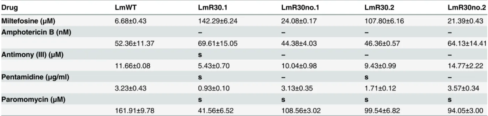
was found in any of the R30 clones or cured lines to amphotericin B, antimony (III) and paro-momycin (Table 2). Interestingly, miltefosine resistance significantly increases the sensitivity of the parasite to treatment with pentamidine 3-fold lower than WT (Table 2). When MIL has been withdrawn, the sensitivity of the parasite to this particular treatment is restored to levels comparable with the wild-type (Table 2), suggesting a potential synergistic mechanism. A simi-lar synergy has been reported for sitamaquine/pentamidine combinations inL.donovani[32], although the use of a combined therapy of miltefosine and pentamidine is hindered by the high toxicity of pentamidine [33]. Lastly, treatment of R30 MIL-resistant cultures with paromomy-cin had a significant effect on the sensitivity (ranging from 2–4 fold lower than WT) of one of the clones (R30.2), indicative of potential clonal variability.
Metacyclogenesis in MIL-resistant parasites
ProcyclicL.majorpromastigotes differentiate into highly virulent metacyclic promastigotes during metacyclogenesis [34]. This process occurs in the midgut of sandflies and can be
Table 1. Identification of point mutations previously identified in MIL-resistantL.donovani(T421N, L856P, W210*, M1) andL.major(G582D, M547del).
T421N*
L.majorwild-type 1256 TCGAAGCCGAC
L.majorR40 clone 1 1256 TCGACGCCGAC
L.majorR40 clone 2 1256 TCGACGCCGAC
L856P*
L.majorwild-type 2562 GGTGGTGCAGT
L.majorR40 clone 1 2562 GTCGGTGGTGC
L.majorR40 clone 2 2562 GTCGGTGGTGC
W210**
L.majorwild-type 625 CACGAGCAAGC
L.majorR40 clone 1 625 CACGAGCAAGC
L.majorR40 clone 2 625 CACGAGCAAGC
M1**
L.majorwild-type 1 ATGCTCGG
L.majorR40 clone 1 1 ATGCTCGG
L.majorR40 clone 2 1 ATGCTCGG
G852D*
L.majorwild-type 2518 GCCAGCTGCAT
L.majorR40 clone 1 2518 GCCAGCTGCAT
L.majorR40 clone 2 2518 GCCAGCTGCAT
M547del* * *
L.majorwild-type 1635 CCATCTGAATCCA
L.majorR40 clone 1 1635 CCATCTGAATCCA
L.majorR40 clone 2 1635 CCATCTGAATCCA
Previously identified mutations were sequenced and are indicated with an asterisk (*) and highlighted in bold font, usingL.majorFVI wild-type as the reference strain. No mutations were detected in any of the resistant lines.
mimickedin vitrowhen acidification occurs in the medium. Due to the lack of phenotypic dif-ferences in our clonal lines we performed the followingin vitroandin vivoexperiments with the R40.2 line. We enriched metacyclic promastigotes by Ficoll 400 step gradient and peanut agglutination, as described in Material and Methods. Analyses of metacyclogenesis showed thatL.majorR40 had higher percentages (2-fold) of metacyclics thanL.majorWT (Fig 3, right panel). qRT-PCR was used to amplify SHERP gene, which is almost exclusively and highly expressed in infective and non-replicative stages of the parasite [35]. SHERP expres-sion was significantly elevated in R40 parasites (Fig 3, left panel), confirming our metacyclic enrichment approaches. Increased metacyclogenesis has been reported in antimony-resistant L.donovaniclinical isolates [36], and metacyclogenesis is regarded as a major contributor to the fitness of the parasite. In New World cutaneous species,L.mexicanaresistant to Glibencla-mide, an ATP-binding-cassette (ABC)-transporter blocker exhibited a reduced expression of the Meta-1 protein [37].
Table 2. MIL resistance inL.majorFVI promastigotes does not confer cross-resistance to alternative antileishmanials.
Drug LmWT LmR30.1 LmR30no.1 LmR30.2 LmR30no.2
Miltefosine (μM) 6.68±0.43 142.29±6.24 24.08±0.17 107.80±6.16 21.39±0.43
Amphotericin B (nM) − − − −
52.36±11.37 69.61±15.05 44.38±4.03 46.36±0.57 64.13±14.41
Antimony (III) (μM) s − − −
11.66±0.08 5.43±0.70 10.04±0.98 9.43±0.99 14.77±2.22
Pentamidine (μg/ml) s − s −
3.23±0.43 0.93±0.10 3.13±0.35 1.71±0.12 3.57±0.34
Paromomycin (μM) s s s s
161.91±9.78 41.56±6.52 108.56±3.02 99.54±6.82 94.05±3.00
1x10⁶parasites/mL were incubated in increasing concentrations of amphotericin B (nM), pentamidine (
μg/mL), paromomycin (μM), or antimony (III) (μM)
for 48 hours at 27°C, using solvent controls where appropriate. Surviving cells were determined through the proliferation Cell Titer Blue assay using a Typhoon FLA-9500 laser scanner. Results are the average of triplicate experiments±SD. (−) indicates no cross-resistance demonstrated as compared to
WT, and (s) indicates an increased susceptibility to treatment.
doi:10.1371/journal.pntd.0003948.t002
Fig 3. Metacyclogenesis in WT and MIL-resistantL.major.L.majorpromastigotes resistant to MIL exhibit increased metacyclogenesis as determined by qRT-PCR of SHERP expression relative to housekeeping gene GAPDH and normalized to WT expression levels (left). 5-day stationary parasites were subjected to peanut agglutination and Ficoll-400 gradients and percentage of metacyclics is shown (right). Results are the average of triplicate experiments±SD. Statistical differences determined with a Student’sttest relative to control values (*p<0.05)
In vitro
and
in vivo
infection studies
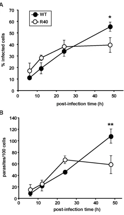
The stationary phase-specific differences of R40 primed us to study their capacity to infect RAW264.7 murine macrophage cells. We routinely passage ourL.majorcell lines through Balb/c mice to compensate for the loss of virulence due toin vitroculture. 5-day stationary cul-tures were subjected to peanut agglutination, and R40 and WT lines were incubated with RAW264.7 cells at a multiplicity of infection of 10 metacyclics per host cell. Intracellular para-site burden was determined by nuclear staining and microscopy at 6, 12, 24, and 48 h postinfec-tion. Initial levels of R40 infections are comparable to the control (Fig 4A). A significant difference in R40 infectivity was apparent 48 hours post infection. This was further corrobo-rated by decreased intracellular proliferation of R40 cells 48 hours post infection by over 20% (Fig 4B). Pentamidine-resistantL.mexicanashowed no differences in thein vitroinfectivity in resident mouse macrophages when compared with the wild-type clone [38].
In contrast, higher metacyclogenesis levels in clinical isolates ofL.donovaniresistant to antimony translated into higherin vitroinfection levels [36].
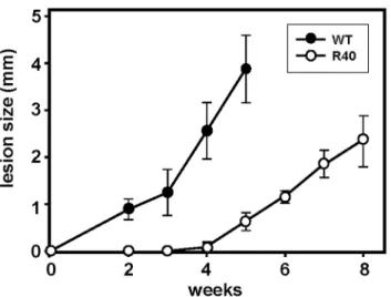
We next investigated the virulence of WT and R40 using an established experimental mouse infection [39]. Control and R40 were normalized for virulence through one passage in Balb/c mice [40]. 105WT and R40 metacyclic parasites were inoculated into the hind footpad of groups of five-six female Balb/c mice. A Vernier caliper was used to monitor lesion forma-tion by measuring the increase in footpad size weekly. Control parasites attained a lesion size of ca. 4 mm, 5 weeks after inoculation and resulted in necrotic lesions (Fig 5). Interestingly, R40 were highly attenuated and lesions were only apparent 4 weeks after infection. Our obser-vationsin vitrowith R40 cells showing a decreased infectivity and intracellular proliferation seem to have extended well to anin vivomouse model. Amphotericin-resistantL.mexicana parasites were able to infect Balb/c mice, but the resulting lesion growth was slower than that after infection with susceptible parasites [41]. In contrast, several clinical isolates ofL.donovani resistant to pentavalent antimonials showed a greater virulence in a mouse model of visceral leishmaniasis [42]. Importantly, our data suggest that metacyclogenesis alone is not a reliable marker of fitness, at least in MIL-resistantL.major, andin vitroandin vivostudies are neces-sary to further assess its competitive fitness. In this scenario, theL.major /MIL combination resembles the reduction in fitness widely observed inPlasmodium falciparumpopulations resistant to chloroquine [43].
Sandfly infection studies
Fitness ofLeishmaniaparasites is linked to transmission success in the natural insect vector, therefore we tested whether MIL resistance would impact the capacity ofLeishmaniato survive in the natural sandfly vector. Three-to-five day old femalePhlebotomus papatasi(Origin: Tur-key, PPTK) sandflies were infected with 1×10⁷logarithmic parasites/mL as described in Mate-rial and Methods. 24h post-blood meal, the presence or absence of blood in the sandfly digestive tract was verified and one week post-blood meal, 9 midguts of blood-fed sandflies infected with WT and 14 midguts from the R40 group were individually dissected. Parasite load per individual midgut was assessed. No significant differences were observed between the two groups (Fig 6) suggesting that MIL resistance does not affect the survival capacity ofL. majorin the natural vector.
the competitive fitness of MIL-resistantL.major, and studies would be further strengthened with the use of recent clinical isolates of both MIL-sensitive and MIL-resistantL.major para-sites. Further studies will attempt to understand the impaired ability of MIL-resistantL.major
Fig 4. Host cell infection assay.Early stages of macrophage invasion are similar betweenL.majorWT and R40, as determined by infection of RAW264.7 murine macrophages. Metacyclic parasites were incubated in the presence of macrophages at a MOI of 10 metacyclic parasites per macrophage and cells were collected at 6h, 12h, 24h, and 48h. Samples were stained and infection was determined through light microscopy.(A)
The percentage of infected macrophages, and(B)the number of parasites/100 cells were recorded. Results are the average of triplicate experiments±SD. Statistical differences determined with a Studentsttest relative to control values (*p<0.05;**p<0.01)
to survive in the mammalian host at the molecular level. Overall, our findings are relevant for current and future antileishmanial chemotherapy strategies.
Supporting Information
S1 Fig. Flow cytometry analysis of MIL-resistantL.majorpromastigotes.WT,L.majorFVI promastigotes grown in 40μM MIL, and R40 promastigotes where the MIL selection has been
withdrawn, using two different apoptotic markers(A)Annexin V and(B)YO-PRO. (TIF)
S1 Table. List of primers used in this study. (DOCX)
Fig 5. Virulence of WT and MIL-resistantL.major.R40 demonstrate attenuated virulencein vivo
compared with WT promastigotes. 1×10⁶WT (n = 5) and R40 (n = 6) metacyclic promastigotes were injected into the footpads of female BALB/c mice. Lesion size was recorded weekly by taking measurements of footpad thickness with a Vernier caliper, results are averages±SD.
doi:10.1371/journal.pntd.0003948.g005
Fig 6. WT and R40L.majorparasites exhibit comparable ability to colonize and survive in the sandfly vector.P.papatasiwere fed with heat-inactivated fresh mouse blood mixed with 1×10⁷parasites/mL of both
L.majorWT and R40. Blood-fed sandflies (WT: n = 9, R40: n = 14) were maintained for one week on a sucrose diet, after which the midgut was dissected. Midguts were placed in 50μL 1X PBS and crushed with a
pestle. Parasite presence in each midgut was assessed by counting with a Neubauer chamber. No significant differences were observed.
Acknowledgments
We thank Dr. Nicholas Geraci for his assistance with macrophage infections.
Author Contributions
Conceived and designed the experiments: KGT MAMo. Performed the experiments: KGT PV MRM MW. Analyzed the data: KGT MAMc MAMo. Contributed reagents/materials/analysis tools: MW MAMc. Wrote the paper: KGT MAMo.
References
1. Herwaldt BL. Leishmaniasis. Lancet. 1999; 354(9185):1191–9. Epub 1999/10/08. doi: 10.1016/S0140-6736(98)10178-2PMID:10513726.
2. Alvar J, Velez ID, Bern C, Herrero M, Desjeux P, Cano J, et al. Leishmaniasis worldwide and global esti-mates of its incidence. PloS one. 2012; 7(5):e35671. Epub 2012/06/14. doi:10.1371/journal.pone. 0035671PMID:22693548; PubMed Central PMCID: PMC3365071.
3. Zilberstein D, Shapira M. The role of pH and temperature in the development of Leishmania parasites. Annual review of microbiology. 1994; 48:449–70. PMID:7826014.
4. Singh N, Kumar M, Singh RK. Leishmaniasis: current status of available drugs and new potential drug targets. Asian Pac J Trop Med. 2012; 5(6):485–97. doi:10.1016/S1995-7645(12)60084-4PMID: 22575984.
5. Croft SL, Sundar S, Fairlamb AH. Drug resistance in leishmaniasis. Clinical microbiology reviews. 2006; 19(1):111–26. PMID:16418526.
6. Dorlo TP, Balasegaram M, Beijnen JH, de Vries PJ. Miltefosine: a review of its pharmacology and thera-peutic efficacy in the treatment of leishmaniasis. The Journal of antimicrobial chemotherapy. 2012; 67 (11):2576–97. Epub 2012/07/27. doi:10.1093/jac/dks275PMID:22833634.
7. Luque-Ortega JR, Rivas L. Miltefosine (hexadecylphosphocholine) inhibits cytochrome c oxidase in Leishmania donovani promastigotes. Antimicrobial agents and chemotherapy. 2007; 51(4):1327–32. doi:10.1128/AAC.01415-06PMID:17283192; PubMed Central PMCID: PMC1855476.
8. Canuto GA, Castilho-Martins EA, Tavares MF, Rivas L, Barbas C, Lopez-Gonzalvez A. Multi-analytical platform metabolomic approach to study miltefosine mechanism of action and resistance in Leish-mania. Analytical and bioanalytical chemistry. 2014; 406(14):3459–76. doi: 10.1007/s00216-014-7772-1PMID:24722876.
9. Marinho FD, Goncalves KCD, de Oliveira SS, de Oliveira ACDC, Bellio M, d'Avila-Levy CM, et al. Milte-fosine induces programmed cell death in Leishmania amazonensis promastigotes. Memorias do Insti-tuto Oswaldo Cruz. 2011; 106(4):507–9. PMID:WOS:000292402300021.
10. Vincent IM, Weidt S, Rivas L, Burgess K, Smith TK, Ouellette M. Untargeted metabolomic analysis of miltefosine action in Leishmania infantum reveals changes to the internal lipid metabolism. Int J Parasi-tol-Drug. 2014; 4(1):20–7. doi:10.1016/j.ijpddr.2013.11.002PMID:WOS:000331708400003.
11. Perez-Victoria FJ, Sanchez-Canete MP, Seifert K, Croft SL, Sundar S, Castanys S, et al. Mechanisms of experimental resistance of Leishmania to miltefosine: Implications for clinical use. Drug Resist Updat. 2006; 9(1–2):26–39. Epub 2006/07/04. doi:10.1016/j.drup.2006.04.001PMID:16814199.
12. Perez-Victoria FJ, Gamarro F, Ouellette M, Castanys S. Functional cloning of the miltefosine trans-porter. A novel P-type phospholipid translocase from Leishmania involved in drug resistance. The Jour-nal of biological chemistry. 2003; 278(50):49965–71. Epub 2003/09/30. doi:10.1074/jbc.M308352200 PMID:14514670.
13. Perez-Victoria FJ, Sanchez-Canete MP, Castanys S, Gamarro F. Phospholipid translocation and milte-fosine potency require both L. donovani miltemilte-fosine transporter and the new protein LdRos3 in Leish-mania parasites. The Journal of biological chemistry. 2006; 281(33):23766–75. Epub 2006/06/21. doi: 10.1074/jbc.M605214200PMID:16785229.
14. Bhandari V, Kulshrestha A, Deep DK, Stark O, Prajapati VK, Ramesh V, et al. Drug susceptibility in Leishmania isolates following miltefosine treatment in cases of visceral leishmaniasis and post kala-azar dermal leishmaniasis. PLoS neglected tropical diseases. 2012; 6(5):e1657. Epub 2012/05/26. doi: 10.1371/journal.pntd.0001657PMID:22629478; PubMed Central PMCID: PMC3358331.
16. Cojean S, Houze S, Haouchine D, Huteau F, Lariven S, Hubert V, et al. Leishmania resistance to milte-fosine associated with genetic marker. Emerg Infect Dis. 2012; 18(4):704–6. doi:10.3201/eid1804. 110841PMID:22469394; PubMed Central PMCID: PMC3309694.
17. Hendrickx S, Boulet G, Mondelaers A, Dujardin JC, Rijal S, Lachaud L, et al. Experimental selection of paromomycin and miltefosine resistance in intracellular amastigotes of Leishmania donovani and L. infantum. Parasitology research. 2014; 113(5):1875–81. doi:10.1007/s00436-014-3835-7PMID: 24615359.
18. Vanaerschot M, Huijben S, Van den Broeck F, Dujardin JC. Drug resistance in vectorborne parasites: multiple actors and scenarios for an evolutionary arms race. FEMS microbiology reviews. 2014; 38 (1):41–55. doi:10.1111/1574-6976.12032PMID:23815683.
19. Vanaerschot M, Decuypere S, Berg M, Roy S, Dujardin JC. Drug-resistant microorganisms with a higher fitness—can medicines boost pathogens? Crit Rev Microbiol. 2013; 39(4):384–94. doi:10.3109/ 1040841X.2012.716818PMID:22950457.
20. Natera S, Machuca C, Padron-Nieves M, Romero A, Diaz E, Ponte-Sucre A. Leishmania spp.: profi-ciency of drug-resistant parasites. International journal of antimicrobial agents. 2007; 29(6):637–42. doi:10.1016/j.ijantimicag.2007.01.004PMID:17353113.
21. Garcia-Hernandez R, Gomez-Perez V, Castanys S, Gamarro F. Fitness of Leishmania donovani Para-sites Resistant to Drug Combinations. PLoS neglected tropical diseases. 2015; 9(4):e0003704. doi:10. 1371/journal.pntd.0003704PMID:25849149.
22. LeBowitz JH. Transfection experiments with Leishmania. Methods Cell Biol. 1994; 45:65–78. PMID: 7707995.
23. Vacchina P, Morales MA. In vitro screening test using Leishmania promastigotes stably expressing mCherry protein. Antimicrobial agents and chemotherapy. 2014; 58(3):1825–8. doi:10.1128/AAC. 02224-13PMID:24395225; PubMed Central PMCID: PMC3957829.
24. Larkin MA, Blackshields G, Brown NP, Chenna R, McGettigan PA, McWilliam H, et al. Clustal W and Clustal X version 2.0. Bioinformatics. 2007; 23(21):2947–8. PMID:17846036.
25. Bookout AL, Cummins CL, Mangelsdorf DJ, Pesola JM, Kramer MF. High-throughput real-time quanti-tative reverse transcription PCR. Curr Protoc Mol Biol. 2006;Chapter 15:Unit 15 8. doi:10.1002/ 0471142727.mb1508s73PMID:18265376.
26. Morales MA, Pescher P, Spath GF. Leishmania major MPK7 protein kinase activity inhibits intracellular growth of the pathogenic amastigote stage. Eukaryotic cell. 2010; 9(1):22–30. Epub 2009/10/06. doi: 10.1128/EC.00196-09PMID:19801421; PubMed Central PMCID: PMC2805286.
27. Ramalho-Ortigao JM, Kamhawi S, Joshi MB, Reynoso D, Lawyer PG, Dwyer DM, et al. Characteriza-tion of a blood activated chitinolytic system in the midgut of the sand fly vectors Lutzomyia longipalpis and Phlebotomus papatasi. Insect Mol Biol. 2005; 14(6):703–12. doi:10.1111/j.1365-2583.2005. 00601.xPMID:16313571.
28. Titus RG, Marchand M, Boon T, Louis JA. A limiting dilution assay for quantifying Leishmania major in tissues of infected mice. Parasite immunology. 1985; 7(5):545–55. PMID:3877902
29. Urbina JA. Mechanisms of action of lysophospholipid analogues against trypanosomatid parasites. Transactions of the Royal Society of Tropical Medicine and Hygiene. 2006; 100 Suppl 1:S9–S16. doi: 10.1016/j.trstmh.2006.03.010PMID:16930650.
30. Hendrickx S, Inocencio da Luz RA, Bhandari V, Kuypers K, Shaw CD, Lonchamp J, et al. Experimental induction of paromomycin resistance in antimony-resistant strains of L. donovani: outcome dependent on in vitro selection protocol. PLoS neglected tropical diseases. 2012; 6(5):e1664. Epub 2012/06/06. doi:10.1371/journal.pntd.0001664PMID:22666513; PubMed Central PMCID: PMC3362622.
31. Perez-Victoria FJ, Castanys S, Gamarro F. Leishmania donovani resistance to miltefosine involves a defective inward translocation of the drug. Antimicrobial agents and chemotherapy. 2003; 47(8):2397–
403. Epub 2003/07/25. PMID:12878496; PubMed Central PMCID: PMC166066.
32. Seifert K, Munday J, Syeda T, Croft SL. In vitro interactions between sitamaquine and amphotericin B, sodium stibogluconate, miltefosine, paromomycin and pentamidine against Leishmania donovani. The Journal of antimicrobial chemotherapy. 2011; 66(4):850–4. doi:10.1093/jac/dkq542PMID:21393188.
33. Olliaro PL, Guerin PJ, Gerstl S, Haaskjold AA, Rottingen JA, Sundar S. Treatment options for visceral leishmaniasis: a systematic review of clinical studies done in India, 1980–2004. The Lancet infectious diseases. 2005; 5(12):763–74. doi:10.1016/S1473-3099(05)70296-6PMID:16310148.
34. da Silva R, Sacks DL. Metacyclogenesis is a major determinant of Leishmania promastigote virulence and attenuation. Infection and immunity. 1987; 55(11):2802–6. PMID:3666964.
36. Ouakad M, Vanaerschot M, Rijal S, Sundar S, Speybroeck N, Kestens L, et al. Increased metacyclo-genesis of antimony-resistant Leishmania donovani clinical lines. Parasitology. 2011; 138(11):1392–9. doi:10.1017/S0031182011001120PMID:21819638.
37. Silva N, Camacho N, Figarella K, Ponte-Sucre A. Cell differentiation and infectivity of Leishmania mexi-cana are inhibited in a strain resistant to an ABC-transporter blocker. Parasitology. 2004; 128(Pt 6):629–34. PMID:15206465.
38. Sereno D, Lemesre JL. In vitro life cycle of pentamidine-resistant amastigotes: stability of the chemore-sistant phenotypes is dependent on the level of resistance induced. Antimicrobial agents and chemo-therapy. 1997; 41(9):1898–903. PMID:9303381; PubMed Central PMCID: PMC164032.
39. Norris-Mullins B, VanderKolk K, Vacchina P, Joyce MV, Morales MA. LmaPA2G4, a homolog of human Ebp1, is an essential gene and inhibits cell proliferation in L. major. PLoS neglected tropical diseases. 2014; 8(1):e2646. doi:10.1371/journal.pntd.0002646PMID:24421916; PubMed Central PMCID: PMC3888471.
40. Moreira D, Santarem N, Loureiro I, Tavares J, Silva AM, Amorim AM, et al. Impact of continuous axenic cultivation in Leishmania infantum virulence. PLoS neglected tropical diseases. 2012; 6(1):e1469. Epub 2012/02/01. doi:10.1371/journal.pntd.0001469PMID:22292094; PubMed Central PMCID: PMC3265455.
41. Al-Mohammed HI, Chance ML, Bates PA. Production and characterization of stable amphotericin-resis-tant amastigotes and promastigotes of Leishmania mexicana. Antimicrobial agents and chemotherapy. 2005; 49(8):3274–80. doi:10.1128/AAC.49.8.3274–3280.2005PMID:16048936; PubMed Central PMCID: PMC1196255.
42. Vanaerschot M, De Doncker S, Rijal S, Maes L, Dujardin JC, Decuypere S. Antimonial resistance in Leishmania donovani is associated with increased in vivo parasite burden. PloS one. 2011; 6(8): e23120. doi:10.1371/journal.pone.0023120PMID:21829701; PubMed Central PMCID: PMC3148249.
43. Felger I, Beck HP. Fitness costs of resistance to antimalarial drugs. Trends in parasitology. 2008; 24 (8):331–3. doi:10.1016/j.pt.2008.05.004PMID:18603474.