Trypanosomatid comparative genomics:
Contributions to the study of parasite biology and different parasitic diseases
Santuza M. Teixeira
1, Rita Márcia Cardoso de Paiva
1, Monica M. Kangussu-Marcolino
2and Wanderson D. DaRocha
21
Departamento de Bioquímica e Imunologia, Universidade Federal de Minas Gerais,
Belo Horizonte, MG, Brazil.
2
Departamento de Bioquímica e Biologia Molecular, Universidade Federal do Paraná, Curitiba, PR, Brazil.
Abstract
In 2005, draft sequences of the genomes of Trypanosoma brucei, Trypanosoma cruzi and Leishmania major, also known as the Tri-Tryp genomes, were published. These protozoan parasites are the causative agents of three dis-tinct insect-borne diseases, namely sleeping sickness, Chagas disease and leishmaniasis, all with a worldwide distribution. Despite the large estimated evolutionary distance among them, a conserved core of ~6,200 trypanoso-matid genes was found among the Tri-Tryp genomes. Extensive analysis of these genomic sequences has greatly increased our understanding of the biology of these parasites and their host-parasite interactions. In this article, we review the recent advances in the comparative genomics of these three species. This analysis also includes data on additional sequences derived from other trypanosmatid species, as well as recent data on gene expression and func-tional genomics. In addition to facilitating the identification of key parasite molecules that may provide a better under-standing of these complex diseases, genome studies offer a rich source of new information that can be used to define potential new drug targets and vaccine candidates for controlling these parasitic infections.
Key words: Trypanosoma brucei,Trypanosoma cruzi, Leishmania major,genome, RNAseq.
Received: August 8, 2011; Accepted: October 18, 2011.
Tri-Tryp Diseases and The Tri-Tryp Genomes
Trypanosoma brucei, Trypanosoma cruzi and
Leishmania majorare unicellular protozoa of considerable medical importance since they are the etiologic agents of sleeping sickness (African trypanosomiasis), Chagas dis-ease (American trypanosomiasis) and leishmaniasis, re-spectively. The geographic range of these parasites is deter-mined by their insect vectors: in the case of sleeping sickness, a blood sucking fly of the genusGlossina, also known as the tsetse fly, for Chagas disease, a reduviid bug known as the “kissing bug” and for leishmaniasis, a phlebo-tomine sandfly. While sleeping sickness occurs in sub-Saharan Africa, Chagas disease is prevalent in Latin Amer-ica. Leishmaniasis is considered to be endemic in 88 coun-tries, 72 of which are developing countries in Asia, South America and Africa. Together, these three parasitic dis-eases represent a huge burden since approximately 0.5 mil-lion people are infected withT. brucei, 10 million withT. cruziand an estimated 12 million with different species of Leishmania. In addition to the two human-infective
subspe-cies,T. brucei gambienseandT. b. rhodesiense, other spe-cies and subspespe-cies of African trypanosomes cause the dis-ease known as nagana in domestic animals, imposing a fur-ther economic burden on several African countries. Different forms of leishmaniasis are caused by at least 20 leishmanial species: cutaneous leishmaniasis, with an esti-mated 1.5 million cases, and visceral leishmaniasis, with about 500,000 new cases annually, are the most common. Although control of the arthropod vectors of these diseases is an achievable goal and has been successful against theT. cruzivector in parts of Latin America, the alarming resur-gence of sleeping sickness in Africa and of leishmaniasis in parts of Asia and Latin America is a constant reminder of the need for better forms of chemotherapy and prevention of these diseases. More detailed information on African and American trypanosomiasis and the different forms of leishmaniasis, including vector distribution, disease control and treatment protocols can be found at http://apps.who.int/tdr/.
Trypanosoma brucei,T. cruzi andLeishmania spp. are hemoflagellates of the family Trypanosomatidae (order Kinetoplastida) that is characterized by the presence of a single flagellum and one mitochondrion containing a uni-que organelle known as the kinetoplast which contains the
Send correspondence to Wanderson Duarte DaRocha. Departa-mento de Bioquímica e Biologia Molecular, Universidade Federal do Paraná, Caixa Postal 19046, 81531-990 Curitiba, PR, Brazil. E-mail: [email protected].
mitochondrial DNA (Simpsonet al., 2006). Each parasite has a complex life cycle that involves humans as one of their various hosts (Figure 1). As some of the earliest diver-gent members of the Eukaryotae (Haaget al., 1998), these parasites have peculiar aspects of gene expression, includ-ing polycistronic transcription of most of their genomes (Martínez-Calvillo et al., 2010), RNA polymerase I-me-diated transcription of protein-coding genes (Gunzlet al., 2003), RNA trans-splicing to generate mature, capped mRNAs (LeBowitzet al., 1993) and extensive RNA edit-ing to generate functional mRNAs transcribed from mito-chondrial genes (Hajduk et al., 1993). Apart from their medical relevance, these peculiar characteristics make these parasites very interesting models for studying ge-nome evolution and other aspects of gege-nome function. On the other hand, the early evolutionary divergence of these organisms has resulted in biochemical characteristics that are not common in higher eukaryotes, such as enzymes re-lated to antioxidant metabolism (Olin-Sandoval et al.,
2010) as well as sterol and glycosylphosphatidylinositol (GPI) biosynthesis (Lepeshevaet al., 2011; Koeller and Heise, 2011) that have been exploited as promising drug targets.
Genome sequencing of Tri-Tryp parasites began in the early 90s with the analyses of 518 expressed sequence tags (ESTs) generated from mRNA isolated from blood-stream forms ofT. b. rhodesiense(El-Sayedet al., 1995). Shortly thereafter, a comparison between EST and genomic sequences showed that sequencing random DNA fragments was as efficient as EST analyses for discovering new genes in the African trypanosome (El-Sayed and Donelson, 1997). In 1996, an EST analysis of cDNA libraries con-structed with mRNA fromL. majorpromastigotes was pub-lished (Levicket al., 1996), and the first EST analysis ofT. cruziepimastigote forms was published in 1997 (Brandão
et al., 1997). During this period, pulsed-field gel electro-phoretic analysis of chromosomes and the sequencing of large DNA fragments from cosmid, bacterial artificial chromosome and yeast artificial chromosome libraries were also undertaken to generate physical maps of Tri-Tryp genomes (Blackwell and Melville, 1999). In 1999, the se-quence of a 257-kilobase region spanning almost the entire chromosome 1 ofL. majorrevealed the unusual distribu-tion of protein-coding genes that was later found to be char-acteristic of all Tri-Tryp genomes. The complete sequence of L. major chromosome 1 revealed 79 protein-coding genes, with the first 29 genes all encoded on one DNA strand and the remaining 50 genes encoded on the opposite strand (Myleret al., 1999).
The Tri-Tryp gene organization is reminiscent of bac-terial operons, with protein coding genes densely packed within directional clusters in one strand separated by strand switch regions (i.e., changes in the coding strand) (Figu-re 2). Experimental evidence suggests that transcription ini-tiates bi-directionally between two divergent gene clusters (Martínez-Calvilloet al., 2003, 2004) to produce polycis-tronic pre-mRNAs that are subsequently processed. Re-markably, with the exception of the spliced leader (SL) promoter, no promoter is recognized by RNA polymerase II and only a few transcription factors have been identified (Cribb and Serra, 2009; Cribbet al., 2010). Even more sur-prisingly, although orthologs of all conserved components of the RNA polymerase II complex were identified in the Tri-Tryp genome (Ivenset al., 2005), the transcription of some trypanosomatid genes such as VSG (Variant Surface Glycoprotein) and the procyclin genes ofT. brucei,as well as several exogenous genes transfected intoT. cruzi, are mediated by RNA polymerase I (Gunzlet al., 2003). Once the polycistronic pmRNA is produced, two coupled re-actions (trans-splicing and poly-adenylation) result in ma-ture monocistronic transcripts.
Trans-splicing means that every mature mRNA has an identical capped sequence of 39 nucleotides, known at the spliced leader (SL), at the 5’ end (Lianget al., 2003).
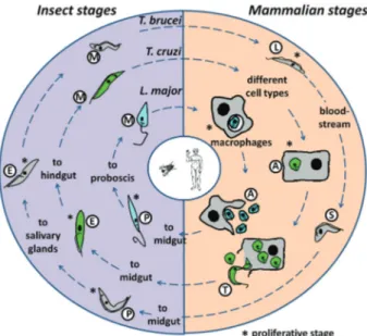
Figure 1- The Tri-Tryp life cycles. Representation of the life cycles of
Leishmania major, Trypanosoma cruzi and T. brucei, the etiological agents of leishmaniasis, Chagas disease and sleeping sickness, respec-tively, are shown, with the parasitic forms that are present in the insect vectors and the mammalian hosts.Leishmania majorproliferates as pro-mastigotes (P) in the sand fly midgut. The parasite is transmitted during bites by this fly and invades mammalian macrophages in the metacyclic promastigote (M) form. Inside the cell, the M form is converted into amastigotes (A) and divides before been released during cell lysis.
Whilst no sequence consensus for polyadenylation or SL addition has been found, several studies have demonstrated that polypyrimidine-rich tracts located within intergenic re-gions guide SL addition and poly-adenylation, resulting in mature mRNAs (LeBowitz et al., 1993) (Figure 3). Intergenic sequences involved in the processing ofT. cruzi,
T. bruceiandLeishmaniamRNA have been thoroughly in-vestigated by comparing mRNA with genomic sequences, initially using EST databases (Benzet al., 2005; Camposet al., 2008; Smith et al., 2008) and, more recently, using high-throughput RNA-sequencing (RNAseq) (Siegelet al., 2010; Kolevet al., 2010; Nilssonet al., 2010). In addition to providing valuable information on the mechanisms of gene expression in these organisms, these analyses also
yielded data that allowed the optimization of transfection vectors used to express foreign genes and genetic manipu-lation in trypanosomatids.
Comparative genomic analyses using the Tri-Tryp se-quences have already provided interesting insights into the genetic and evolutionary bases of the distinct and shared lifestyles of these parasites. Probably the most striking finding is that the three genomes display high levels of synteny and share a conserved set of ~6,200 genes, 94% of which are arranged in syntenic directional gene clusters (El-Sayedet al., 2005a). Alignment of the deduced protein sequences of the majority of the clusters of orthologous genes across the three organisms reveals an average 57% identity betweenT. cruziandT. bruceiand 44% identity
tween T. cruzi and L. major that reflected the expected phylogenetic relationships (Lukeset al., 1997; Haaget al., 1998; Stevenset al., 1999; Wrightet al., 1999). The major-ity of species-specific genes occurs on non-syntenic chro-mosomes and consists of members of large surface antigen families. Structural RNAs, retroelements and gene family expansion are also often associated with breaks in the con-servation of gene synteny (El-Sayedet al., 2005a). Multi-gene family expansions are Multi-generally species-specific and most pronounced in theT. cruzigenome. As discussed be-low, a number ofT. cruzimulti-gene families encode sur-face proteins, such as trans-sialidases, mucin-associated surface proteins (MASP) and mucins TcMUC and GP63 that likely play important roles in host-parasite interactions (Di Noiaet al., 1995; Vargaset al., 2004; Baidaet al., 2006; Bartholomeuet al., 2009). Based on their location in re-gions of synteny breaks these arrays may be subject to ex-tensive rearrangements during the parasite’s evolution and
are thus directly associated with the specificities of each of the three parasitic diseases.
The Genetic Diversity of T. Cruzi and the
Genomes of Different Parasite Strains
Chagas disease, caused by T. cruzi, is endemic in more than 20 Latin American countries, where an estimated 10 million people are infected and the “domiciliation” of the triatomines exposes at least 90 million individuals to the risk of infection. With no vaccine or effective drug treat-ment available, the main strategy for control must rely on the prevention of transmission by the insect vectors and blood transfusions. The parasite proliferates in the midgut of several species of a triatomid hematophagous vector. After reaching the insect’s hindgut, epimastigote forms differentiate into non-dividing, infective metacyclic trypo-mastigotes that are excreted in the insect’s feces. Trypo-mastigotes can infect a mammalian host by passing through
mucous membranes or skin lesions during feeding by the insect. Once inside the mammalian host, trypomastigotes invade different types of cells where they transform into proliferative intracellular amastigotes. After a number of cell divisions in the host cell cytoplasm, amastigotes differ-entiate into trypomastigotes that are released into the bloodstream after host cell rupture and, after being taken up by an insect during a blood meal, they start a new cycle (Brener, 1973) (Figure 1). The highly heterogenousT. cruzi
population consists of a large number of strains with dis-tinct characteristics related to morphology, growth rate, parasitemia curves, virulence, pathogenicity, drug sensitiv-ity, antigenic profile, metacyclogenesis and tissue tropism (Buscaglia and Di Noia, 2003).
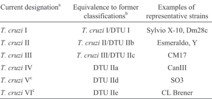
Despite the broad genetic diversity observed among different strains and isolates, early studies based on differ-ent genotyping strategies iddiffer-entified two major lineages in the parasite population, namedT. cruzi I and T. cruziII (Souto et al., 1996; Momen 1999). These divergent lin-eages occupy distinct ecological environments, namely, the sylvatic cycle (T. cruziI) and the domestic cycle (T. cruzi
II) of Chagas disease (Zingaleset al., 1998), as well as dis-tinct sylvatic host associations (Buscaglia and Di Noia, 2003). Further analyses led some authors to propose the sub-division ofT. cruziII into five sub-groups:T. cruziIIa, IIb, IIc, IId and IIe (Brisseet al., 2000). Phylogenetic analy-ses of theT. cruzistrains became more confusing when ad-ditional data indicated the existence of not just two, but three major groups in theT. cruzipopulation, in addition to hybrid strains (Miles et al., 1978; Augusto-Pinto et al., 2003; de Freitaset al., 2006). After intense debate, in 2009 an international consensus recognized the existence of six major strains, also known as discrete typing units (DTUs) I-VI (Zingaleset al., 2009) (Table 1). Since Chagas disease spawns a variety of clinical forms, these studies are highly relevant: understanding the genetic variation among strains can potentially explain differences in disease pathogenesis, host preferences and, most importantly, provides essential information for the identification of new drug targets and good antigenic candidates for better diagnosis and vaccine development. For instance,T. cruziII strains and the hybrid
strains belonging toT. cruziV and VI are the predominant causes of human disease in South America (Zingaleset al., 2009), whereasT. cruziI strains are more abundant among wild hosts and vectors. Although detailed analysis of the bi-ological and molecular factors underlyingT. cruzi popula-tion structure and the epidemiology of Chagas disease are beyond the scope of this review, one must keep in mind that the genetic variability found in theT. cruzipopulation is an essential aspect to be considered when analyzing this para-sites genome.
CL Brener, a clone derived from a hybrid T. cruzi
strain belonging toT. cruziVI, was chosen as a reference strain for the initialT. cruzigenome project. The hybrid na-ture of the CL Brener clone became clear only after the ge-nome sequencing had begun, when analyses of nuclear and mitochondrial sequences showed that this strain resulted from a fusion event that had occurred between ancient ge-notypes corresponding to strains belonging toT. cruziII and III groups (El-Sayedet al., 2005a; de Freitas et al., 2006). Prior to this knowledge, the choice of the clone CL Brener, initially classified as a member of sub-group IIe, was based on five characteristics: (1) it was isolated from the domiciliary vectorTriatoma infestans, (2) its pattern of infectivity in mice was very well known, (3) it had prefer-ential tropism for heart and muscle cells, (4) it showed a clear acute phase in accidentally infected humans, and (5) it was susceptible to drugs used to treat Chagas disease (Zin-galeset al., 1997). In addition, several genomic studies had previously used this strain for karyotype analyses (Branche
et al., 2006) and the generation of physical maps and ESTs from all three stages of the parasite life cycle (Canoet al., 1995; Henrikssonet al., 1995; Brandãoet al., 1997; Verdun
et al., 1998; Porcelet al., 2000; Cerqueiraet al., 2005). TheT. cruziCL Brener haploid genome, estimated to be 55 Mb, was sequenced using the WGS (whole genome shotgun) strategy. Because of its hybrid nature and the high level of allelic polymorphism, a 14X coverage, much higher than the usual 8-10X coverage, was required to dis-tinguish the ambiguities derived from allelic variations from those produced by sequencing errors. In contrast to the other two Tri-Tryp genomes, theT. cruzidraft sequence (El-Sayedet al., 2005b) was published as an assembly of 5,489 scaffolds built by 8,740 contigs. Four years later, based on synteny maps for theT. brucei chromosomes, Weatherlyet al.(2009) assembled theT. cruzicontigs and scaffolds initially in 11 pairs of homologous “T. brucei -like” chromosomes and, ultimately, in 41T. cruzi chromo-somes. Since trypanosomatid chromosomes do not conden-sate during mitosis and are therefore not visualized in metaphasic cells the predicted number ofT. cruzi chromo-somes was based on studies of pulsed-field gel electropho-resis (PFGE) analyses (Brancheet al., 2006), which turned out to be similar to the number of assembled chromosomes. As mentioned above, the genome organization inT. cruziis largely syntenic with the other Tri-Tryp (T. bruceiandL.
Table 1- Classification ofT. cruzistrains.
Current designationa Equivalence to former classificationsb
Examples of representative strains
T. cruziI T. cruziI/DTU I Sylvio X-10, Dm28c
T. cruziII T. cruziII/DTU IIb Esmeraldo, Y
T. cruziIII T. cruziIII/DTU IIc CM17
T. cruziIV DTU IIa CanIII
T. cruziVc DTU IId SO3
T. cruziVIc DTU IIe CL Brener
DTU = discrete typing unit.aZingaleset al.(2009),bMomem (1999) (T. cruziI and II classification), Brisseet al.(2000) (DTU I, IIa-e), de Freitas
major) genomes, with most species-specific genes, such as surface protein gene families, occurring in internal and subtelomeric regions of non-syntenic chromosome (El-Sayedet al., 2005a).
Because of its hybrid nature, the CL Brener genome is represented by a redundant dataset since homologous re-gions displaying a high level of polymorphism were assem-bled separately, generating two set of contigs, each corres-ponding to one haplotype. To identify the two haplotypes, reads from the genome of the cloned Esmeraldo strain, a member of T. cruzi II, and representing one of the CL Brener parental strain (de Freitaset al., 2006), were gener-ated. Thus, in the annotation data of the CL Brener genome, the two haplotypes are referred to as “Esmeraldo-like” or “non-Esmeraldo-like” sequences (Aslettet al., 2010).
The haploid CL Brener genome has an estimated 12,000 genes. As with the other Tri-Tryps, the T. cruzi
genes are organized in long polycistronic clusters that are transcribed by RNA polymerase II and processed into monocistronic mRNAs that accumulate differentially dur-ing the various stages of the parasite life cycle. As indicated before, one of the main characteristics revealed by the com-plete sequence of theT. cruzigenome was the dramatic ex-pansion of families encoding surface proteins (El-Sayedet al., 2005a). Compared toT. bruceiandL. major,T. cruzi
has the largest set of multi-gene families, perhaps because of its unique capacity to invade and multiply within differ-ent types of host cells. Long terminal repeat (LTR) and non-LTR retroelements and other sub-telomeric also con-tribute to the large proportion of repetitive sequences (50% of the genome) in this genome. The largest protein gene family encodes a group of surface proteins known as trans-sialidases (TS), with 1,430 members. TSs are surface mole-cules identified as virulent factors ofT. cruzithat are re-sponsible for transferring sialic acid from host sialogly-coconjugates to the terminal ß-galactose on T. cruzi
mucins. Mucin-associated surface proteins (MASP) are the second largestT. cruzigene family, with a total of 1,377 members. Although MASP sequences correspond to ~6% of the parasite diploid genome, they were only identified during annotation of the T. cruzi genome. MASPs are glycosylphosphatidylinositol (GPI)-anchored surface pro-teins that are preferentially expressed in trypomastigotes; these proteins are characterized by highly conserved N- and C-terminal domains and a strikingly variable and repetitive central region (Bartholomeuet al., 2009). Together with the mucin and GP63 gene families, these four gene families account for ~17% of all protein-coding genes and are orga-nized as dispersed clusters of tandem and interspersed re-peats.
Other large families consist of the previously de-scribed RHS and DGF-1 genes whose functions are un-known and which, like the TS genes, occur mostly at sub-telomeric locations. Examples of other gene families with more than 10 members also present in theT. cruzi
ge-nome include glycosyltransferases, protein kinases and phosphatases, kinesins, amino acid transporters and heli-cases, in addition to several gene families encoding hypo-thetical proteins (El-Sayedet al., 2005a). The collapse of nearly identical repeats in some gene families, such as the gene cluster encodinga- andb-tubulins, meant that not all copies of the family were included in the original genome assembly.
Arneret al.(2007) described an analysis of the total genomic repetitive content of protein coding sequences and concluded that 18% of all protein coding sequences existed in 14 or more copies. In addition to the need to evade the host immune system, the existence of highly repetitive gene families in theT. cruzigenome in which a large number of gene copies can lead to the enhanced expression of various proteins may help to overcome a major problem in this ge-nome, namely, the lack of strong promoters capable of gen-erating high levels of mRNA from single copy genes. It is also likely that many of the striking polymorphisms among
T. cruziisolates that are reflected in several epidemiologi-cal and pathologiepidemiologi-cal aspects of Chagas disease are partly at-tributable to variability within regions containing gene families. Whole genome comparisons of distinctT. cruzi
lineages are beginning to improve our understanding of this question.
Soon after the CL Brener genome was completed sev-eral groups began sequencing the genome of representative strains of other majorT. cruzilineages. As indicated above, the hybrid nature of the CL Brener genome provided data for two genomes, with “Esmeraldo-like” and “non-Esme-raldo” contigs making it possible to distinguish information fromT. cruziII and III groups, respectively (see Table 1). Recently, Franzénet al.(2011) published a draft genome sequence of Sylvio X10, a strain belonging toT. cruziI group, which is the predominant agent of Chagas disease in Central America and in the Amazon. Although rarely iso-lated from humans in endemic areas in southern countries of Latin America where most cases of Chagas disease with mega-syndromes occur,T. cruziI strains are highly abun-dant among wild hosts and vectors (Zingaleset al., 1998; Buscaglia and Di Noia, 2003). Thus, the distinct ecological niches occupied byT. cruziI and II strains, together with the fact these strains are highly divergent in terms of phylo-genetic analysis, prompted Franzén et al. (2011) to se-quence the genome of a representative ofT. cruziI group and to undertake a comparative analysis with the CL Brener genome.
In agreement with previous analyses, the Sylvio X10 genome was estimated to be ~44 Mb in size,i.e., smaller than the CL Brener genome. Indeed, smaller genomes seems to be a general feature ofT. cruziI strains (Branche
polycistronic transcription. As with the CL Brener genome, the presence of repetitive sequences meant that the Sylvio X10 genome was represented as fragmented contigs. The technical difficulties associated with the assembly of repet-itive sequences meant that only about 49% of the generated Sylvio X10 sequence data was incorporated into contigs, leaving 710,109 reads that were not included in the assem-bly. Consequently, the draft genome of Sylvio X10 was as-sembled into 7,092 contigs, which is slightly less than the number of contigs reported for the draft genome of CL Brener. The alignment of these contigs to both CL Brener haplotypes showed that the mean nucleotide identity was greater between Sylvio X10 and non-Esmeraldo (98.2%) than between Sylvio X10 and Esmeraldo (97.5%). This finding agrees with previous phylogenetic analyses indicat-ing that sequences fromT. cruziI strains are more closely related toT. cruziIII (represented by the non-Esmeraldo CL Brener haplotype) than toT. cruziII (represented by the Esmeraldo-like haplotype) (Cerqueiraet al., 2008; Ruval-caba-Trejo and Sturm, 2011).
In contrast to the hybrid CL Brener genome, for which the amount of heterozygosity in the core genome was estimated to be 5.5% (El-Sayedet al., 2005a), the dip-loid Sylvio X10 genome was homozygous (< 0.08% heterozygosity). Most importantly, analysis of the core gene content of CL Brener and Sylvio X10 revealed six open reading frames that were missing in the Sylvio X10 genome. Besides these six genes, estimations based on total sequence reads indicated that several multicopy gene fami-lies, including DGF, mucin, MASP and GP63 contained substantially fewer genes in Sylvio X10 than in CL Brener. A 5.9 Mb size difference between the Sylvio X10/1 and CL Brener genomes largely reflected the expansion of these gene families. However, the extent to which these genomic variations are related to strain differences in host prefer-ence and the ability to cause Chagas disease remains to be determined.
The advent of next-generation sequencing technolo-gies has ushered in a new era in comparative sequencing by allowing the exploration of a wide range of evolutionary and pathological questions within theT. cruzilineage. Sev-eral groups have initiated sequencing analyses of additional
T. cruziisolates. A consortium of laboratories funded by the National Institutes of Health/National Institutes of Al-lergy and Infectious Diseases (NIAID) and the National Human Genome Research Institute (NHGRI) is sequencing
T. cruzistrains representative of each one of the six main groups, such as Esmeraldo (T. cruziII), 3869 (T. cruziIII), Can III (T. cruziIV), NRcl3 (T. cruziV) and Tula cl2 (T. cruziVI) (N. El-Sayed, personal communication). Our lab-oratory has been involved in the sequencing of anotherT. cruziI strain (Dm28c) and CL-14, a non-virulent strain that belongs to theT. cruziVI group (S. Teixeira, unpublished). In contrast to CL Brener, BALB/c mice injected with CL-14 trypomastigotes showed no parasitemia but
devel-oped high resistance against a lethal challenge with virulent trypomastigotes from the CL Brener or Y strains (Limaet al., 1995). Our goal in this work is to use comparative anal-yses of the CL Brener and CL-14 genomes to identify po-tential sequences that can restore the virulence of CL-14 and then test these in transfection protocols.
In addition to investigations of the nuclear genome, several studies have examined the mitochondrial genome of kinetoplastids which contains a mass of concatenated DNA known as kinetoplast DNA (kDNA) that is easily identified near the insertion of the flagellum (Brener, 1973). InT. cruzi,kDNA consists of a highly structured disk-shaped network of thousands of concatenated mini-circles 0.5-10 kb in size and dozens of concatenated maxicircles 20-40 kb in size. Whereas minicircle sequences are present exclusively in kinetoplastids, maxicircles are the homologues of mtDNA molecules found in other euka-ryotes (Lukeset al., 1997). Following publication of theT. cruzi genome, Westenberger et al. (2006) described the complete sequences of maxicircle DNAs corresponding to groupsT. cruziII (from sequences of the Esmeraldo strain) and III (from CL Brener sequences). As with other trypano-somatid mitochondrial genes, sequence analyses showed thatT. cruzimaxicircle DNA contained frameshift errors in most of its genes that were corrected at the RNA level by a complex U-insertion/deletion process known as RNA edit-ing (Hajduket al., 1993). Key elements of this repair pro-cess include gRNAs (guide RNAs) which are encoded mainly by minicircles, although a few gRNA sequences are also present in maxicircles. The gRNAs hybridize to the 3’ end of a target message and undertake direct U insertion and deletion by the so-called editosome machinery (Stuart and Panigrahi, 2002).
The complete sequences of the 25 kbT. cruzi maxi-circles revealed 18 tightly clustered mitochondrial pro-tein-coding genes and two rRNA genes that were syntenic with previously sequenced maxicircles of T. brucei and
Leishmania tarentolae. Fifteen of the 18 protein-coding genes were edited. Outside the coding region, strain-spe-cific repetitive regions and a variable region that was unique for each strain were identified (Westenbergeret al., 2006). More recently, comparative analyses of the mito-chondrial genomes ofT. cruziI, II and III were reported af-ter Ruvalcaba-Trejo and Sturm (2011) generated the se-quence of the coding region of the maxicircle from Sylvio X10. In agreement with the nuclear genomic analysis, phylogenetic analysis of the maxicircle coding regions sup-ported a close evolutionary relationship betweenT. cruziI and III. Based on their mitochondrial DNA analyses, these authors proposed a model in which an ancestral strain be-longing toT. cruziI provided the maxicircle for the progeny of a TcI-TcII hybridization event that resulted in the gener-ation ofT. cruziIII andT. cruziIV strains. A subsequent ‘back-cross’ hybridization betweenT. cruziII andT. cruzi
CL Brener, that carry the maxicircle from theirT. cruziIII ancestor.
Comparative Genomics of Leishmania Species
That Cause Distinct Forms of Leishmaniasis
Leishmania spp.are parasitic protozoa transmitted by the bites of phlebotomine sand flies that are endemic in tropical and subtropical regions worldwide. More than 20 species are responsible for a wide spectrum of diseases, known as leishmaniasis (Murrayet al., 2005). Parasites in this genus are classified into two subgenera according to the part of the sandfly gut where colonization and develop-ment occur: the subgenusLeishmania (Leishmania) con-sists of parasites with mid and foregut development, whereas the subgenus Leishmania (Viannia) consists of parasites that undergo hindgut development (Lainsonet al., 1977; Bates, 2007). Depending on the species of
Leishmania, infection of humans may result in diverse clin-ical forms of leishmaniasis with symptoms ranging from self-healing cutaneous lesions (L. major/L. tropica/L. mexicana) to fatal visceral leishmaniasis (L. donovani/L. infantum/L. chagasi). Infection byLeishmaniacan also re-sult in mucosal leishmaniasis (mainly caused by L. braziliensis) and diffuse cutaneous leishmaniasis (mainly caused by L. amazonensis/L. guyanensis/L. aethiopica) (Desjeux, 1996). In addition to the species ofLeishmania, other factors such as the genetic variability of the human host may determine the disease tropism and clinical mani-festations in leishmaniasis (Blackwell et al., 2009; Sakthianandeswarenet al., 2009).
The World Health Organization (WHO) estimates that there are over two million new cases of leishmaniasis each year, with more than 360 million people at risk of con-tracting this disease in 88 countries on five continents (Asia, Africa, Europe, North America and South America) (www.who.int/tdrdiseases/leish). As part of their life cycle,
Leishmaniaspp. alternate between the alimentary tract of the sandfly vector, where they grow as extracellular flagel-lated promastigotes and differentiate into infective non-dividing metacyclic forms, and the phagolysosome of the vertebrate host macrophages, where they differentiate into aflagellated, replicative amastigotes (Figure 1). There is no effective vaccine against Leishmania and the available therapeutic arsenal is extremely limited (Mauel, 2002). Thus, completion of the genome sequences of several
Leishmania species (Ivens et al., 2005; Peacock et al., 2007) represents a long awaited aspiration for groups in-volved in the discovery and development of new drugs and vaccine targets.
Leishmania major Friedlin was chosen as the
Leishmaniareference strain for the Tri-Tryp genome pro-ject. TheL. majorhaploid genome (~32.8 Mb) is distrib-uted among 36 relatively small chromosomes ranging from 0.28 to 2.8 Mb in size (Wincker, 1996) and was sequenced
after shotgun cloning of large DNA fragments derived from chromosomal bands separated in agarose gels. Prior to pub-lication of the Tri-Tryp genome sequence, the complete se-quences of chromosomes 1 and 3 from L. major were published (Myleret al., 1999; Wortheyet al., 2003) and an optical map of the entire genome was generated (Zhouet al., 2004). In 2007, the complete genomes of two other
Leishmaniaspecies,L. infantumandL. braziliensis, were also described (Peacock et al., 2007). Leishmania infantum, also known asL. chagasiin Latin America, was chosen as the secondLeishmania species to have its ge-nome sequenced on the basis of its virulence in animals, transmissibility in sandflies and adaptability to laboratory experimentation (Deniseet al., 2006). This species is the causative agent of visceral leishmaniasis, the most serious form of the disease and frequently fatal if left untreated. The New World speciesL. braziliensis, within the subge-nusL. (Viannia), is the third and most divergent species se-quenced. The L. infantum and L. braziliensis genome sequences were obtained by the whole-genome shotgun ap-proach with five- and six-fold coverage, respectively (Pea-cocket al., 2007). Importantly, the three complete genomes are from strains that cause distinct types of leishmanial dis-eases, are adapted for maintenance and manipulation in the laboratory and are also frequently used in studiesin vitro
and in animal models of infection (Laurentinoet al., 2004; Ivenset al., 2005; Deniseet al., 2006). The complete se-quences of all threeLeishmaniagenomes can be accessed in the Tri-Tryp database; sequencing of the genome from a fourth species (L. mexicana) is in progress.
Thus far, the only gene for which there is experimen-tal evidence indicating direct involvement in the differen-tial tropism amongLeishmaniadiseases is the A2 locus. Initially identified as an amastigote-specific gene family in
L. donovani, A2 has been shown to play a major role in par-asite virulence and visceralization (Zhang et al., 2003). Multiple copies of the A2 gene alternating with a distinct gene termed the A2rel gene are found in the genome of sev-eral species of theL. donovanigroup that is responsible for visceral diseases. Although its precise function is still un-known, the product of the A2 gene, an endoplasmic reticu-lum protein with a large repetitive domain, may be related to the parasite stress response (McCall and Matlashewski, 2010). In L. major, which does not cause visceral leishmaniasis, the A2 gene is a pseudogene and introduc-tion of theL. donovaniA2 gene intoL. majorenhanced the ability ofL. majorto survive in visceral organs of suscepti-ble BALB/c mice (Zhanget al., 2003). More recently, the expression of A2 inL. tarentolae, a lizard parasite that is not pathogenic in mammals, significantly increased the infectivity of this species and enhanced its ability to survive in the liver of BALB/c mice (Mizbaniet al., 2011). In addi-tion to experiments suggesting a possible role for the A2 gene in the differential tropism of cutaneous and visceral leishmania parasites, the A2 gene has been used as a prom-ising vaccine candidate and diagnostic antigen (Fernandes
et al., 2008).
Another locus possibly involved in macrophage inva-sion and that varies considerably among the three
Leishmaniagenomes is the GP63 locus. Also known as the major surface protease (MSP), GP63 constitutes a family of surface metalloproteases expressed in all trypanosomatids examined so far (Yaoet al., 2003). GP63 sequences identi-fied in the threeLeishmaniagenomes and in theT. cruzi
andT. bruceigenomes vary in their gene copy number, and this may have implications for differences in the disease phenotype (Vothet al., 1998). A similar conclusion may apply to the amastin multi-gene family that encodes a fam-ily of amastigote-specific, highly glycosylated hydropho-bic surface proteins also present inT. cruzibut which has been greatly expanded in the genusLeishmania(Teixeiraet al., 1995; Jackson, 2010). Interestingly, in theT. brucei ge-nome, which does not have an intracellular stage, only two copies of a highly divergent amastin sequence are present (Jackson, 2010). The identification of these species-specific genes represents an initial step towards the charac-terization of parasite factors that may determine the speci-ficities of each type of parasitic infection. On the other hand, antigens common to allLeishmaniaspecies could be used as potential vaccine candidates (Peacocket al., 2007). However, apart from differences in gene sequences, it is possible that the distinct clinical manifestations observed in the different parasitic diseases may be a consequence of differential gene expression that occurs throughout the var-ious stages of life cycle in each parasite species.
As discussed above,Leishmaniagenes are arranged in the genome as directional gene clusters that resemble prokaryotic polycistronic transcription units (Martínez-Calvilloet al., 2004). This type of gene organization and polycistronic transcription have profound implications on the regulation of gene expression, which must rely on post-transcriptional mechanisms (Boucher et al., 2002; Myunget al., 2002; Holzeret al., 2006; Leifsoet al., 2007). Since the dependency on promoter-based transcription ini-tiation mechanisms for the control of mRNA levels is greatly reduced, greater emphasis is placed on post-transcriptional regulatory mechanisms controlling mRNA stability and translation, as well as protein turnover (Clay-ton, 2002). Despite significant differences in life stage mor-phology, biochemical properties and disease phenotypes, comparative gene expression studies have revealed surpris-ingly few differences in gene expression when mRNA lev-els in different life-cycle stages or in the same stage but in differentLeishmaniaspecies were compared (Peacock et al., 2007). Global interspecies analyses inL. majorandL. infantumhave shown that only 10%-12% of differentially expressed genes are unique to each species (Rochetteet al., 2008; Depledgeet al., 2009). A more careful examination of the protein expression patterns and the elucidation of regulatory mechanisms will provide unique insights into this important aspect of the parasite’s biology. This infor-mation will also improve our understanding of the host-parasite interaction and lead to the development of new strategies for contolling leishmaniasis.
Eukaryotic genomes contain an abundance of re-peated DNA and some of these rere-peated sequences are mo-bile elements. Transposable elements (TEs) are defined as DNA sequences that are able to move from one location to another in the genome and have been identified in all organ-isms (prokaryotic and eukaryotic) examined so far. TEs can occupy a high proportion of a species’ genome. Retro-posons, also known as non-long-terminal-repeat (LTR) retrotransposons, are ubiquitous elements that transpose through an RNA intermediate and are found in the genomes of most eukaryotes (Ivens et al., 2005; Peacock et al., 2007).Trypanosoma bruceiandT. cruzicontain long au-tonomous retroposons of the ingi clade (Tbingi and L1Tc, respectively) and short nonautonomous truncated versions (TbRIME and NARTc, respectively), as well as degenerate ingi-related retroposons devoid of coding capacity (DIREs) that represent the most abundant transposable elements in these genomes (< 3% of the nuclear genome).
In contrast,L. majorcontains only remnants of ex-tinct retroposons (LmDIREs) and short nonautonomous heterogenous elements (LmSIDERs). Recently, small de-generate retroposons (< 0.55 kb) containing the “79-bp sig-nature” known as LmSIDERs (for short interspersed
braziliensis, the site-specific non-LTR retroposon SLACS/CZAR, which is associated with tandemly re-peated spliced leader sequences in an arrangement similar to that of the SLACS or CZAR element inT. bruceiorT. cruzi, respectively, is fully active (Aksoy et al., 1987; Villanuevaet al., 1991). However, in contrast to the Afri-can trypanosome genomes and similar toL. major(Ivenset al., 2005) andL. infantum, no potentially active ingi-related retroposons were detected in theL. braziliensis genome (Bringaudet al., 2009).
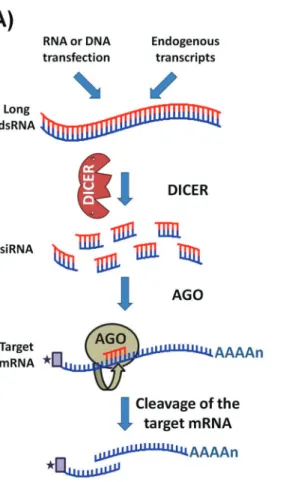
Mobile elements are involved in creating mutations and genomic rearrangements and, in many eukaryotes, these effects can be regulated through an RNA silencing mechanism such as RNA interference (RNAi) (Shiet al., 2004b; Girard and Hannon, 2008). Since first reported in 1998 (Fireet al., 1998), RNAi has swept through all fields of eukaryotic biology and has proven to be a very useful tool for analyzing gene function in a variety of organisms in which the introduction or expression of short double-stranded RNAs leads to the rapid destruction of cognate
mRNAs (Figure 4A) (Ngoet al., 1998). This approach has a number of advantages: it is fast, requires very little se-quence information, and reduces the expression of multiple gene copies, an action that is especially advantageous in asexual diploid organisms (LaCountet al., 2000; Shiet al., 2000; Wanget al., 2000).
Soon after it was described inC. elegans, RNAi was rapidly identified inT. brucei (Ngoet al., 1998). As dis-cussed below, RNAi has proven to be a powerful new tool for functional genomic studies inT. brucei.Unexpectedly, experimental evidence as well as searches of genome data-bases quickly showed that RNAi is absent inL. majorand
T. cruzi(Robinson and Beverley, 2003; DaRocha et al., 2004). It came as a surprise when Peacocket al.(2007) re-vealed that the genome ofL. braziliensisretained key genes involved with RNAi. These authors showed that L. braziliensisgenome contains orthologs carrying domains characteristic of the Dicer protein as well as genes with the typical argonaute domains PAZ and PIWI, the latter con-taining conserved amino acid residues that are essential for
functional TbAGO1 (Shi et al., 2004a). In addition, an N-terminal RGG domain, present in TbAGO1 and shown to be essential for its association with polyribosomes, is also present in theL. braziliensis Ago1 gene (Shiet al., 2004a). More recently, Lyeet al.(2010) demonstrated that the RNAi pathway is functional inL. braziliensisand in other species within theLeishmaniasubgenusViannia(L. guyanensis and L. panamensis) (Figure 4B). Thus, this di-vergent species ofLeishmaniaappear to have retained not only the mechanisms for (RNAi)-mediated regulation but also potentially active retroposons (Peacocket al., 2007), which might have assisted to create the greater divergence within theL. braziliensisgenome compared with the other
Leishmania species. For molecular parasitologists, these findings came as very good news since, as shown inT. brucei, the efficacy of RNAi knockdown as a tool for sys-tematic analysis of gene function is now also applicable in some species ofLeishmania.
Functional Genomics of T. Brucei and Gene
Expression Studies in a High Throughput Era
Infection byT. bruceioccurs after metacyclic trypo-mastigotes are injected into the bloodstream by the bite of a tsetse fly. In the mammalian host, the parasite differentiates into long slender trypomastigotes (bloodstream form -BSF) that divide, colonize the body fluids and can trans-form into a nonproliferative bloodstream trans-form, also known as the short stump form. After a blood meal, trypomas-tigotes acquired by the insect vector differentiate into pro-cyclic trypomastigotes in the gut, replicate and then migrate to salivary glands where they transform into metacyclic trypomastigotes (Matthews, 2005) (Figure 1). The ability of the BSF to invade the central nervous system leads to the neurological manifestations associated with sleeping sick-ness.
In contrast toT. cruziandLeishmania,T. brucei de-velops extracellularly throughout its entire life cycle. Its di-rect exposure to a strong antibody response in the bloodstream requires a sophisticated immune evasion pro-tocol, known as variant surface protein (VSG) switching (Payset al., 2007). VSGs are bloodstream-specific surface proteins with a hypervariable N-terminal domain that is ex-posed extracellularly and a more conserved C-terminal do-main buried in the parasite’s surface coat (Van der Ploeget al., 1982; Turner and Barry, 1989). Since they are tightly packed at the surface, VSGs are able to shield other invari-ant surface proteins from attack by the immune system. The tactic for evasion is based on antigenic variation in which the expression of a specific VSG is replaced by another gene from a supply of almost thousand copies at a rate of approximately one event per 100 cell divisions (Van der Ploeget al., 1982; Turner and Barry, 1989). VSG genes are thus a family ofT. brucei-specific genes whose monoallelic expression distinguishes African trypanosomes from the
other two groups of pathogenic trypanosomatids; the lack of antigenic variation in T. cruzi and Leishmania spp. means that these species must evade the host’s immune sys-tem by entering and multiplying inside host cells.
After completion of theT. b. bruceigenome, it was found that of the 9,068 predicted genes, including 904 pseudogenes, there were 1,700 T. brucei-specific genes, 806 of them (or 9% of the whole genome) corresponding to VSGs (El-Sayedet al., 2005b). Several VSG genes occur within hundreds of mini-chromosomes (50-150 kb in size) that are also part of theT. brucei genome. Surprisingly, analyses of all VSG sequences showed that only 57 are fully functional genes and have all the recognizable fea-tures of a typical VSG (Berrimanet al., 2005). It has been long known that VSG variability can be generated inT. bruceiby creating mosaic genes that include pseudogenes (Rothet al., 1989). Besides producing variability in VSG epitopes, these rearrangements may have the capacity to generate protein with new functions, as in the case of the SRA gene discussed below (Vanhammeet al., 2003).
As part of their “life in the bloodstream”, African trypanosomes that are pathogenic to humans have devel-oped a mechanism to withstand other types of attack from the host immune system. In contrast toT. brucei brucei, which do not infect humans,T. b. gambiense and T. b. rhodesienseare resistant to trypanolytic factors (TLF) pres-ent in normal human serum (NHS). TLF activity has been attributed to apolipoprotein L1 (ApoL1) and haptoglobin (Hp)-related protein found in high density lipoprotein (for a review, see Wheeler, 2010). NHS resistance in T. b. rhodesienseis conferred by a truncated VSG known as se-rum resistance associated (SRA) protein which is located in endosomes and binds and neutralizes TLF (De Greef and Hamers, 1994; De Greef et al., 1992; Vanhamme et al., 2003; Pérez-Morga et al., 2005; Wheeler, 2010).
Trypanosoma b. gambienselacks the SRA gene but still in-fects humans. The killing ofT. b. bruceirequires the bind-ing of TLF-1 and traffickbind-ing to the parasite acidic lysosome. It has been proposed that, inT. b. gambiense, changes in the receptor that binds TLF, as well as decreased expression of this gene, are responsible for the resistance of thisT. bruceisub-species to attack by the human innate im-mune system (Kieftet al., 2010).
SinceT. b. brucei is harmless to humans and T. b. gambienseis the most clinically relevant subspecies, Jack-sonet al.(2010) sequenced the genome ofT. b. gambiense
very few differences in their genomes. One of these differ-ences involved a gene encoding a putative iron-ascorbate oxidoreductase that is specific to a fewT. b. bruceistrains and is also absent in other trypanosomatids (L. majorandT. cruzi). Thus, it seems that not only differences in gene con-tent per se, but also individual single nucleotide poly-morphisms (indels) and variations in gene expression, or a combination of these factors, may contribute to this pheno-typic variation (Jacksonet al., 2010).
Coordinated changes in gene expression are vital for trypanosomes since they must deal with rapid changes in their environment (nutrient availability, temperature, host defenses, the presence of drug, etc.) and be able to dissemi-nate by alternating through different hosts. In addition, unique mechanisms for regulating gene expression are re-quired since there is an almost complete absence of trans-criptional control at the level of initiation. The lack of transcriptional regulatory elements, including a typical RNA polymerase II promoter, led to the conclusion that most factors involved in controlling gene expression act at the post-transcriptional level. By using approaches such as differential display, RNA fingerprinting, differential screening of cDNA libraries, random sequencing of cDNA clones and DNA microarrays, several groups have shown that significant changes in mRNA levels occur during the life cycle of all Tri-Tryps (Teixeiraet al., 1995; El-Sayedet al., 1995; Mathieu-Daudéet al., 1996; Diehlet al., 2002; Cerqueiraet al., 2005; Kabaniet al., 2009). Experimental evidence also indicates that, in addition to mRNA stability, changes in polysomal mobilization constitute an important mechanism for regulating gene expression (Alveset al., 2010). With the advent of next generation sequencing tech-nologies such as RNAseq, a global description of gene ex-pression patterns in T. brucei has finally been achieved (Kolevet al., 2010; Nilssonet al., 2010; Siegelet al., 2010; Veitch et al., 2010). This approach has provided much more complete information about mRNA structure and ex-pression levels, including the patterns of spliced leader (SL) and poly-A additions, as well as alternative pre-mRNA processing. Moreover, these global gene expression analyses have confirmed that only two T. brucei genes (poly-A polymerase and DNA/RNA helicase) contain introns.
Nilssonet al. (2010) showed that 2,500 alternative splicing events occur during processing ofT. bruceigenes, with a large number of these being regulated during the life cycle (a total of 600 genes have transcripts with more than one trans-spliced variant). The alternative splicing reac-tions can alter the message dramatically, as shown by Nilsson et al. (2010) and represented in Figure 3, with changes in the SL addition site leading to alterations in reg-ulatory elements in the 5’UTR (resulting in the modifica-tion of gene expression) or the initiator AUG (generating a different N-terminus or even causing a complete change in the translated ORF). Aminoacyl tRNA synthetase
tran-scripts are interesting examples of trans-spliced variants since differences in the enzyme N-terminus result in changes in the protein targeting signal: distinct mRNAs produce proteins that are directed to mitochondria or re-main in the cytoplasm (Nilssonet al., 2010). Likewise, al-ternative splicing is a potential mechanism for dual localization of theT. cruziLYT1 gene (Benabdellahet al., 2007). In addition to providing a mechanism for generating changes in the proteome, such flexibility in the selection of splicing sites is compatible with evidence that polycistronic transcription may also initiate at internal sites in gene clus-ters since correct processing of the 5’ end of mRNA can oc-cur at various points in the primary transcript (Kolevet al., 2010).
The mechanisms involved in transcription initiation in trypanosomes have always been an intriguing question. Recently, chromatin immunoprecipitation in combination with conventional Sanger sequencing (Respuela et al., 2008) or next-generation sequencing technology (Siegelet al., 2009; Thomaset al., 2009; Wrightet al., 2010) has pro-vided a much awaited analysis of the distribution patterns of modified histones throughout theT. bruceigenome. As summarized in Figure 3, the divergent strand-switch re-gions (SSR) have been found to contain an enrichment of histone variants (H2AZ and H2BV), acetylated histone 4 at lysine 10 (H4K10ac), histone 3 acetylated at residues K9 and K14 and trimethylated at K4, the bromodomain factor BDF3, and the transcription factors TRF4 and SNAP50 (Respuelaet al., 2008; Siegelet al., 2009; Thomaset al., 2009; Wrightet al., 2010). The histone variants H3V and H4V are present at transcription termination sites (Siegelet al., 2009). Similar patterns of chromatin modifications from transcription start sites (TSS) located at the beginning of polycistronic units were also found internally in the same polycistronic unit, suggesting the presence of internal TSS, in agreement with RNAseq data (Siegelet al., 2009; Kolev
et al., 2010; Wrightet al., 2010).
The increasing number of published genomes and transcriptomic data, partly as a consequence of the intro-duction of new sequencing platforms, has created enor-mous challenges for the field of functional genomics. One of the most useful techniques that has been used to identify gene function inT. bruceiis a combination of the inducible system mediated by T7 RNA polymerase and the tetracy-cline repressor (Wirtzet al., 1999) in combination with RNAi gene knockdown. As indicated before, in contrast to
T. cruziandL. major, RNAi is functional inT. brucei(Ngo
et al., 1998; Robinson and Beverley 2003; DaRochaet al., 2004; Lye et al., 2010). The first large scale functional genomics study using RNAi was reported by Morriset al.
but were unable to bind lectin, these authors identified clones with a reduced glycosylation of surface proteins and showed that these clones had lower expression of hexo-kinase (Morriset al., 2002).
A few years later, the functions of 197 ORFs fromT. b. brucei chromosome I were tested by RNAi. RNAi-induced parasites were tested for growth, nuclear and kinetoplast abnormalities by DAPI staining, and a pleio-tropic phenotype, morphology and motility. At least one of these phenotypes was found in 68 individual knockdowns (Subramaniamet al., 2006). In a similar approach, Portman
et al. (2009) identified novel components of the para-flagellar rod (PFR) structure by comparing proteomic pro-files before and after ablating the expression of individual PFR proteins. The silencing of PFR1 and PFR15 was evalu-ated by two-dimensional difference gel electrophoresis, and the spots with a two-fold change in volume were sub-jected to tandem MS protein identification. Proteomic anal-ysis combined with RNAi knockdown led to the identifica-tion of 30 proteins as potential PFR components, 20 of which were novel proteins. High-throughput cloning sys-tems also enhance functional analyses. By using the Gate-way®technology to create yeast two-hybrid vectors, eight non-redundant protein-protein interactions were detected among proteins with a PFR structure (Lacomble et al., 2009). These authors were able to construct a map showing the complex interaction of PFR proteins.
The availability of efficient methods for genetic ma-nipulation and for testing gene function through RNAi knockdown has led to major advances in genomic, trans-criptomic and proteomic analyses ofT. brucei, and has made this species a model organism for studying basic aspects of trypanosomatid biology. More recently, with the discovery of functional RNAi machinery inL. braziliensisand other
Leishmaniaspecies, gene function studies can now be con-ducted in this group and will soon provide valuable new in-formation aboutLeishmania-specific genes. However, as is becoming increasingly apparent from the data generated by comparative genomic analyses, each of the trypanosomatid species has its peculiarities. Researchers thus face the chal-lenge of developing new protocols specific for studying each parasite and its corresponding diseases. With our current knowledge of each Tri-Tryp disease and the new research methods that are being developed we can expect many new studies to emerge from hypotheses based on the Tri-Tryp genomic data. As a consequence of these new findings, better methods of controlling and preventing these diseases will follow.
Acknowledgments
The work from SMT and RMCP was funded by Conselho Nacional de Desenvolvimento Científico e Tec-nológico (CNPq), Fundação de Amparo a Pesquisa do Estado de Minas Gerais (FAPEMIG), Instituto Nacional de Ciencia e Tecnologia de Vacinas (INCTV) and the Howard
Hughes Medical Institute (HHMI). The work from WDR and MMKM was supported by FAPEMIG, Fundação Araucária de Apoio ao Desenvolvimento Científico e Tec-nológico do Paraná (Fundação Araucária), CAPES/Reuni, PPSUS/MS and CNPq.
References
Aksoy S, Lalor TM, Martin J, Van der Ploeg LH and Richards FF (1987) Multiple copies of a retroposon interrupt spliced
leader RNA genes in the African trypanosome,
Trypanosoma gambiense. EMBO J 6:3819-3826.
Alves LR, Avila AR, Correa A, Holetz FB, Mansur FC, Manque PA, de Menezes JP, Buck GA, Krieger MA and Goldenberg S (2010) Proteomic analysis reveals the dynamic association of proteins with translated mRNAs inTrypanosoma cruzi. Gene 452:72-78.
Araujo PR, Burle-Caldas GA, Silva-Pereira RA, Bartholomeu DC, DaRocha WD and Teixeira SM (2011) Development of a dual reporter system to identify regulatory cis-acting
ele-ments in untranslated regions of Trypanosoma cruzi
mRNAs. Parasitol Int 60:161-169.
Arner E, Kindlund E, Nilsson D, Farzana F, Ferella M, Tammi
MT and Andersson B (2007) Database of Trypanosoma
cruzirepeated genes: 20,000 additional gene variants. BMC Genomics 8:e391.
Aslett M, Aurrecoechea C, Berriman M, Brestelli J, Brunk BP, Carrington M, Depledge DP, Fischer S, Gajria B, Gao X,et al.(2010) TriTrypDB: A functional genomic resource for the Trypanosomatidae. Nucleic Acids Res 38:D457-D462. Augusto-Pinto L, Teixeira SM, Pena SD and Machado CR (2003)
Single-nucleotide polymorphisms of theTrypanosoma cruzi
MSH2 gene support the existence of three phylogenetic lin-eages presenting differences in mismatch-repair efficiency. Genetics 164:117-126.
Baida RC, Santos MR, Carmo MS, Yoshida N, Ferreira D, Fer-reira AT, El Sayed NM, Andersson B and da Silveira JF (2006) Molecular characterization of serine-, alanine-, and proline-rich proteins ofTrypanosoma cruziand their possi-ble role in host cell infection. Infect Immun 74:1537-1546. Bartholomeu DC, Cerqueira GC, Leão AC, DaRocha WD, Pais
FS, Macedo C, Djikeng A, Teixeira SM and El-Sayed NM (2009) Genomic organization and expression profile of the mucin-associated surface protein (masp) family of the
hu-man pathogen Trypanosoma cruzi. Nucleic Acids Res
37:3407-3417.
Bates PA (2007) Transmission ofLeishmaniametacyclic promas-tigotes by phlebotomine sand flies. Int J Parasitol 37:1097-1106.
Benabdellah K, González-Rey E and González A (2007) Alterna-tive trans-splicing of the Trypanosoma cruziLYT1 gene transcript results in compartmental and functional switch for the encoded protein. Mol Microbiol 65:1559-1567. Benz C, Nilsson D, Andersson B, Clayton C and Guilbride DL
(2005) Messenger RNA processing sites inTrypanosoma brucei. Mol Biochem Parasitol 143:125-134.
Berriman M, Ghedin E, Hertz-Fowler C, Blandin G, Renauld H, Bartholomeu DC, Lennard NJ, Caler E, Hamlin NE, Haas B,
et al. (2005) The genome of the African trypanosome
Blackwell JM and Melville SE (1999) Status of protozoan
ge-nome analysis: Trypanosomatids. Parasitology 118
(Suppl):S11-14.
Blackwell JM, Fakiola M, Ibrahim ME, Jamieson SE, Jeronimo SB, Miller EN, Mishra A, Mohamed HS, Peacock CS, Raju M, et al. (2009) Genetics and visceral leishmaniasis: Of mice and man. Parasite Immunol 31:254-266.
Boucher N, McNicoll F, Dumas C and Papadopoulou B (2002) RNA polymerase I-mediated transcription of a reporter gene integrated into different loci ofLeishmania. Mol Biochem Parasitol 119:153-158.
Branche C, Ochaya S, Aslund L and Andersson B (2006) Compar-ative karyotyping as a tool for genome structure analysis of
Trypanosoma cruzi. Mol Biochem Parasitol 147:30-38. Brandão A, Urmenyi T, Rondinelli E, Gonzalez A, de Miranda
AB and Degrave W (1997) Identification of transcribed se-quences (ESTs) in theTrypanosoma cruzigenome project. Mem Inst Oswaldo Cruz 92:863-866.
Brener Z (1973) Biology ofTrypanosoma cruzi. Annu Rev Mi-crobiol 27:347-382.
Bringaud F, Berriman M and Hertz-Fowler C (2009) Trypa-nosomatid genomes contain several subfamilies of ingi-related retroposons. Eukaryot Cell 8:1532-1542.
Brisse S, Dujardin JC and Tibayrenc M (2000) Identification of sixTrypanosoma cruzilineages by sequence-characterised amplified region markers. Mol Biochem Parasitol 111:95-105.
Buscaglia CA and Di Noia JM (2003)Trypanosoma cruziclonal diversity and the epidemiology of Chagas’ disease. Mi-crobes Infect 5:419-427.
Campos PC, Bartholomeu DC, DaRocha WD, Cerqueira GC and Teixeira SM (2008) Sequences involved in mRNA process-ing inTrypanosoma cruzi. Int J Parasitol 38:1383-1389. Cano MI, Gruber A, Vazquez M, Cortés A, Levin MJ, González
A, Degrave W, Rondinelli E, Zingales B, Ramirez JL,et al.
(1995) Molecular karyotype of clone CL Brener chosen for the Trypanosoma cruzi genome project. Mol Biochem Parasitol 71:273-278.
Cerqueira GC, DaRocha WD, Campos PC, Zouain CS and Tei-xeira SM (2005) Analysis of expressed sequence tags from
Trypanosoma cruziamastigotes. Mem Inst Oswaldo Cruz 100:385-389.
Cerqueira GC, Bartholomeu DC, DaRocha WD, Hou L, Frei-tas-Silva DM, Machado CR, El-Sayed NM and Teixeira SM (2008) Sequence diversity and evolution of multigene
fami-lies in Trypanosoma cruzi. Mol Biochem Parasitol
157:65-72.
Clayton CE (2002) Life without transcriptional control? From fly to man and back again. EMBO J 21:1881-1888.
Cribb P and Serra E (2009) One- and two-hybrid analysis of the interactions between components of theTrypanosoma cruzi
spliced leader RNA gene promoter binding complex. Int J Parasitol 39:525-532.
Cribb P, Esteban L, Trochine A, Girardini J and Serra E (2010)
Trypanosoma cruzi TBP shows preference for C/G-rich DNA sequencesin vitro. Exp Parasitol 124:346-349. DaRocha WD, Otsu K, Teixeira SM and Donelson JE (2004)
Tests of cytoplasmic RNA interference (RNAi) and con-struction of a tetracycline-inducible T7 promoter system in
Trypanosoma cruzi. Mol Biochem Parasitol 133:175-186. de Freitas JM, Augusto-Pinto L, Pimenta JR, Bastos-Rodrigues L,
Gonçalves VF, Teixeira SM, Chiari E, Junqueira AC,
Fer-nandes O, Macedo AM,et al.(2006) Ancestral genomes, sex, and the population structure of Trypanosoma cruzi. PLoS Pathog 2:e24.
De Greef C and Hamers R (1994) The serum resistance-associated (SRA) gene ofTrypanosoma brucei rhodesienseencodes a variant surface glycoprotein-like protein. Mol Biochem Parasitol 68:277-284.
De Greef C, Chimfwembe E, Kihang’a Wabacha J, Bajyana Songa E and Hamers R (1992) Only the serum-resistant bloodstream forms ofTrypanosoma brucei rhodesiense ex-press the serum resistance associated (SRA) protein. Ann Soc Belg Med Trop 72(Suppl 1):13-21.
Denise H, Poot J, Jimenez M, Ambit A, Hermman DC, Ver-meulen AN, Coombs GH and Mottram JC (2006) Studies on the CPA cysteine peptidase in theLeishmania infantum ge-nome strain JPCM5. BMC Mol Biol 7:42.
Depledge DP, Evans KJ, Ivens AC, Aziz N, Maroof A, Kaye PM and Smith DF (2009) Comparative expression profiling of
Leishmania: Modulation in gene expression between spe-cies and in different host genetic backgrounds. PLoS Negl Trop Dis 3:e476.
Desjeux P (1996) Leishmaniasis. Public health aspects and con-trol. Clin Dermatol 14:417-423.
Di Noia JM, Sánchez DO and Frasch AC (1995) The protozoan
Trypanosoma cruzihas a family of genes resembling the mucin genes of mammalian cells. J Biol Chem 270:24146-24149.
Diehl S, Diehl F, El-Sayed NM, Clayton C and Hoheisel JD (2002) Analysis of stage-specific gene expression in the bloodstream and the procyclic form ofTrypanosoma brucei
using a genomic DNA-microarray. Mol Biochem Parasitol 123:115-123.
El-Sayed NM and Donelson JE (1997) A survey of the
Trypanosoma brucei rhodesiensegenome using shotgun se-quencing. Mol Biochem Parasitol 84:167-178.
El-Sayed NM, Alarcon CM, Beck JC, Sheffield VC and Donelson JE (1995) cDNA expressed sequence tags ofTrypanosoma brucei rhodesienseprovide new insights into the biology of the parasite. Mol Biochem Parasitol 73:75-90.
El-Sayed NM, Myler PJ, Blandin G, Berriman M, Crabtree J, Aggarwal G, Caler E, Renauld H, Worthey EA, Hertz-Fowler C,et al.(2005a) Comparative genomics of trypa-nosomatid parasitic protozoa. Science 309:404-409. El-Sayed NM, Myler PJ, Bartholomeu DC, Nilsson D, Aggarwal
G, Tran AN, Ghedin E, Worthey EA, Delcher AL, Blandin G, et al.(2005b) The genome sequence ofTrypanosoma cruzi, etiologic agent of Chagas disease. Science 309:409-415.
Fernandes AP, Costa MM, Coelho EA, Michalick MS, de Freitas E, Melo MN, Luiz Tafuri W, Resende DM, Hermont V, Abrantes CF,et al.(2008) Protective immunity against chal-lenge withLeishmania(Leishmania)chagasiin beagle dogs vaccinated with recombinant A2 protein. Vaccine 26:5888-5895.
Fire A, Xu S, Montgomery MK, Kostas SA, Driver SE and Mello CC (1998) Potent and specific genetic interference by
dou-ble-stranded RNA in Caenorhabditis elegans. Nature
391:806-811.