Seroprevalence of Brucellosis, Leptospirosis,
and Q Fever among Butchers and
Slaughterhouse Workers in South-Eastern
Iran
Saber Esmaeili1,2,3, Saied Reza Naddaf4, Behzad Pourhossein1,5, Abdolrazagh Hashemi Shahraki1,2, Fahimeh Bagheri Amiri1,6, Mohammad Mehdi Gouya7, Ehsan Mostafavi1,2*
1Department of Epidemiology, Pasteur Institute of Iran, Tehran, Iran,2National Reference Laboratory for Plague, Tularemia and Q fever, Research Centre for Emerging and Re-emerging Infectious Diseases, Pasteur Institute of Iran, Akanlu, Kabudar Ahang, Hamadan, Iran,3Department of Bacteriology, Faculty of Medical Sciences, Tarbiat Modares University, Tehran, Iran,4Department of Parasitology, Pasteur Institute of Iran, Tehran, Iran,5Department of Virology, School of Public Health, Tehran University of Medical Sciences, Tehran, Iran,6Department of Epidemiology, Faculty of Veterinary Medicine, University of Tehran, Tehran, Iran,7Centre of Disease Control (CDC), Ministry of Health, Tehran, Iran
*mostafavi@pasteur.ac.ir
Abstract
Zoonotic diseases can be occupational hazards to people who work in close contact with animals or their carcasses. In this cross-sectional study, 190 sera were collected from butchers and slaughterhouse workers in different regions of the Sistan va Baluchestan province, in Iran in 2011. A questionnaire was filled for each participant to document per-sonal and behavioural information. The sera were tested for detection of specific IgG anti-bodies against brucellosis, leptospirosis, and Q fever (phase I and II) using commercial enzyme-linked immunosorbent assays (ELISA). The seroprevalence of brucellosis was 7.9%, leptospirosis 23.4%, and phase I and II of Q fever were 18.1% and 14.4%, respec-tively. The seroprevalence of Q fever and leptospirosis, but not brucellosis, varied among regions within the province (p= 0.01). Additionally, a significant relationship was found
between seropositivity of Q fever and camel slaughtering (p= 0.04). Reduced seropositivity
rate of brucellosis was associated with use of personal protective equipment (PPE) (p=
0.004). This study shows that brucellosis, leptospirosis and Q fever occur among butchers and slaughterhouse workers in this area.
Introduction
Many zoonotic diseases and human pathogens are occupational hazards faced by individuals who come into close contact with animals or their carcasses. The probability of contact with zoonotic pathogens while working depends upon various factors, such as the health status of the animals, the type of work performed, the frequency of contact with live animals, carcasses
OPEN ACCESS
Citation:Esmaeili S, Naddaf SR, Pourhossein B, Hashemi Shahraki A, Bagheri Amiri F, Gouya MM, et al. (2016) Seroprevalence of Brucellosis,
Leptospirosis, and Q Fever among Butchers and Slaughterhouse Workers in South-Eastern Iran. PLoS ONE 11(1): e0144953. doi:10.1371/journal. pone.0144953
Editor:Jonas Waldenström, Linneaus University, SWEDEN
Received:November 13, 2014
Accepted:November 25, 2015
Published:January 5, 2016
Copyright:© 2016 Esmaeili et al. This is an open access article distributed under the terms of the Creative Commons Attribution License, which permits unrestricted use, distribution, and reproduction in any medium, provided the original author and source are credited.
Data Availability Statement:All relevant data are within the paper and its Supporting Information file.
and tissues of slaughtered animals, the use of personal and environmental protective measures, and the attitudes and levels of knowledge of the people at risk [1]. Butchers and slaughterhouse workers are at high risk of contracting zoonotic diseases. In Iran, previous studies have identi-fied zoonoses like brucellosis, leptospirosis and Q fever as potential occupational hazards for slaughterhouse workers [2–5].
Brucellosis is an important zoonosis worldwide affecting both livestock and humans; it is listed among the top ten pathogens at the wildlife-livestock interface [6,7]. It can be transmitted to humans through direct contact with infected tissues (especially genital organs and birth products), inhalation of aerosols, and ingestion of raw milk and dairy products from infected animals [8]. Brucellosis in humans is characterized mainly by intermittent fever, with manifes-tations such as gastrointestinal, cardiovascular, genitourinary, hematopoietic, nervous, skeletal, pulmonary, cutaneous, and ocular involvement [9].
Leptospirosis is a wide spread zoonotic disease that also affects both humans and animals [10]. The etiological agent,Leptospiraspp., can be transmitted to humans through broken skin or mucous membranes during contact with tissues, body fluids, and organs from infected ani-mals, or by consumption of food or water contaminated with the urine of infected animals [11]. In humans, leptospirosis cause a wide range of symptoms including fever, myalgia, con-junctivitis, jaundice, kidney failure, meningitis, myocarditis, meningoencephalitis and pulmo-nary haemorrhage with respiratory failure, which sometimes results in death[10]. The disease is an occupational hazard for farmers, sewer workers, miners, dairy and slaughterhouse work-ers, and fish industry workers [11].
Q fever is also a significant zoonotic disease caused by the rickettsia-like bacteriumCoxiella burnetii[12]. This disease is considered to be an occupational hazard for livestock handlers, farmers, veterinarians, and butchers and slaughterhouse workers [13]. Livestock such as cattle, sheep and goats are among the main sources of human infection. In animals, Q fever is mostly asymptomatic or subclinical [12,14]. The disease is mainly transmitted to humans through inhalation of infectious agents, consumption of unpasteurized contaminated milk and dairy products, contact with infectious tissues, and, rarely, via tick bites [15]. About 60% of people infected with Q fever are asymptomatic. The symptoms of acute Q fever in humans may include severe headache, prolonged fever, pneumonia, hepatitis, myalgia, arthralgia, cough, cardiac failure and neurological disorders [12]. Patients with chronic Q fever have symptoms such as endocarditis, vascular infection, and fatigue and have a higher likelihood of abortion and stillbirth [15]. Phase I and II antibodies are detectable in patients with chronic Q fever, but antibodies against phase II are indicative of acute Q fever [12].
As butchers and slaughterhouse workers are in close contact with animals or their body flu-ids and tissues they are at high risk of contracting zoonotic diseases [16]. Imports of large num-bers of livestock from eastern neighbouring countries, Afghanistan and Pakistan, to the Sistan va Baluchestan province in Iran [17], and a recent report of brucellosis and Q fever outbreak in Afghanistan [18] prompted us to evaluate the seroprevalence of brucellosis, leptospirosis and Q fever among butchers and slaughterhouse workers in this province. We also evaluated the risk factors related to these diseases among these individuals.
Materials and Methods
Study area
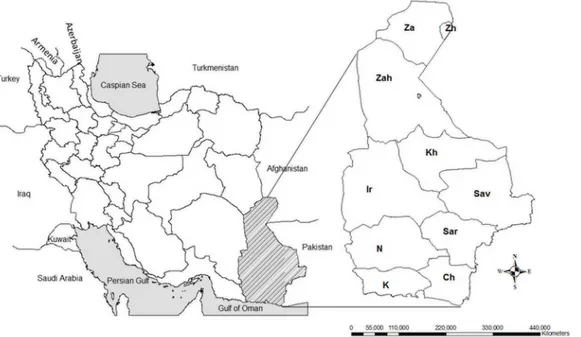
This study was carried out in the Sistan va Baluchestan province in south-eastern Iran in 2011. This province, the largest in the country, covers an area of 187,502 km2and has a population of about 2.5 million people. Sistan va Baluchestan is bordered by the Oman Sea to the south, by Afghanistan and Pakistan in the east, by South Khorasan province to the north, and by the Competing Interests:The authors have declared
Kerman and Hormozgan provinces in the west (Fig 1). The climate of this province is semi-arid and experiences long, hot summers and short winters.
Ethical considerations
The ethical committee of the Pasteur Institute of Iran approved the consent procedure, the pro-posal and protocol of this study, covering all the samples taken (blood), questionnaire and ver-bal informed consent as most of the participants were either illiterate or had a primary education.
Samples collection
In this cross-sectional study, blood samples were obtained from butchers and slaughterhouse workers after obtaining their informed consent. All official slaughterhouses in this province were recruited and participants were selected randomly among the employees. Sampling was carried out in slaughterhouses in the north, centre, and south of the province. The inclusion criteria were being over 18 and working as a butcher or slaughterhouse worker for a minimum of 6 months. Information of each participant, including demographic characteristics (age and gender), work history, exposure to risks during work, use of personal protective equipment (PPE) (including mask, gloves, overalls and boots), and their knowledge, attitude, and practices about common zoonotic diseases was collected by means of a researcher-developed question-naire (Table 1). A 10 mL blood sample was collected from each participant after obtaining informed consent. Sera were kept at -20°C and transferred to the Department of Epidemiology of the Pasteur Institute of Iran (Tehran).
Serological tests
Detection of brucellosis antibody (IgG). The commercial enzyme-linked immunosor-bent assay (ELISA) kit (IBL, Hamburg, Germany), was used for brucellosis antibody (IgG) detection. Briefly, 1μL of the sera was diluted 1:100, and after washing with buffer-washing, 100μL of horseradish peroxidase-conjugated anti-human IgG was added. The mixture was incubated for 30 minutes at room temperature and then treated with tetra methyl benzidine (TMB) for 20 minutes. ABrucellaantibody-antigen reaction was indicated by a blue coloration. Subsequently, a TMB stop solution was added, and the optical density (OD) of the well was measured at 450 nm by a micro plate reader (ELx808, BioTek Instruments Inc., Winooski, VT, USA). Antibody activities were calculated using a standard curve according to the manufac-turer's guidelines.
The positive and borderline sera detected using the ELISA method were confirmed with the standard tube agglutination (STA) test as a gold standard test for brucellosis diagnosis. We used a locally prepared antigen and a STA test produced by the Pasteur Institute of Iran. Sera were serially diluted from 1:20 to 1:1280, mixed with the standard tube agglutination antigen and then incubated at 37°C for approximately 24 hours. Each batch of the test included a posi-tive control and a negaposi-tive control. Serum titers1:80 were considered positive [19,20].
Detection ofCoxiella burnetiiantibodies (IgG I and II). IgG antibodies againstC.
burne-tiiwere detected using a commercial ELISA kit (Serion/Verion Co., Germany, Kit number ESR 1312 G) according to the manufacturer’s instructions. Phase I and II antibodies were identified in separate assays. The plates were read at 405 nm using a microplate reader (ELx808, BioTek Instruments Inc., Winooski, VT, USA). For phase I antibodies, the sample was considered pos-itive when the serum OD was>10% above the OD cut-off value. For phase II, antibody
activi-ties in IU/mL were calculated using a standard curve which was incorporated in the kit following the manufacturer's guidelines.
Statistical analysis
The data were analysed using SPSS software (Version 16, SPSS Inc, Chicago, Ill). Chi-square, Fisher's exact and logistic regression tests were used to compare the variables during analysis. All results were considered statistically significant if thep-value was equal to or less than 0.05, and marginally significant if the p-value was between 0.05 and 0.1.
Results
In this study, 190 blood samples was taken from butchers and slaughterhouse workers residing in 11 counties of the Sistan va Baluchistan province, including Zahak and Zabol in the north, Iranshahr, Zahedan and Khash in the centre and Chabahar, Sarbaz, Saravan and Konarak in the south of the province. The median (maximum, minimum) age and work experience of par-ticipants in this study were 33.5 (18, 86) and 8 (1, 44) years, respectively. All parpar-ticipants were male and 96.8% of them claimed to be satisfied with their occupation. Detailed information about the all participants is shown inS1 Dataset.
In total, 162 (85.3%) participants were directly involved in animal slaughtering, 43 (22.6%) in transportation and handling of animal residues, and 2 (1.1%) only inspected the carcases. A
Fig 1. Map of Sistan va Baluchestan province in southeast Iran.Sampling from butchers and slaughterhouse workers was performed in the different counties of Sistan va Baluchestan province including: Zahak (Zh) and Zabol (Za) in the north, Zahedan (Zah), Iranshahr (Ir) and Khash (Kh) in the centre, and Saravan (Sav), Sarbaz (Sar), Nikshahr(N), Konarak (K) and Chabahar (Ch) in the south.
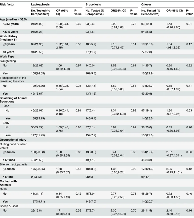
Table 1. Analysis of risk factors associated with the seroprevalence of leptospirosis, brucellosis and Q fever among butchers and slaughterhouse workers in south-eastern Iran in 2011.
Risk factor Leptospirosis Brucellosis Q fever
No. Tested (% Seropositive)
OR (95% CI)
P-value
No. Tested (% Seropositive)
OR(95% CI) P-value
No. Tested (% Seropositive)
OR (95% CI) P-value
Age (median = 33.5)
33.5 years 91(21.98) 1.20(0.61,
2.38) 0.60 93(8.6) 0.99(0.91,1.08) 0.78 93(19.4) 1.43(0.76,2.86) 0.31
>33.5 years 91(25.27) 93(7.5) 94(25.5)
Work History (median = 8)
8 years 82(21.95) 1.22(0.61,
2.45) 0.58 105(5.7) 2.18(0.74,6.42) 0.14 102(18.6) 1.64(.081,3.32) 0.17
>8 years 94(25.53) 77(11.7) 77(27.3)
Work Type
Slaughtering
No 13(23.08) 1.06
(0.29,4.98) 0.97 14(0.0) 1.53(0.23,35.95) 0.61 14(35.7) 0.50(0.16,1.60) 0.32
Yes 158(24.05) 162(9.3) 160(21.9)
Transportation of the remaining livestock
No 129(26.36) 0.56(0.21,
1.34) 0.21 133(7.5) 1.62(0.52,5.03) 0.53 131(23.7) 0.85(0.37,1.97) 0.71
Yes 42(16.67) 43(11.6) 43(20.9)
Splashing of Animal Secretions
Face
No 46(23.91) 0.96(0.44,
2.18)
0.91 47(6.4) 1.34
(0.362,4.98)
0.99 47(19.1) 1.30
(0.57,2.97) 0.53
Yes 138(23.19) 143(8.4) 140(23.6)
Body
No 36(22.22) 1.09(0.46,
2.76) 0.86 37(8.1) 0.97(0.26,3.64) 0.99 36(25.0) 0.85(0.36,1.98) 0.70
Yes 147(31.25) 152(7.9) 150(22.0)
Occupational Injury
Cutting hand or other organs
5 times 130(23.08) 1.20
(0.55,2.55)
0.63 136(8.8) 0.44
(0.09,2.04)
0.36 134(19.4) 2.07 (0.97,4.341)
0.06
>5 times 49(26.53) 49(4.1) 48(33.3)
Bite from ectoparasite
5 times 175(22.85) 1.68
(0.33,7.07) 0.48 181(8.3) 1.35(0.06,9.21) 0.92 178(21.3) 2.95(0.75,11.51) 0.12
>5 time 9(33.33) 9(0.0) 9(44.4)
Contact with Animals
Cattle
No 45(31.11) 0.54
(0.25,1.19)
0.12 45(8.9) 0.77
(0.23,2.59)
0.75 45(26.7) 0.72
(0.33,1.56) 0.40
Yes 137(19.71) 143(7.0) 140(20.7)
Sheep & Goat
No 26(15.8) 1.71
total of 161 (84.7%) participants were in contact with sheep and goats, 143 (75.3%) with cattle including calves, and 80 (42.1%) with camels during their daily activities. Moreover 75.3% of the workers reported a history of being splashed with animal fluids and viscera more than once onto their face and 80.0% on other parts of their bodies. In addition 25.8% of all individuals had a history of cutting their hands or other parts of their bodies at least once during their work, and 17.4% recalled ectoparasite bites within the last year. The obtained data showed that 39.7% of participants did not use any PPE (masks, gloves, overalls and boots), while 22.8% always used it. Also 83.6% of participants had never applied chemical disinfectants to their kni-ves and hands, while 79.9% knew they were at risk of zoonotic infections.
ELISA results revealed that 8.4% of participants were positive and 4.2% at borderline for anti-BrucellaIgG; the seroprevalence of brucellosis among the participants was 7.9% using the standard tube agglutination test (STAT).
Furthermore, 23.4% and 15.8% of participants were positive and borderline, respectively, for anti-LeptospiraIgG using ELISA.
Seroprevalence of Q fever antibodies, of phases I and II (IgG) were 18.1% and 14.4%, respec-tively, and 13.9% and 8.4% of participants had a borderline titre for phase I and phase II anti-bodies, respectively. The overall seroprevalence of Q fever (participants having antibodies of phase I and/or phase II) was 22.5%.
Table 1. (Continued)
Risk factor Leptospirosis Brucellosis Q fever
No. Tested (% Seropositive)
OR (95% CI)
P-value
No. Tested (% Seropositive)
OR(95% CI) P-value
No. Tested (% Seropositive)
OR (95% CI) P-value
Yes 156(28.05) 161(8.1) 159(23.9)
Camel
No 105(21.90) 1.09
(0.53,2.20) 0.81 108(8.3) 0.73(0.24,2.28) 0.60 106(17) 2.01(0.99,4.09) 0.049
Yes 77(23.38) 80(6.2) 79(29.1)
All 3 group of animals
No 112(20.53) 1.33
(0.65,2.72)
0.42 115(8.7) 0.61
(0.18,2.02)
0.41 112(17.9) 1.86 (0.92,3.74)
0.08
Yes 70(25.71) 73(5.5) 73(28.8)
Attitude and Practice (Personal Protection)
See themselves at risk for zoonotic diseases
No 35(20.00) 1.28
(0.53,3.41) 0.60 38(7.9) 0.99(.27,3.71) 0.99 37(10.8) 0.35(.012,1.07) 0.06
Yes 148(24.32) 151(7.9) 149(25.5)
Personal Protection (median = 12)*
12 91(26.37) 1.41
(0.71,2.84)
0.33 92(2.2) 0.14
(0.03,0.64)
0.004 91(25.3) 1.41 (0.70,2.83)
0.34
>12 91(19.78) 95(13.7) 93(19.4)
*Total scores earned by each participant in the use of PPE was considered as the performance of each participant. The median of the performance for all participants was 12.
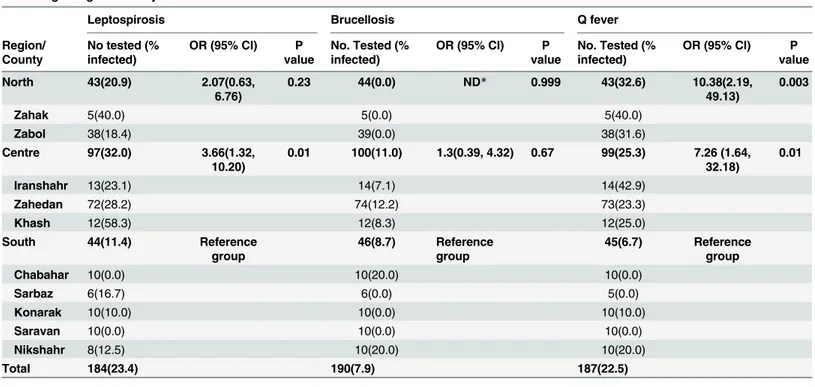
The highest brucellosis seroprevalence was observed in Chabahar (20.0%) and Nikshahr (20.0%), and the highest Q fever seroprevalence was in Zahak (40.0%) and Iranshahr (42.9%). Leptospirosis seroprevalence was highest in Khash (58.3%) and Zahak (40.0%). The partici-pants from Saravan County in the south of the province had no antibodies to any of the three pathogens (Table 1).
Participants from central and northern regions were 7.26 (OR: 7.26, 95%CI: 1.64, 32.18) (p= 0.01) and 10.38 (OR: 10.38, 95%CI: 2.19, 49.13) (p= 0.003) times more likely to be sero-positive, respectively, for Q fever than those from southern regions. A significant difference in leptospirosis seropositivity was seen between southern (11.4%) and central (32%) regions (OR: 3.66, 95%CI: 1.32, 10.2,p= 0.01), but no significant difference was found between southern and northern regions (OR: 2.07, 95%CI: 0.63, 6.76,p= 0.23). Brucellosis seroprevalence did not differ significantly among regions (Table 2).
There were no associations between the age of the investigated persons and the seropreva-lence rates of leptospirosis, Q fever or brucellosis. There was, however, a significant association between seroprevalence of Q fever and camels slaughtering (p= 0.05, OR: 2.01, 95%CI: 0.99, 4.09). The use of PPE was negatively associated with the seroprevalence of brucellosis (p= 0.004, OR: 0.14, 95% CI: 0.03, 0.64). A marginally significant positive correlation was found between seroprevalence of Q fever and having a history of cutting hands (p= 0.06, OR: 2.07, 95% CI: 0.97, 4.34) and the attitude of the workers (considering themselves at risk of zoo-notic disease, because of their job:p= 0.06, OR: 0.35, 95% CI: 0.12, 1.07;Table 1).
Variables such as work history, work type, splashing of animal secretions on face or body and occupational injury were not significantly associated with seroprevalence rates of leptospi-rosis, brucellosis or Q fever.
Discussion
In this study we obtained the seroprevalence of antibodies to three zoonotic bacterial diseases among slaughterhouse workers and butchers in the Sistan va Baluchestan province in south-eastern Iran.
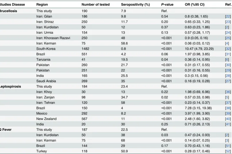
Brucellosis is an endemic disease in Iran and has been reported from different parts of the country [21]. In our study, the seroprevalence of brucellosis among butchers and slaughter-house workers was 7.9%. Different similar studies were conducted among slaughterslaughter-house workers and general population [2,4,22–27]. This rate was lower than those obtained from similar studies conducted on butchers and slaughterhouse workers from Khorasan Razavi province in the northeast of Iran (48%) [25], Kerman city in the south of Iran (58.6%) [4], Saudi Arabia (35%) [27], India (25.5%) [28], Pakistan (22%) and (21.7%) [29,30], and Tanza-nia (19.5%) [6] and was higher than what was reported from Brazil (4.2%) [31], and South Korea (0.8%) [32] (Table 3). Collectively, these studies indicate that butchers and slaughter-house workers might face different levels of risk to zoonotic infectious diseases in different areas, possibly due to variation in infection rates among animals, differences in human lifestyle and use of PPE. Taken together, working in a slaughterhouse in Iran seems to be a risk factor for contracting brucellosis, which is further supported by a previous screening among blood donors in the general population in the Bushehr province, southern Iran, that estimated brucel-losis seroprevalence to 0.057% [26].
brucellosis in Iran include the consumption of fresh cheese, contact with animal skins and con-suming undercooked meat or raw milk [33]. Brucellosis is also an occupational hazard for cer-tain professions in health settings, such as veterinary and laboratory personnel’s [33].
The environment and the socioeconomic conditions along the Caspian Sea littoral zone in the north of Iran are more favourable for the survival and transmission ofLeptospiraspp. spi-rochetes than in other areas of Iran [34,35]. The dry and desert climates in the Sistan va Balu-chestan are believed to be less suitable for environmental survival ofLeptospiraspp.
spirochetes. This disease can be transmitted to people coming into close contact with animals. Different studies were conducted among slaughterhouse workers and butchers in Iran [5,36– 38]. In our study, the seroprevalence of leptospirosis was 23.4% among butchers and slaughter-house workers in the province. In other studies carried out on butchers and slaughterslaughter-house workers in Iran, the seroprevalence of leptospirosis was higher in Zanjan province (northwest of Iran)) 34.7% ([5] and in Tehran province (northern Iran)) 58% ([37]. However, in similar studies in other countries, the seroprevalence of leptospirosis was lower than this study: Brazil (4%) [38], Mexico (8.2%) [39] and New Zealand (11%) [40] (Table 3). The comparatively high seropositivity rate in this study is interesting, as no human leptospirosis case has been reported from Sistan va Baluchestan and that none of the seropositive individuals in this study had clini-cal symptoms registered in their mediclini-cal history. However, this disease has been reported from South Khorasan and Afghanistan which share borders with Sistan va Baluchestan [42,43]. A study in Pakistan in 2010–2011 revealed a 44% seropositivity rate among veterinarians, pet-owners and livestock holders [44].
The last human case of Q fever in Iran was reported in 1973 [45]. Recently, this infection was reported in livestock in different regions of the country [46–48], and anti-C.burnetii anti-bodies were detected in febrile patients in the Kerman province, west of Sistan va Baluchestan
Table 2. Seroprevalence of Q fever, brucellosis and leptospirosis among butchers and slaughter workers in Sistan va Baluchestan province according to region and city.
Leptospirosis Brucellosis Q fever
Region/ County
No tested (% infected)
OR (95% CI) P value
No. Tested (% infected)
OR (95% CI) P value
No. Tested (% infected)
OR (95% CI) P value
North 43(20.9) 2.07(0.63,
6.76)
0.23 44(0.0) ND* 0.999 43(32.6) 10.38(2.19,
49.13)
0.003
Zahak 5(40.0) 5(0.0) 5(40.0)
Zabol 38(18.4) 39(0.0) 38(31.6)
Centre 97(32.0) 3.66(1.32,
10.20)
0.01 100(11.0) 1.3(0.39, 4.32) 0.67 99(25.3) 7.26 (1.64, 32.18)
0.01
Iranshahr 13(23.1) 14(7.1) 14(42.9)
Zahedan 72(28.2) 74(12.2) 73(23.3)
Khash 12(58.3) 12(8.3) 12(25.0)
South 44(11.4) Reference
group
46(8.7) Reference group
45(6.7) Reference group
Chabahar 10(0.0) 10(20.0) 10(0.0)
Sarbaz 6(16.7) 6(0.0) 5(0.0)
Konarak 10(10.0) 10(0.0) 10(10.0)
Saravan 10(0.0) 10(0.0) 10(0.0)
Nikshahr 8(12.5) 10(20.0) 10(20.0)
Total 184(23.4) 190(7.9) 187(22.5)
*ND: Not Defined
[49]. It is known that antibodies against antigens of phase I and II persist for months or years after primary infection. Since the clinical diagnosis is difficult, in most cases, diagnosis of Q fever depends on serological tests [50]. In our study, the seroprevalence rate of phase I and II antibodies in butchers and slaughterhouse workers of Sistan va Baluchestan were 18.1% and 14.4%, respectively.
In other studies carried out on butchers and slaughterhouse workers, the seroprevalence of phase I and II IgG antibodies for Q fever was higher in Kurdistan province, western Iran (38%) [2], Kerman province, south-eastern Iran (68%) [3], Brazil (29%) [51] and Turkey (50.9%) [52] (Table 3). In the present study, the seropositivity of Q fever among butchers and slaughter-house workers in the central and northern regions of Sistan va Baluchestan was significantly higher than the southern regions of the province, which may be related to the recent human outbreak of Q fever in Afghanistan (Bamyan province in 2011) [18], and/or to larger livestock population in this region and high number of animals imported from Afghanistan. In this study, we observed an association between Q fever seropositivity and camel slaughtering, a finding which is in agreement with other studies reporting Q fever infection in camels in south-eastern Iran [14,53]. Also, in a study in Chad, Africa, camel breeding was a significantly associated with Q fever seropositivity [54].
Table 3. The comparison of the seroprevalence surveys of brucellosis, leptospirosis and Q fever carried out in different areas among butchers and slaughter workers.
Studies Disease Region Number of tested Seropositivity (%) P-value OR (%95 CI) Ref.
Brucellosis This study 190 7.9 Ref.
Iran: Gilan 186 9.8 0.54 0.8 (0.38, 1.65) [22]
Iran: Shiraz 250 11.7 0.20 0.65 (0.33, 1.25) [23]
Iran: Kurdistan 50 12 0.37 0.63 (0.23, 1.86) [2]
Iran: Urmia 154 13 0.13 0.57 (0.28, 1.17) [24]
Iran: Khorasan Razavi 250 48 <0.001 0.9 (0.05, 0.16) [25]
Iran: Kerman 75 58.6 <0.001 0.06 (0.03, 0.12) [4]
South-Korea 1482 0.8 <0.001 10.47 (4.79, 23.29) [32]
Brazil 551 4.2 0.06 1.97 (0.98, 3.85) [31]
Tanzania 41 19.5 0.04 0.36 (0.14, 0.95) [6]
Pakistan 260 21.7 <0.001 0.31 (0.17, 0.55) [30]
Pakistan 251 22 <0.001 0.31 (0.16, 0.55) [29]
India 165 25.5 <0.001 0.3 (0.15, 0.56) [28]
Saudi Arabia 269 35 <0.001 0.16 (0.19, 0.28) [27]
Leptospirosis This study 184 23.4 Ref.
Iran: Khoy 30 13 0.22 1.98 (0.69, 6.96) [36]
Iran: Zanjan 98 34.7 0.02 0.57 (0.33, 0.98) [5]
Iran: Tehran 120 58 <0.001 0.23 (0.14, 0.37) [37]
Brazil 150 4 <0.001 7.28 (3.15, 19.38) [38]
Mexico 292 8.2 <0.001 3.97 (1.99, 3.90) [39]
New Zealand 567 11 <0.001 2.48 (1.60, 3.82) [40]
India 20 30 0.25 0.71 (0.26, 2.13) [41]
Q Fever This study 187 22.5 Ref.
Iran: Kurdistan 50 38 0.03 0.47 (0.24, 0.93) [2]
Iran: Kerman 75 68 <0.001 0.14 (0.07, 0.25) [3]
Brazil 144 29 0.17 0.70 (0.43, 1.16) [51]
Turkey 118 50.9 <0.001 0.28 (0.17, 0.46) [52]
Apart from occupation, other factors like access to safe water supplies and basic sanitation can contribute to infection risk. The risk of these three bacterial zoonotic diseases to butchers and slaughterhouse workers could have been evaluated more precisely if we had included the general population sera as a control group, which unfortunately was not feasible at the time of the study. Another, drawback was lack of access to the gold standard tests for confirmation of leptospirosis and Q fever.
The current study provided some valuable information on health status of butchers and slaughterhouse workers from south-eastern Iran, which can be useful for health policy makers in their future planning.
Supporting Information
S1 Dataset. Full data set.
(XLSX)
Acknowledgments
We like to express our gratitude to Dr Behzad Esfandiari, from Pasteur institute of Iran, and the staff of Zahedan and Zabol University of Medical Sciences for their help in sampling and Ms Manijeh Yousefi Behzadi, who assisted us in data entry.
Author Contributions
Conceived and designed the experiments: MMG EM. Performed the experiments: SE SRN BP EM. Analyzed the data: SE FBA EM. Contributed reagents/materials/analysis tools: SE BP EM FBA. Wrote the paper: SE EM SRN BP ÁHS FBA MMG. Critically revised the manuscript for intellectual content, read and approved the final manuscript: SE EM SRN BP AHS FBA MMG.
References
1. Battelli G. Zoonoses as occupational diseases. Veterinaria Italiana. 2008; 44: 601–609. PMID: 20411487
2. Esmaeili S, Pourhossein B, Gouya MM, Amiri FB, Mostafavi E. Seroepidemiological Survey of Q Fever and Brucellosis in Kurdistan province, Western Iran. 2014; Vector-Borne and Zoonotic Diseases 14: 41–44. doi:10.1089/vbz.2013.1379PMID:24359427
3. Khalili M, Mosavi M, Diali HG, Mirza HN. Serologic survey forCoxiella burnetiiphase II antibodies among slaughterhouse workers in Kerman, southeast of Iran. Asian Pacific Journal of Tropical Biomed-icine. 2014; 4: S209–S212. doi:10.12980/APJTB.4.2014C1268PMID:25183082
4. Khalili M, Sami M, Aflatoonian MR, Shahabi-Nejad N. Seroprevalence of brucellosis in slaughterhouse workers in Kerman city, Iran. Asian Pacific Journal of Tropical Disease. 2012; 2: 448–450.
5. Majd NS, Darian EK, Khaki P, Bidhendi SM, Yahaghi E, Mirnejad R. Epidemiological patterns of Lep-tospira spp. among slaughterhouse workers in Zanjan–Iran. Asian Pacific Journal of Tropical Disease. 2012; 2: 550–552.
6. Swai E, Schoonman L. Human brucellosis: seroprevalence and risk factors related to high risk occupa-tional groups in Tanga Municipality, Tanzania. Zoonoses and Public Health. 2009; 56: 183–187. doi: 10.1111/j.1863-2378.2008.01175.xPMID:18811674
7. Wiethoelter AK, Beltrán-Alcrudo D, Kock R, Mor SM. Global trends in infectious diseases at the wild-life–livestock interface. Proceedings of the National Academy of Sciences. 2015; 112: 9662–9667.
8. Franco MP, Mulder M, Gilman RH, Smits HL. Human brucellosis. The Lancet Infectious Diseases. 2007; 7: 775–786. PMID:18045560
9. Aygen B, Doğanay M, Sümerkan B, Yildiz O, KayabaşÜ. Clinical manifestations, complications and
treatment of brucellosis: a retrospective evaluation of 480 patients. Medecine et Maladies Infectieuses. 2002; 32: 485–493.
11. Adler B, De La Pena Moctezuma A.Leptospiraand leptospirosis. Veterinary Microbiology. 2010; 140: 287–296. doi:10.1016/j.vetmic.2009.03.012PMID:19345023
12. Angelakis E, Raoult D. Q fever. Veterinary Microbiology. 2010; 140: 297–309. doi:10.1016/j.vetmic. 2009.07.016PMID:19875249
13. Van den Brom R, Vellema P. Q fever outbreaks in small ruminants and people in the Netherlands. Small Ruminant Research. 2009; 86: 74–79.
14. Doosti A, Arshi A, Sadeghi M. Investigation of Coxiella burnetii in Iranian camels. Comparative Clinical Pathology. 2014; 23(1): 43–46.
15. Angelakis E, Raoult D. Emergence of Q fever. Iranian Journal of Public Health. 2011; 40: 1–18.
16. Omokhodion FO, Adebayo A. Occupational hazards and self-reported health problems of butchers in Ibadan, southwest Nigeria. Journal of Public Health. 2013; 21(2): 131–134.
17. Alavi-Naini R, Moghtaderi A, Koohpayeh HR, Sharifi-Mood B, Naderi M, Metanat M, et al. Crimean-Congo hemorrhagic fever in Southeast of Iran. Journal of Infection. 2006; 52: 378–382. PMID: 16182370
18. Saeed K, Ansari J, Asghar R, Ahadi J. Concurrent brucellosis and Q fever infection: A case control study in Bamyan province, Afghanistan in 2011. International Journal of Infectious Diseases. 2012; 16 (1): e37.
19. Amini B, Baghchesaraie H, Jelodar DT. Seroprevalence ofBrucellaantibody titer in rural population of Abhar, Iran. Archives of Clinical Infectious Diseases 2010; 5: 152–155.
20. Roushan MRH, Amiri MJS, Laly A, Mostafazadeh A, Bijani A. Follow-up standard agglutination and 2-mercaptoethanol tests in 175 clinically cured cases of human brucellosis. International Journal of Infec-tious Diseases. 2010; 14: e250–e253.
21. Mostafavi E, Asmand M. Trends of brucellosis (malta fever) in Iran during the period 1991–2008. Jour-nal of Epidemiology and Community Health. 2011; 65: A136–A137.
22. Nikokar I, Hosseinpour M, Asmar M. Seroprevalence of Brucellosis among high risk individuals in Gui-lan, Iran. Journal of Research in Medical Sciences: The Official Journal of Isfahan University of Medical Sciences. 2011; 16: 1366–1371.
23. Karimi A, Alborzi A, Rasooli M, Kadivar M, Nateghian A. Prevalence of antibody toBrucellaspecies in butchers, slaughterers and others. East Mediterr Health Journal. 2003; 9: 178–184.
24. Taravati M, Salari S, Khalili F, Kheiri A. Seroepidemiogical study of brucellosis among slaughter house, veterinary staff in Urmia. Urmia Medical Journal. 2007; 18: 459–450
25. Parizadeh SMJ, Seyednozadi M, Erfanian MR, Nezhad MA. A Survey on Antibody Levels among Indi-viduals at Risk of Brucellosis in Khorasan Razavi province, Iran. Pakistan Journal of Nutrition. 2009; 8: 139–144.
26. Rabbani Khorasgani M, Esmaeili H, Pourkarim M, Mankhian A, Zahraei Salehi T. Anti-Brucella antibod-ies in blood donors in Boushehr, Iran. Comparative Clinical Pathology 2008; 17: 267–269.
27. Al-Sekait MA. Seroepidemiological survey of brucellosis antibodies in Saudi Arabia. Annals of Saudi Medicine. 1999; 19: 219–222. PMID:17283457
28. Barbuddhe S, Kumar P, Malika S, Singh D, Gupta L. Seropositivity for intracellular bacterial infections among abattoir associated personnels. The Journal of Communicable Diseases. 2000; 32: 295–299. PMID:11668941
29. Mukhtar F. Brucellosis in a high risk occupational group: seroprevalence and analysis of risk factors. JPMA-Journal of the Pakistan Medical Association. 2010; 60: 1031–1034.
30. Mukhtar F, Kokab F.Brucellaserology in abattoir workers. Journal of Ayub Medical College Abbotta-bad. 2008; 20: 57–61.
31. Ramos TRR, Pinheiro JW Junior, Moura Sobrinho PA, Santana VLA, Guerra NR, Melo LEH, et al. Epi-demiological aspects of an infection by Brucella abortus in risk occupational groups in the microregion of Araguaína, Tocantins. Brazilian Journal of Infectious Diseases. 2008; 12: 133–138. PMID: 18641850
32. Yoo SJ, Choi YS, Lim HS, Lee K, Park MY, Chu C, et al. Seroprevalence and risk factors of brucellosis among slaughterhouse workers in Korea. Journal of Preventive Medicine and Public Health. 2009; 42: 237–242. doi:10.3961/jpmph.2009.42.4.237PMID:19675400
33. Alavi SM, Motlagh ME. A review of epidemiology, diagnosis and management of brucellosis for general physicians working in the Iranian health network. Jundishapur Journal of Microbiology. 2012; 5(2): 384–387.
35. Zakeri S, Sepahian N, Afsharpad M, Esfandiari B, Ziapour P, Dinparast-jadid N. Molecular epidemiol-ogy of leptospirosis in northern Iran by nested polymerase chain reaction/restriction fragment length polymorphism and sequencing methods. The American journal of Tropical Medicine and Hygiene. 2010; 82: 899–903. doi:10.4269/ajtmh.2010.09-0721PMID:20439973
36. Imandar M, Hassanpour A, Asgarlou S, Abdollahpour G, Sadeghi Z, Khakpoor M. Seroprevalence of leptospirosis in industrial livestock slaughterhouse workers in Khoy City. Scientific Journal of Kurdistan University of Medical Sciences. 2011; 16: 77–85.
37. Vande-yousefi J, Moradi BidHendi S, Aarabi A, Adeli M, Charkhkar S. Serological study of Leptospiro-sis in humans and livestock. Third National congress of diseases transmitted between humans and ani-mals. 1997: 59–60.
38. Gonçalves DD, Teles PS, Reis CRd, Lopes FMR, Freire RL, Navarro IT, et al. Seroepidemiology and occupational and environmental variables for leptospirosis, brucellosis and toxoplasmosis in slaughter-house workers in the Paraná State, Brazil. Revista do Instituto de Medicina Tropical de São Paulo.
2006; 48: 135–140. PMID:16847502
39. Rodríguez-Parra ME, Bocanegra-Alonso A, Casar-Solares A, Acosta-González RI, Cruz-Hernández N, Flores-Gutiérrez GH, et al. Epidemiological patterns ofLeptospira interrogansamong slaughter-house workers from the Eastern United States-Mexico border region. African Journal of Microbiology Research. 2012; 6: 1584–1590.
40. Dreyfus A, Benschop J, Collins-Emerson J, Wilson P, Baker MG, Heuer C. Sero-prevalence and risk factors for leptospirosis in abattoir workers in New Zealand. International journal of Environmental Research and Public Health. 2014; 11: 1756–1775. doi:10.3390/ijerph110201756PMID:24503973
41. Sharma S, Vijaiachari P, Sugunan AP, Natrajaseenivasan K, Sehgal SC. Seroprevalence of leptospiro-sis among high-risk population of Andaman Islands, India. The American Journal of Tropical Medicine and Hygiene. 2006; 74: 278–283. PMID:16474084
42. Sakhaee E. Detection ofLeptospiralantibodies by microscopic agglutination test in north–east of Iran. Asian Pacific Journal of Tropical Biomedicine 2011; 1: 227–229. doi: 10.1016/S2221-1691(11)60032-4PMID:23569764
43. Wallace MR, Hale BR, Utz GC, Olson PE, Earhart KC, Thornton SA, et al. Endemic infectious diseases of Afghanistan. Clinical Infectious Diseases. 2002; 34: S171–S207. PMID:12019465
44. Saleem MH, Khan MS, Durrani AZ, Hassan A, Ijaz M, Ali MM, et al. Leptospirosis: An Emerging Zoono-sis in Pakistan. Pakistan Journal of Zoology. 2013; 45(4): 909–912.
45. Mostafavi E, Rastad H, Khalili M. Q Fever: An Emerging Public Health Concern in Iran. Asian Journal of Epidemiology. 2012; 45(3): 66–74.
46. Khalili M, Sakhaee E. An update on a serologic survey of Q fever in domestic animals in Iran. The Amer-ican journal of tropical medicine and hygiene 2009; 80: 1031–1032. PMID:19478271
47. Esmaeili S, Mostafavi E, Shahdordizadeh M, Mahmoudi H. A seroepidemiological survey of Q fever among sheep in Mazandaran province, northern Iran. Annals of Agricultural and Environmental Medi-cine. 2013; 20: 708–710. PMID:24364439
48. Esmaeili S, Bagheri Amiri F, Mostafavi E. Seroprevalence Survey of Q Fever among Sheep in North-western Iran. Vector-Borne and Zoonotic Diseases. 2014; 14: 189–192. doi:10.1089/vbz.2013.1382 PMID:24575713
49. Khalili M, Shahabi-Nejad N, Golchin M. Q fever serology in febrile patients in southeast Iran. Transac-tions of the Royal Society of Tropical Medicine and Hygiene. 2010; 104: 623–624. doi:10.1016/j. trstmh.2010.04.002PMID:20627331
50. Missailidis S, Godoy D, Mares-Guia M, Favacho A, Lemos E (2014) Current and Future Trends in the Clinical Diagnosis of Rickettsioses Sensu Lato. International Journal of Tropical DiseaseIS & Health 4: 147–181.
51. RiemannI HP, Brant PC, Behyber DE, Franti CE.Toxoplasma gondiiandCoxiella burnetiantibodies among Brazilian slaughterhouse employees. American Journal of Epidemiology. 1975; 102: 386–393. PMID:1200023
52. Berktas M, Ceylan E, Yaman G, Çiftci H. Seroprevalence of Coxiella burnetii Antibodies in High Risk Groups in Eastern Turkey. Turkiye Klinikleri Journal of Medical Sciences 2011; 31: 45–50.
53. Rahimi E, Ameri M, Karim G, Doosti A. Prevalence of Coxiella burnetii in Bulk Milk Samples from Dairy Bovine, Ovine, Caprine, and Camel Herds in Iran as Determined by Polymerase Chain Reaction. Food-borne Pathogens and Disease. 2011; 8: 307–310. doi:10.1089/fpd.2010.0684PMID:21091216