shigelloides
by Loop-Mediated Isothermal Amplification
of the
hugA
Gene
Shuang Meng, Jianguo Xu, Yanwen Xiong, Changyun Ye*
State Key Laboratory for Infectious Disease Prevention and Control, National Institute for Communicable Disease Control and Prevention, Chinese Center for Disease Control and Prevention, Changping, Beijing, People’s Republic of China
Abstract
Plesiomonas shigelloides is one of the causative agents of human gastroenteritis, with increasing number of reports describing such infections in recent years. In this study, thehugAgene was chosen as the target to design loop-mediated isothermal amplification (LAMP) assays for the rapid, specific, and sensitive detection ofP. shigelloides. The performance of the assay with reference plasmids and spiked human stools as samples was evaluated and compared with those of quantitative PCR (qPCR). No false-positive results were observed for the 32 non-P. shigelloidesstrains used to evaluate assay specificity. The limit of detection forP. shigelloideswas approximately 20 copies per reaction in reference plasmids and 56103CFU per gram in spiked human stool, which were more sensitive than the results of qPCR. When applied in human stool samples spiked with 2 low levels ofP. shigelloides, the LAMP assays achieved accurate detection after 6-h enrichment. In conclusion, the LAMP assay developed in this study is a valuable method for rapid, cost-effective, and simple detection of
P. shigelloidesin basic clinical and field laboratories in the rural areas of China.
Citation:Meng S, Xu J, Xiong Y, Ye C (2012) Rapid and Sensitive Detection ofPlesiomonas shigelloidesby Loop-Mediated Isothermal Amplification of thehugA
Gene. PLoS ONE 7(10): e41978. doi:10.1371/journal.pone.0041978
Editor:Stefan Bereswill, Charite´-University Medicine Berlin, Germany
ReceivedMay 30, 2012;AcceptedJune 27, 2012;PublishedOctober 15, 2012
Copyright:ß2012 Meng et al. This is an open-access article distributed under the terms of the Creative Commons Attribution License, which permits unrestricted use, distribution, and reproduction in any medium, provided the original author and source are credited.
Funding:This work was supported by the National Science and Technology Key Project on Major Infectious Diseases (2008ZX10004-001, 2009ZX10004-101, 2011ZX10004-001). The funders had no role in study design, data collection and analysis, decision to publish, or preparation of the manuscript.
Competing Interests:The authors have declared that no competing interests exist.
* E-mail: [email protected]
Introduction
Plesiomonas shigelloidesis a motile, oxidase-positive, facultatively anaerobic, gram-negative rod bacterium, which is presently classified in the family Vibrionaceae [1]. P. shigelloides has been isolated from a variety of environmental sources, primarily aquatic [2–4], and is distributed worldwide. Moreover, P. shigelloides has been associated with seafood-associated outbreaks [5].P. shigelloides has been implicated as an agent of human gastroenteritis for many years, with an increasing number of reports describing such infections during the recent years [6]. This bacterium is also of considerable clinical importance as the etiological agent respon-sible for different types of opportunistic infections [7].
Although extra-intestinal infections such as septicemia, cellulitis, and meningitis caused byP. shigelloidesare rarely reported, it has been associated with secondary infections in immunocompromised patients [8–10]. Salerno et al [12] also described an infection ofP. shigelloides with a fatal outcome in a newborn. Since most laboratories concentrate on recovery ofSalmonella,Shigella,E. coli and other classical enteropathogens, P. shigelloides may be overlooked during routine culture of stool samples. The lack of routine analysis forP. shigelloidesin cases of gastroenteritis leads to only sporadic and occasional identification of this bacterium [11]. However, the greatest challenge to clinicians and epidemiologist is the lack of a rapid, early, and accurate diagnostic method for the detection of P. shigelloides as an emerging infectious disease in China.
Several methods such as culture studies and biochemical assays have been developed for detection and identification of P. shigelloides. Despite their effectiveness and accuracy, these assays are time consuming, usually requiring up to 5 days to complete. The isolation ofP. shigelloidesfrom clinical samples has often been unsuccessful owing to the fastidious nature of the organism and the low level of transient bacteremia associated with the disease process. Rapid, specific, and sensitive nucleic acid amplification tests (NAATs) such as standard and real-time PCR have been developed to detectP. shigelloidesby targeting genes encoding for major virulence factors [5,13–14]. The major limitation to the widespread use of these assays is the fact that a sophisticated thermal cycler is an indispensable requirement of such tests, thereby limiting their wide applicability.
Materials and Methods Ethics statement
Feces samples were acquired with the written informed consent from a healthy donor. This study was reviewed and approved by the ethics committee of the National Institute for Communicable Disease Control and Prevention, China CDC, according to the medical research regulations of the Ministry of Health, China.
Bacterial strains and culture conditions
A total of 52 strains (20P. shigelloidesand 32 non-P. shigelloides strains, as described in Table 1) was used for specificity testing. The bacterial load of the strains used for specificity evaluation was 105pg/mL, which is high enough to avoid the false-negative amplification. Strain ATCC 51903 was used for the assay optimization , sensitivity evaluation, and simulating human stool samples. P. shigelloidesandEnterobacteriaceaewere cultured at 37uC overnight on brain heart infusion agar (BHI; BD Diagnostic Systems, Sparks, MD, USA). Non-Enterobacteriaceae strains were grown on blood agar, except forVibriostrains, for which trypticase soy agar (TSA) supplemented with 2% NaCl was used. Campylo-bacterstrains were grown under microaerophilic conditions (85% N2, 10% CO2, and 5% O2).
LAMP primers and reaction conditions
A set of 6 primers targeted toward thehugAgene of the species P. shigelloides were designed using PrimerExplorer V4 software (Eiken Chemical Co. Ltd., Tokyo, Japan) based on the conserved sequences determined by the alignment of thehugAgene sequences obtained from GenBank. The primers shown in Table 2 were synthesized by Sangon Biotech (Shanghai, China). The primer sequences and their positions in the expression site of thehugAgene are shown in Fig. 1. All LAMP reactions were performed with the Loopamp Kit (Eiken Chemical Co. Ltd., Tokyo, Japan) in a 25-mL mixture containing 1.6mM FIP and BIP primers (each), 0.8mM LF and LB primers (each), 0.2mM F3 and B3 primers (each), 20 mM Tris-HCl (pH 8.8), 10 mM KCl, 8 mM MgSO4, 10 mM
(NH4)2SO4, 0.1% Tween 20, 0.8 M betaine, 1.4 mM
deoxynu-cleoside triphosphates (dNTPs; each), and 1mL of Bst DNA polymerase (8 U/mL). The reaction mixture was incubated in a real-time turbidimeter LA320 (Teramecs, Tokyo, Japan) at 65uC for 60 min, followed by 80uC for 5 min to terminate the reaction. Positive and negative samples were distinguished from one another by a turbidity cutoff value of 0.1. After amplification, the LAMP products were detected by electrophoresis on 2% agarose gels with ethidium bromide staining or were determined by visual inspection after adding 1mL of 1,0006SYBR green I.
Reference plasmid
To determine the sensitivity of the LAMP assay, a recombinant plasmid containing the target sequence of thehugAgene from the P. shigelloidesstrain (ATCC 51903) was constructed as follows: 1) A pair of primers was designed to span the sequences between the F3
and B3 primers; forward primer hugA-F (59
-GCGGTCTCCGGTTTCAAAT-39) and reverse primer hugA-R (59-GTTACCGGGTCTGCGTTATG-39); 2) the PCR prod-ucts (259 bp) were cloned into the pEASY-T1 vector using the pEASY-T1 Cloning Kit (Transgen, Beijing, China); 3) the recombinant plasmid was quantified with a NanoPhotometer (Implen, Munich, Germany) and was serially diluted (to concen-trations of 16106, 105, 104, 103, 102, 101, and 100copies/mL) in order to evaluate the limit of detection and the reproducibility of
Evaluation of the sensitivity, specificity, and reproducibility of the LAMP assay
To compare the sensitivities of the LAMP assay and quantita-tive PCR (qPCR), the serially diluted reference plasmids (at concentrations of 16106, 105, 104, 103, 102, 101, and 100copies/
mL) containing the target DNA were used to define the limit of detection. The qPCR assay was performed with the primers and probe in Table 2. qPCR amplification was performed in a 20-mL reaction volume containing 0.25mM primer (each), 0.18mM probe, 16Premix (Takara Bio, Inc., Otsu, Japan) Ex TaqTM, and 2mL of DNA template. The assays were conducted using the PCR settings of pre-denaturation at 95uC for 30 s, 40 cycles of denaturation at 94uC for 5 s, and extension at 60uC for 34 s in an ABI PRISM system (Applied Biosystems, Carlsbad, CA, US). Fluorescence readings were acquired using the
6-carboxyfluor-Table 1.Strains used in this study.
Latin name Strain Strains number
Plesiomonas shigelloides ATCC51903 1 Isolated strains 19
Enteropathogenic E. coli Isolated strain 1
Enterotoxigenic E. coli Isolated strain 1
Enteroinvasive E. coli Isolated strain 1
Enterohemorrhagic E. coli EDL933 1
Enteroaggregative E. coli Isolated strain 1
Salmonella enteric ATCC14028 1
Shigella flexneri Isolated strain 1
Shigella sonnei ATCC25931 1
Salmonella typhi H98125 1
Klebsiella pneumoniae ATCC700603 1
Proteus vulgaris Isolated strain 1
Aeromonas veronii 1.2205 1
Clostridium perfringens Isolated strain 1
Enterobacter cloacae Isolated strain 1
Serratia marcescens Isolated strain 1
Vibrio parahaemolyticus ATCC17802 1
Staphylococcus aureus ATCC6538 1
Streptococcus pneumoniae Isolated strain 1
Streptococcus pyogenes Isolated strain 1
Streptococcus sanguis Isolated strain 1
Streptococcus salivarius Isolated strain 1
Streptococcus bovis Isolated strain 1
Enterococcus faecalis ATCC35667 1
Yersinia enterocolitica ATCC23715 1
Pseudomonas aeruginosa ATCC15442 1
Aeromonas hydrophila ATCC7966 1
Listeria monocytogenes 54003 2
Enterobacter sakazakii ATCC51329 1
Campylobacter jejuni ATCC33291 1
Vibrio minicus Isolated strain 1
Vibrio vulnificus Isolated strain 1
Vibrio fluvialis Isolated strain 1
Genomic DNA of the 32 non-P. shigelloidesstrains were detected by LAMP to determine the specificity of thehugALAMP assay. All detection assays were performed in triplicate.
A set of 3 reference plasmids with varying concentrations (106, 104, and 102copies/mL) was amplified in two ways (10 times on 1 day and once on each of 10 days) to evaluate the reproducibility of the LAMP assay. The intra-assay coefficient of variation (CVi) and inter-assay coefficient of variation (CVo) were analyzed at the time of peak precipitation, as measured by turbidity on a real-time turbidimeter. Statistical analyses were conducted using SAS software version 9.1.
LAMP application in simulated human stools
Human stool specimens were obtained from a healthy donor and immediately processed. We have screened the human stool specimens by culture assay and PCR, noP. shigelloidesstrain was detected and no positive amplification ofP. shigelloidesDNA was observed. To determine the detection limit of LAMP in human stool, serial 10-fold dilutions of a mid-log-phase culture of P. shigelloides grown in BHI were prepared in phosphate-buffered saline (PBS) and quantified using the standard plating method, resulting in spiking levels between 56107and 56101CFU/g stool. Aliquots (0.2 g) of the stools were removed for DNA extraction with a QIAamp DNA stool mini kit (QIAGEN, Venlo, Nether-lands). This experiment was independently repeated 3 times, and the supernatants (2mL) were used for both LAMP and qPCR. In addition, the capability of the LAMP assay to detect low levels of P. shigelloidesin human stool was evaluated. For this application, simulated human stool samples were spiked with P. shigelloides cultures at 2 levels: 1 to 2 and 10 to 20 CFU/0.5 g. The samples
were homogenized with buffered peptone water (BPW; BD Diagnostic Systems) supplemented with 50mg/mL ampicillin (Sigma-Aldrich, St. Louis, MO, USA), followed by incubation at 37uC for up to 10 h. Aliquots (1 mL) of the enrichment broth were removed at 4, 6, 8, and 10 h, and processed similarly by QIAamp DNA stool mini kit (QIAGEN). Two microliters of the sample DNA extracts were subjected to both LAMP and qPCR assays, which were repeated twice for each sample.
Results
Specificity of the LAMP assay
The specificity of the LAMP assay targeting hugA gene was tested with 52 bacterial strains (Table 1). Positive amplifications were observed in 20 P. shigelloides strains within a 60-min incubation period. By contrast, 32 non-P. shigelloides strains were not amplified after a 60-min incubation period. This result indicates that no false-positive amplifications were observed with these heterologous species in the LAMP assay.
Sensitivity of LAMP assay
The limit of detection of LAMP (Fig. 2A) and qPCR for the hugA gene were 20 and 200 copies/reaction, respectively. This result indicates that the LAMP assay is more sensitive than qPCR for detectingP. shigelloidesDNA. The LAMP products could also be detected by electrophoresis (Fig. 2C) and visual inspection after adding 1mL of 1,0006SYBR green I (Fig. 2D).
Figure 1. Names and locations of target sequences used as primers for the expression site ofhugALAMP.
doi:10.1371/journal.pone.0041978.g001
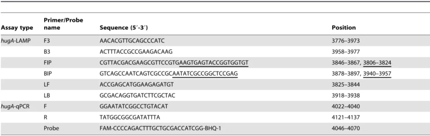
Table 2.LAMP and qPCR primers used in this study to detectP. shigelloides.
Assay type
Primer/Probe
name Sequence (59-39) Position
hugA-LAMP F3 AACACGTTGCAGCCCATC 3776–3973
B3 ACTTTACCGCCGAAGACAAG 3958–3977
FIP CGTTACGACGAAGCGTTCCGTGAAGTGAGTACCGGTGGTGT 3846–3867, 3806–3824
BIP GTCAGCCAATCAGTCGCCGCAATATCGCCGGCTCCGAG 3878–3897, 3940–3957
LF ACCGAGCATGGAAGAGATGT 3825–3844
LB GCGACAGGTGATCTTCGCTAC 3918–3938
hugA-qPCR F GGAATATCGGCCTGTACAT 4022–4040
R TATGGCGGCGATATTTA 4121–4137
Probe FAM-CCCCAGACTTTGCTGCGACCATCGG-BHQ-1 4046–4070
Reproducibility of LAMP assay
The CVi was assessed by testing 3 reference plasmids with varying concentrations (106, 104, and 102copies/mL), 10 times in a single run, whereas the CVo was assessed by testing the same plasmids 10 times in 10 separate runs. The CVi ranged from 1.21% to 1.54%, while the CVo ranged from 2.17% to 3.23%.
Evaluation of LAMP assay in simulated human stool The detection limit of LAMP in simulated human stool was also examined. The LAMP assays detected the presence ofP. shigelloides strains down to as little as 56103CFU/g. By comparison, the qPCR assays had a detection limit of 56104CFU/g forhugAgene in simulated human stool samples (data not shown). Table 3 Figure 2. Real-time sensitivity and detection limit ofhugA-LAMP.(A) Real-time sensitivity ofhugA-LAMP as monitored by the measurement of turbidity (optimal density at 650 nm). A turbidity of.0.1 was considered to be positive forhugA-LAMP. The detection limit was 20 copies/reaction. (B) The relation between the threshold time (Tt) of each sample and the log copies/reaction. The standard curve was drawn on the basis of 3 independent repeats and the linear relationship R2 =0.9787. (C) Sensitivities of electrophoretic analysis ofhugA-LAMP amplified products. Lane M:
DL2000 marker; lane 1: 26106copies/reaction; lane 2: 26105copies/reaction; lane 3: 26104copies/reaction; lane 4: 26103copies/reaction; lane 5:
26102copies/reaction; lane 6: 26101copies/reaction; lane 7: 26100copies/reaction; lane 8: no template. (D) SYBR green I fluorescent dye-mediated
monitoring ofhugA-LAMP assay amplification. The original orange color of the SYBR Green I changed to green in case of positive amplification, whereas the original orange color was retained for a negative control with no amplification.
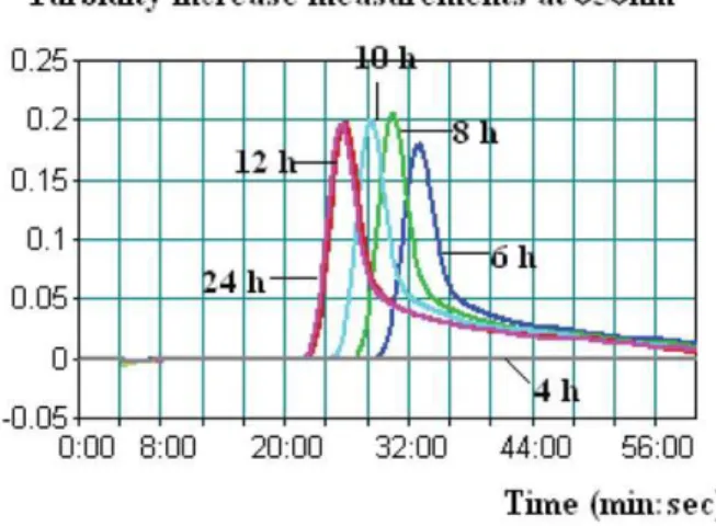
summarizes LAMP and qPCR results in human stool samples spiked with 2 low levels (1 to 2 and 10 to 20 CFU/0.5 g) ofP. shigelloides strains after various enrichment periods. A typical LAMP judgment graph generated for human stool enrichment samples is shown in Fig. 3. Regardless of spiking levels, none of the 4-h-enrichment samples tested positive forP. shigelloidesby either LAMP or qPCR. We observed positive results with LAMP at 6 h with significantly higher threshold time (Tt) values, while for samples enriched for 8, 10, 12, and 24 h, lower and stable Tt values were observed. A similar trend of detection was observed for qPCR (Table 3). In addition, qPCR results were presented by cycles, which were approximately 1 min/cycle. Therefore, an additional 20–40 min of amplification time was needed for qPCR when testing the same enrichment sample.
Discussion
Some reports have suggested thatP. shigelloidesmay cause enteric diseases in normal hosts [5,17]. Moreover, septicemia, cellulitis, meningitis, and cholecystitis due to P. shigelloides have also been documented among immunocompromised patients or patients with other underlying conditions [8–10]. The mortality rate associated with Plesiomonas-induced septicemia is high [18]. Individuals with serious infections are faced with the lack of a rapid and sensitive diagnostic method and inappropriate antimi-crobial therapy, and therefore, they, often, cannot receive timely treatment, leading to diseases and fatal outcomes.
It is well known that the bacteriological methods available for the isolation and identification of P. shigelloides are tedious and
lengthy. Modified PCR techniques such as nested PCR and real-time PCR are complicated and require a high-precision thermal cycler, and therefore, they are not adapted to diagnosing P. shigelloidesin basic clinical and field laboratories in rural areas. In contrast, the LAMP assay reported in this study is advantageous because of the following 3 features: rapid reaction, simple operation, and easy detection. The LAMP assay does not require sophisticated and expensive equipment, maintaining a constant temperature of 60uC–65uC for 1 h is sufficient for the reaction [16]. These features demonstrate that the LAMP assay is suitable for the detection of P. shigelloides in basic clinical and field laboratories in rural areas.
Although the pathogenesis ofP. shigelloides-associated gastroen-teritis has not yet been elucidated, a number of potential virulence factors have been described [19,20]. Acquisition of iron is known to be involved in the virulence of a variety of bacterial pathogens [21,22]. Heme is the most abundant source of iron in the body, and many pathogenic bacteria possess heme transport systems. The hugA gene, one of the characterized genes encoded in the heme iron utilization system of P. shigelloides, encodes an outer membrane receptor that is required for heme iron utilization.
In this study, allhugAgene sequences ofP. shigelloidesrecorded in the GeneBank were aligned, and the LAMP primers were designed on the basis of the conserved regions. We tested 32 non-P. shigelloides strains to evaluate the specificity of the hugA LAMP assay for the bacteria, with the results showing that the specificity of the LAMP assay was 100%.
To the best of our knowledge, this is the first study applying the novel LAMP technology for the detection ofP. shigelloidesin human stool. Previously, spiked samples were usually enriched overnight without characterizing the effects of different enrichment times on the detection outcomes [23,24]. In this study,P. shigelloidesstrain ATCC 51903 was used in experiments with simulated human stool samples, with the LAMP assays having a detection limit of 56103CFU/g stool. Positive detection occurred after a 6-h period of enrichment, and consistently thereafter, for the human stool samples spiked with 2 low levels (1 to 2 and 10 to 20 CFU/0.5 g) of ATCC 51903. We observed that the LAMP assay performed better than qPCR with respect to detection limit and assay speed in spiked human stool. In general, molecular level-based detection methods such as PCR and LAMP are subjected to a variety of inhibitors present in clinical samples. Some researchers have reported that the Bst polymerase in LAMP is less sensitive to the presence of inhibitors than the Taq polymerase used in classic PCR [25,26]. Our results showed that the LAMP assay is more accurate and sensitive than qPCR methods using simulated human stool samples, and proved markedly faster than qPCR by at least 20 min, thereby significantly shortening the total assay time.
In conclusion, the LAMP assay was successfully validated in this study for rapidity, sensitivity, specificity, and robustness; thus, this assay may serve as an effective means for screeningP. shigelloidesin Figure 3. A typical LAMP amplification graph generated when
testing human stool samples spiked with the low level of P. shigelloides strain after various enrichment periods (4, 6, 8, 10, 12, and 24 h).In this graph, the human stool sample was spiked with 1.3 CFU of Strain ATCC 51903.
doi:10.1371/journal.pone.0041978.g003
Table 3.Comparison of LAMP and qPCR assays in human stool samples spiked with low levels ofP. shigelloides.
Cell level (no. of CFU/
0.5 g) LAMP Tt (min) after enrichment qPCR CT (cycles) after enrichment
4 h 6 h 8 h 10 h 12 h 24 h 4 h 6 h 8 h 10 h 12 h 24 h
1–2 Not available 30.3 27.1 25.2 22.7 22.4 Not available 36.5 33.2 30.8 29.6 29.1
10–20 Not available 28.5 25.3 23.8 21.5 21.3 Not available 33.7 29.1 27.7 26.3 25.8
clinical samples. We proved that the LAMP assay demonstrated superior performance to qPCR in simulated human stool samples, and may facilitate rapid and reliable diagnosis of P. shigelloides infections in basic clinical and field laboratories in rural areas.
Author Contributions
Conceived and designed the experiments: SM CY JX. Performed the experiments: SM. Analyzed the data: SM YX. Contributed reagents/ materials/analysis tools: SM YX. Wrote the paper: SM CY.
References
1. Garrity GM, Bell JA, Lilburn TG (2003) Taxonomic outline of the procaryotes. Bergey’s Manual of Systematic Bacteriology, 2nd edn. Release 4.0 Available: http://141.150.157.80/bergeysoutline/main.htm.
2. Krovacek K, Eriksson LM, Gonza´lez-Rey C (2000) Isolation, biochemical and serological characterisation ofPlesiomonas shigelloidesfrom freshwater in Northern Europe. Comp Immunol Microbiol Infect Dis 23:45–51.
3. Reinhardt JF, George WL (1985) Plesiomonas shigelloides-associated diarrhea. JAMA 253:3294–3295.
4. Escobar JC, Bhavnani D, Trueba G (2012) Plesiomonas shigelloides infection, Ecuador, 2004–2008. Emerg Infect Dis 18:322–324.
5. Gonza´lez-Rey C, Svenson SB, Bravo L (2000) Specific detection ofPlesiomonas shigelloidesisolated from aquatic environments, animals and human diarrhoeal cases by PCR based on 23S rRNA gene. FEMS Immunol Med Microbiol 29:107–113.
6. Wouafo M, Pouillot R, Kwetche PF (2006) An acute foodborne outbreak due to
Plesiomonas shigelloidesin Yaounde, Cameroon. Foodborne Pathog Dis 3:209–211. 7. Miller WA, Miller MA, Gardner IA (2006)Salmonellaspp.,Vibriospp.,Clostridium perfringens, andPlesiomonas shigelloidesin marine and freshwater invertebrates from coastal California ecosystems. Microb Ecol 52:198–206.
8. Schneider F, Lang N, Reibke R (2009)Plesiomonas shigelloidespneumonia. Med Mal Infect 39:397–400.
9. Ozdemir O, Sari S, Terzioglu S (2010) Plesiomonas shigelloides sepsis and meningoencephalitis in a surviving neonate. J Microbiol Immunol Infect 43:344–346.
10. Auxiliadora-Martins M, Bellissimo-Rodrigues F, Viana JM (2010) Septic shock caused byPlesiomonas shigelloidesin a patient with sickle beta-zero thalassemia. Heart Lung 39:335–339.
11. Chan SS, Ng KC, Lyon DJ (2003) Acute bacterial gastroenteritis: a study of adult patients with positive stool cultures treated in the emergency department. Emerg Med J 20:335–338.
12. Salerno A, Cizˇna´r I, Krovacek K (2010) Phenotypic characterization and putative virulence factors of human, animal and environmental isolates of
Plesiomonas shigelloides. Folia Microbiol (Praha) 55:641–647.
13. Herrera FC, Santos JA, Otero A (2006) Occurrence ofPlesiomonas shigelloidesin displayed portions of saltwater fish determined by a PCR assay based on the hugA gene. Int J Food Microbiol 108:233–238.
14. Gu W, Levin RE (2006) Quantitative detection ofPlesiomonas shigelloidesin clam and oyster tissue by PCR. Int J Food Microbiol 111:81–86.
15. Mori Y, Notomi T (2009) Loop-mediated isothermal amplification (LAMP): a rapid, accurate, and cost-effective diagnostic method for infectious diseases. J Infect Chemother 15:62–69.
16. Notomi T, Okayama H, Masubuchi H (2000) Loop-mediated isothermal amplification of DNA. Nucleic Acids Res 28:E63.
17. Paul R, Siitonen A, Ka¨rkka¨inen P (1990)Plesiomonas shigelloidesbacteremia in a healthy girl with mild gastroenteritis. J Clin Microbiol 28:1445–1446. 18. Lee AC, Yuen KY, Ha SY (1996)Plesiomonas shigelloidessepticemia: case report
and literature review. Pediatr Hematol Oncol 13:265–269.
19. Janda JM, Abbott SL (1993) Expression of hemolytic activity byPlesiomonas shigelloides. J Clin Microbiol 31:1206–1208.
20. Santos JA, Gonza´lez CJ, Lo´pez TM (1999) Hemolytic and elastolytic activities influenced by iron inPlesiomonas shigelloides. J Food Prot 62:1475–1477. 21. Villarreal DM, Phillips CL, Kelley AM (2008) Enhancement of recombinant
hemoglobin production inEscherichia coliBL21(DE3) containing the Plesiomonas shigelloides heme transport system. Appl Environ Microbiol 74:5854–5856. 22. Oldham AL, Wood TA, Henderson DP (2008) Plesiomonas shigelloides hugZ
encodes an iron-regulated heme binding protein required for heme iron utilization. Can J Microbiol 54:97–102.
23. Ohtsuka K, Tanaka M, Ohtsuka T (2010) Comparison of detection methods for
Escherichia coliO157 in beef livers and carcasses. Foodborne Pathog Dis 7:1563– 1567.
24. Hara-Kudo Y, Niizuma J, Goto I (2008) Surveillance of Shiga toxin-producing
Escherichia coli in beef with effective procedures, independent of serotype. Foodborne Pathog Dis 5:97–103.
25. Kaneko H, Kawana T, Fukushima E (2007) Tolerance of loop-mediated isothermal amplification to a culture medium and biological substances. J Biochem Biophys Methods 70:499–501.