for Rapid Detection of
Trypanosoma brucei rhodesiense
Zablon Kithinji Njiru1,2*, Andrew Stanislaw John Mikosza3, Tanya Armstrong3, John Charles Enyaru4,
Joseph Mathu Ndung’u5, Andrew Richard Christopher Thompson3
1School of Nursing, Murdoch University, Mandurah, Western Australia, Australia,2Trypanosomiasis Research Centre, Kenya Agricultural Research Institute, Kikuyu, Kenya,
3WHO Collaborating Centre for the Molecular Epidemiology of Parasitic Infections, School of Veterinary and Biomedical Sciences, Murdoch University, Murdoch, Western Australia, Australia,4Department of Biochemistry, Faculty of Science, Makerere University, Kampala, Uganda,5Foundation for Innovative New Diagnostics (FIND), Cointrin, Switzerland
Abstract
Loop-mediated isothermal amplification (LAMP) of DNA is a novel technique that rapidly amplifies target DNA under isothermal conditions. In the present study, a LAMP test was designed from theserum resistance-associated(SRA) gene of Trypanosoma brucei rhodesiense, the cause of the acute form of African sleeping sickness, and used to detect parasite DNA from processed and heat-treated infected blood samples. TheSRAgene is specific toT. b. rhodesienseand has been shown to confer resistance to lysis by normal human serum. The assay was performed at 62uC for 1 h, using six primers that recognised eight targets. The template was varying concentrations of trypanosome DNA and supernatant from heat-treated infected blood samples. The resulting amplicons were detected using SYTO-9 fluorescence dye in a real-time thermocycler, visual observation after the addition of SYBR Green I, and gel electrophoresis. DNA amplification was detected within 35 min. TheSRALAMP test had an unequivocal detection limit of one pg of purified DNA (equivalent to 10 trypanosomes/ ml) and 0.1 pg (1 trypanosome/ml) using heat-treated buffy coat, while the detection limit for conventionalSRAPCR was ,1,000 trypanosomes/ml. The expected LAMP amplicon was confirmed through restriction enzymeRsaI digestion, identical melt curves, and sequence analysis. The reproducibility of the SRA LAMP assay using water bath and heat-processed template, and the ease in results readout show great potential for the diagnosis ofT. b. rhodesiensein endemic regions.
Citation:Njiru ZK, Mikosza ASJ, Armstrong T, Enyaru JC, Ndung’u JM, et al. (2008) Loop-Mediated Isothermal Amplification (LAMP) Method for Rapid Detection of Trypanosoma brucei rhodesiense. PLoS Negl Trop Dis 2(2): e147. doi:10.1371/journal.pntd.0000147
Editor:Serap Aksoy, Yale University School of Medicine, United States of America
ReceivedMay 25, 2007;AcceptedNovember 14, 2007;PublishedFebruary 6, 2008
Copyright:ß2008 Njiru et al. This is an open-access article distributed under the terms of the Creative Commons Attribution License, which permits unrestricted use, distribution, and reproduction in any medium, provided the original author and source are credited.
Funding:Funds were received from Murdoch University, Australia, and the Foundation for New Diagnostics (FIND), Geneva. The funders had no role in the study design, data collection and analysis, decision to publish, or preparation of the manuscript.
Competing Interests:The authors have declared that no competing interests exist.
*E-mail: [email protected] or [email protected]
Introduction
Human African trypanosomiasis is endemic in tropical Africa. In eastern and southern Africa the disease is caused byTrypanosoma brucei rhodesiense, while T. b. gambiense infections are common in central and West Africa.T. b. rhodesiensecauses an acute form of disease, whereas T. b. gambiense causes a more chronic form. Moreover, the treatment regimen for the two infections is different, expressing the need for a specific diagnostic test for each trypanosome. The geographical demarcation of T. b. rhodesienseandT. b. gambiense to a large extent forms the basis of trypanosome identification and treatment. In East Africa the introduction of T. b. rhodesiense into the T. b. gambiense region is certain to occur due to the closeness of the two disease foci and continuous movement of the livestock-reservoir host for T. b. rhodesiense. This prospect further obligates the development of test kits that can differentiate the two parasites. The serum resistance-associated (SRA) gene [1,2] is conserved and specific to T. b. rhodesiense[3–5] and therefore provides unequivocal identification of this parasite. It is a low-copy gene, therefore the polymerase chain reaction (PCR) test is inadequate to amplify this target reliably in clinical samples without recourse to parasite multipli-cation in mice. Besides, available molecular methods of parasite detection require elaborate precision instruments [3–7], which
make their use under field conditions unfeasible. There is therefore a need for a simplified method of amplification and product detection that would compliment the available tests and make feasible molecular diagnosis for case detection and confirmation of cure in the regions that are endemic for sleeping sickness.
Recently, a technique called loop-mediated isothermal ampli-fication (LAMP) of DNA has been developed [8]. The technique uses four to six primers that recognise six to eight regions of the target DNA, respectively, in conjunction with the enzyme Bst
dumbbell-shaped DNA structures [8]. The stem-loop thus formed acts as a template, and subsequently one inner primer hybridises to the loop on the product and initiates the displacement DNA synthesis, forming the original stem loop and a new stem loop that is twice as long [13]. The final products are stem-loop DNAs with several inverted repeats of the target DNA, and cauliflower-like structures bearing multiple loops [8].
A number of LAMP tests to detect parasitic protozoa have been designed and used successfully [14–16]. The rapidity, specificity, and simplicity of the technique make it appealing for use in trypanosomiasis-endemic regions. The purpose of the present study was to develop a LAMP test for detection ofT. b. rhodesiense
based on the SRA gene and compare it with PCR test that is specific for T. b. rhodesiense. Our results indicate that the SRA
LAMP is sensitive and specific and has the potential to be developed into a field-friendly diagnostic test.
Materials and Methods Ethical clearance
Institutional Ethical Clearance for the collection of human samples had been obtained from the Livestock Health Research Institute (LIRI), Tororo, Uganda, and the Uganda National Council of Science and Technology (UNCST), Kampala, Uganda, which records and regulates all research activities in the country. At Murdoch University, Perth, Australia, the use of mice was approved by Murdoch University Animal Ethics Committee (AEC).
Preparation of template
The trypanosome DNA samples used in this study are shown in Table 1. The samples which most had been passaged in mice were chosen to ensure a wide geographical representation, different times of isolation, and hosts (Table 1). Six samples designated as JE (three each from blood and cerebrospinal fluid [CSF]) were direct isolates from human hosts. The DNA had been prepared using several methods (see footnotes in Table 1). The samples for studying analytical sensitivity and tolerance of LAMP were obtained from the blood of mice infected with T. b. rhodesiense
and divided into two portions. The first portion was centrifuged at 3,000 rpm for 10 min and the buffy coat was collected, and the second portion was divided into aliquots of 10ml. Then each of the two portions was mixed with 40ml of ultrapure water, boiled for 3 min, and centrifuged at 14,000gfor 5 min. Samples of 10–15ml of supernatant were recovered and stored at220uC for later use.
Polymerase chain reaction
Trypanosomes belonging to the subgenus Trypanozoon were analysed using TBR1 and 2 primers [7]. Furthermore T. b. rhodesiensewas detected by a PCR specific for theSRAgene [3]), whereasT. b. gambiense was detected using a PCR for the T. b. gambiense-specific glycoprotein (TgsGP)gene [17].
LAMP reaction
LAMP reactions of 25ml were standardised for optimal reagent concentrations, temperature, and time conditions using T. b. rhodesienseisolate LVH 56 and following the Taguchi design [18]. Briefly, the FIP and BIP were varied from 0.8mM to 2.4mM, dNTPs from 100mM to 400mM, betaine from 0.2 M to 0.8 M, and MgSO4from 0 to 4 mM. The FIP, BIP, F3, and B3 primers were
designed using the PrimerExplorer v3 software (http:/primerex-plorer.jp/lamp) based on the SRA gene sequence (GenBank accession number Z37159) (Table 2). Loop primers [loop forward (LF) and loop backward (LB)] were designed manually. The reactions were optimised at 2.0mM for FIP and BIP primers, 0.8mM for loop primer (LF and LB), 0.2mM for F3 and B3 outer primers, 200mM for each dNTP, 0.8 M betaine (Sigma), 20 mM Tris-HCl (pH 8.8), 10 mM KCl, 10 mM (NH4)2SO4, 2 mM
MgSO4, 0.1% Triton X-100, and 8 U of Bst DNA polymerase
large fragment (New England Biolabs). For real-time reactions 3.34mM SYTO-9 fluorescence dye (Molecular Probes) was added. The template was,100 pg for trypanosome lysate DNA samples and 2ml of buffy coat and supernatant prepared from boiled blood. To find the optimum temperature for the LAMP test, the reactions were carried out for 1 h at 58, 60, 62, and 64uC using the Rotor-Gene 3000 thermocycler (Corbett Research) or in a water bath at the same temperature settings. The reaction was terminated by increasing the temperature to 80uC for 4 min.
Detection of LAMP product
Three methods were used to analyse DNA amplification, and included electrophoresis in 1.5% agarose gels stained with ethidium bromide, direct visual inspection of the LAMP product after addition of 1ml of 1/10 dilution of SYBR Green I (Invitrogen), and by monitoring fluorescence of the double-stranded DNA (dsDNA)-specific dye SYTO-9 [19] in a Rotor-Gene 3000 thermocycler. Real-time fluorescence data was obtained on the FAM channel (excitation at 470 nm and detection at 510 nm) [19]. Three approaches were used to confirm that the
SRALAMP test amplified the correct target: (1) the product was digested with restriction enzyme RsaI (New England Biolabs) at 37uC for 3 h, followed by electrophoresis in 3% agarose gel; (2) following amplification, the DNA melting curves were acquired on the FAM channel using 1uC steps, with a hold of 30 s, from 62 to 96uC [19]; and (3) some of the LAMP amplicon bands were excised from an agarose gel and cloned into a TOPO-TA vector (Invitrogen), transformed inE. coliand inserts sequenced using an automated DNA 3730 analyser (Applied Biosystems). The resulting sequences were aligned with the target sequence using the DNAman computer software version 5.0 (Lynnon Biosoft).
Sensitivity and specificity of LAMP
10-fold dilutions were made from infected mouse blood containing 1.06106trypanosomes/ml and from 100 ng of purified T. b. rhodesienseDNA, and used to determine the analytical sensitivity ofSRALAMP and PCR tests. The reactions were done in triplicates and repeated after 2 wks. The LAMP test was carried out using both cold and heated templates. The specificity of the tests were assessed with DNA from human, tsetse fly, bovine,Plasmodium falciparum, and Author Summary
Table 1.Trypanosome Isolates and Amplification Results
Species/Subspecies
Identification
Code Origin
Year of Isolation
Original
Host Specific PCR Results SRA-LAMP Results
TBRa SRA
Geneb TgsGPc SYBRGreen I Gel RealTime
T. b. rhodesiense ATCC 30027 Tanganyika 1934 Human + + 2 + + + T. b. rhodesiense Gambella IId Ethiopia 1968 Human
+ + 2 + + +
T. b. rhodesiense LVH 058d Luangwa Valley, Zambia 1974 Human
+ + 2 + + +
T. b. rhodesiense LVH 56d Lambwe Valley, Kenya 1978 Human
+ + 2 + + +
T. b. rhodesiense LVH 108d Lambwe Valley, Kenya 1980 Human
+ + 2 + + +
T. b. rhodesiense TMRS 010d Kasulu, Tanzania 1991 Human
+ + 2 + + +
T. b. rhodesiense TMRS 127d Mpanda, Tanzania 1994 Human
+ + 2 + + +
T. b. rhodesiense UTRO 2509d Uganda N/A Human
+ + 2 + + +
T. b. rhodesiense WB56d Uganda N/A Human
+ + 2 + + +
T. b. rhodesiense KETRI 2355d Busoga, Uganda 1977 Human
+ + 2 + + +
T. b. rhodesiense KETRI 3739e Busia, Kenya 2001 Dog
+ + 2 + + +
T. b. rhodesiense KETRI 1900e Lambwe Valley, Kenya 1971 Hyena
+ + 2 + + +
T. b. rhodesiense KETRI 2492e Lambwe Valley, Kenya 1980 Tsetse fly
+ + 2 + + +
T. b. rhodesiense KETRI 2532e Lambwe Valley, Kenya 1980 Cow
+ + 2 + + +
T. b. rhodesiense KETRI 3007e Busia, Kenya 1987 Pig
+ + 2 + + +
T. b. rhodesiense JE1 Busoga, Uganda 1990 Human + 2 2 + + + T. b. rhodesiense JE2f Tororo, Uganda 1991 Human
2 2 2 + + +
T. b. rhodesiense JE3f Tororo, Uganda 2005 Human 2 2 2
+ + +
T. b. rhodesiense JE4f Tororo, Uganda 2002 Human
+ 2 2 + + +
T. b. rhodesiense JE5 Serere, Uganda 2001 Human + + 2 + + + T. b. rhodesiense JE6 Serere, Uganda 2001 Human + + 2 + + + T. b. rhodesiense JE8g Tororo, Uganda 2001 Human 2 2 2
+ + +
T. b. rhodesiense JE9g Tororo, Uganda 2001 Human
+ 2 2 + + +
T. b. rhodesiense JE10g Tororo, Uganda 2001 Human
+ 2 2 + + +
T. b. rhodesiense JE12 Serere, Uganda 2003 Human + + 2 + + + T. b. rhodesiense JE13 Serere, Uganda 2003 Human + + 2 + + + T. b. rhodesiense JE14 Serere, Uganda 2001 Human + + 2 + + + T. b. rhodesiense STIB849h Uganda 1991 Human nd nd nd
+ + +
T. b. rhodesiense AnTat25.1h Rwanda 1971 Human nd nd nd
+ + +
T. b. rhodesiense AnTat12.1h Rwanda 1991 Human nd nd nd
+ + +
T. b. rhodesiense JE15 Serere, Uganda 2003 Human + + 2 + + + T. b. rhodesiense EATRO 149 Nyanza, Kenya 1961 Human + 2 2 + + + T. b. rhodesiense KETRI 2473 Nyanza, Kenya 1970 Human + + 2 + + + T. b. rhodesiense EATRO 2636 Mozambique 1983 Human + 2 2 + + + T. b. rhodesiense KETRI 3537 Bugoma, Kenya 1998 Human + + 2 + + + T. b. rhodesiense KETRI 3624 Busia, Kenya 1998 Human + + 2 + + + T. b. rhodesiense KETRI 3639 Busia, Kenya 1999 Human + + 2 + + + T. b. rhodesiense TMRS 51a Kibondo, Tanzania 2004 Human + + 2 + + + T. b. rhodesiense TMRS 51b Kibondo, Tanzania 2004 Human + + 2 + + +
T. b. rhodesiense TMRS 51c Kibondo 2005 Human + + 2 + + +
trypanosomes belonging to other species (Trypanosoma brucei brucei,T. b. gambiense, T. b. evansi, Trypanosoma congolensesavannah,T. c.kilifi,T. c. forest, Trypanosoma simiae, T. s. tsavo, Trypanosoma godfreyi, Trypanosoma vivax,andTrypanosoma lewisi).
Results LAMP test
The results of theSRALAMP assay are shown in Figures 1–4 and Table 1. When the test was carried out without loop primers a
product was detected after 50 min. The inclusion of loop primers reduced the reaction time from an average of 50 min down to between 20 and 25 min and increased the sensitivity 100-fold. The best results were obtained when the reaction temperature was maintained at 62uC. All the positive LAMP reactions produced a characteristic ladder of multiple bands on an agarose gel (Figure 1A and 1B), indicating that stem-loop DNA with inverted repeats of the target sequence was produced. Positive reactions turned green on addition of SYBR Green I, while the negative ones remained orange (Figure 3). RsaI restriction enzyme digestion and Species/Subspecies
Identification
Code Origin
Year of Isolation
Original
Host Specific PCR Results SRA-LAMP Results
TBRa SRA
Geneb TgsGPc
SYBR
Green I Gel
Real Time
T. b. rhodesiense TMRS JM Kasulu, Tanzania 2001 Human + 2 2 + + + T. b. rhodesiense TMRS 58 Mpanda, Tanzania 2006 Human + + 2 + + + T. b. rhodesiense TMRS 4M Urambo, Tanzania 2006 Human 2 2 2 + + + T. b. gambiense MOSd Mbam, Cameroon 1974 Human
+ 2 + 2 2 2
T. b. gambiense PT16d Ivory Coast 1992 Human
+ 2 + 2 2 2
T. b. gambiense Boulad Bouenza, Congo 1989 Human
+ 2 + 2 2 2
T. b. gambiense NW2d Uganda 1992 Human
+ 2 + 2 2 2
T. b. gambiense Dal 972d Daloa, Ivory Coast 1978 Human
+ 2 + 2 2 2
T. b. brucei LUMP 266d Kiboko, Kenya 1969 Fly,
G. pallidipes
+ 2 2 2 2 2
T. b. brucei KP2Nd Kouassi-Perita, Ivory Coast 1982 Fly,G.
palpalis
+ 2 2 2 2 2
T. b. brucei B8/18d Nsukka, Nigeria 1962 Pig
+ 2 2 2 2 2
T. b. brucei J10d Luangwa Valley, Zambia 1973 Hyena
+ 2 2 2 2 2
T. b. brucei STIB 215d Serengeti, Tanzania 1971 Lion
+ 2 2 2 2 2
T. b. brucei Kateremad Uganda 1990 Cow
+ 2 2 2 2 2
T. evansi SA17 Isiolo, Kenya 2003 Camel + 2 2 2 2 2
T. evansi KETRI 2426 Ukunda, Kenya 1978 Camel + 2 2 2 2 2
T. evansi KETRI 3093 Colombia, South America 1979 Horse + 2 2 2 2 2
T. evansi SA263 Samburu, Kenya 2003 Camel + 2 2 2 2 2
T. evansi KETRI 243 Kulal, Kenya 1979 Camel + 2 2 2 2 2
T. evansi KETRI 3565 Athi River, Kenya 1994 Camel + 2 2 2 2 2
T. congolenseforest Cam 22d Mbetta, Cameroon 1984 Goat 2 2 2 2 2 2
T. c.kilifi WG5d Kenya 1980 Sheep 2 2 2 2 2 2
T. c.savannah KETRI 1869 Kenya — — 2 2 2 2 2 2
T. simiae Ken 4d Keneba, The Gambia 1988 Fly 2 2 2 2 2 2
T. simiaetsavo KETRI 1864d Kenya — Fly 2 2 2 2 2 2
T. godfreyi Ken 7d Kenya 1988 Fly,
G. morsitans
2 2 2 2 2 2
T. vivax Y58 Nigeria — — 2 2 2 2 2 2
JE samples are from Uganda and were processed using Sigma Genomic DNA extraction kit, followed by precipitation with 3 M sodium acetate and absolute alcohol. TMRS samples were from Tanzania and the DNA was prepared using Qiagen DNA extraction kit.
aSubgenus
TrypanozoonPCR test [7].
bT. b. rhodesiensePCR test [3]. cT. b. gambiensePCR test [17].
dSource: Wendy Gibson, University of Bristol, UK.
eThe samples were processed using the saponin lysis method [5]. fDNA prepared directly from human blood.
gDNA prepared directly from human CSF.
hSource: Philippe Bu¨scher, Institute of Tropical Medicine Antwerp, Belgium. DNA prepared with Qiagen DNA extraction kit.
nd, not done in this study (identification reported [17]). doi:10.1371/journal.pntd.0000147.t001
Table 2.Nucleotide Sequences for theSRALAMP Primers
Primer Type Sequencea(59–39) Length Amplicon Sizeb Target
SRA-F3 F3 GCGGAAGCAAGAATGACC 18 162 SRAgene
SRA-B3 B3 TCTTACCTTGTGACGCCTG 19 — —
SRA-FIP FIP (F1c+F2) GGACTGCGTTGAGTACGCATCCGCAAGCACAGACCACAGC 40 — —
SRA-BIP BIP (B1c+B2) CGCTCTTACAAGTCTTGCGCCCTTCTGAGATGTGCCCACTG 41 — —
SRA-LF LF CGCGGCATAAAGCGCTGAG 19 — —
SRA-LB LB GCAGCGACCAACGGAGCC 18 — —
aAccession number Z37159.
bThe length between F2 and B2 is 162 bp. However, the amplified amplicon sizes will be more than 162 bp since the FIP and BIP primers consist of F1c (21 bp) and B1c
(21 bp), respectively.
doi:10.1371/journal.pntd.0000147.t002
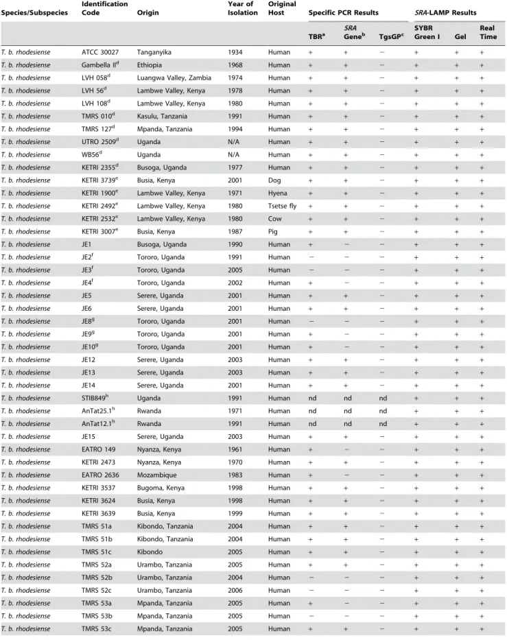
Figure 1. Analytical Sensitivity ofSRALAMP and Restriction Enzyme Digestion Results.(A) Sensitivity of theSRALAMP assay using DNA lysate fromT. b. rhodesienseisolate LVH 56. Sensitivity results for 10-fold dilutions from infected mouse blood showed identical results. The lysate/ supernatant was heated at 96uC for 1 min before being added to the reaction mixture. The reactions which were done in triplicates showed detection limit of<1 pg an equivalent of dilution 1025. M, 100 bp marker, TMRS 127,T. b. rhodesiense, neat (100 ng), 1021(10 ng), 1022(1 ng), 1023(100 pg),
1024(10 pg), 1025(1 pg), 1026(100 fg), 1027 (10 fg), and NC, negative control. The detection limit for SRAPCR was 1022(100 pg <1,000
trypanosomes/ml). (B) Electrophoresis results for isolates TMRS 127 (lane 1) and LVH 56 (lane 3), and theirRsaI restriction enzyme digests (lane 2 for TMRS 127 and lane 4 for LVH 56). M, 100 bp marker.
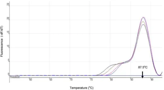
electrophoresis gave the predicted sizes of 90 bp and 114 bp (Figure 1B). TheSRALAMP amplicons showed reproducible melt curves with a Tmof,87.5uC, suggesting amplicons of the same
sequence (Figure 4). The cloned sequence showed 100% identity with the target sequence, and revealed that the length varied with sequence repeats of primers and there complementary sequences. The analytical sensitivity of SRA LAMP assay improved from a dilution of 1024to 1026when a template (DNA or supernatant) was preheated before being added to a reaction (Figure 2), with the best detection limit of dilution 1027recorded with supernatant prepared from the buffy coat. The classical PCR based on the same gene [3] showed a detection limit of dilution 1024. The
SRALAMP detected all the 49 (100%)T. b. rhodesiense(including the six samples isolated directly from HAT patients), while TBR1 and 2 primers detected 39 out of 46 (84.8%) and SRA PCR detected 31 out of 46 (67.4%) samples (Table 1). TheSRALAMP test was specific and no cross-reaction was recorded with nontarget DNA.
Discussion
In the present study we were able to demonstrate the successful amplification ofT. b. rhodesiense DNA within 20–25 min at 62uC using theSRALAMP assay. However, we set the optimal time at 35 min to amplify DNA at low concentrations. The results of the
SRA LAMP assay were identical when either a water bath or a thermocycler was used to maintain the temperature at 62uC, demonstrating its robustness. Preheating of the template increased the efficiency of the assay by shortening the duration (Figure 2) and increasing sensitivity of the test. DNA amplification is preceded by strand separation under isothermal conditions using betaine, which destabilises the DNA helix [8]. It would appear that preheating of the sample produced a faster and/or a greater amount of strand separation, which translated into a far more rapid assay. All positive samples detected by gel electrophoresis or in real-time using SYTO-9 fluorescence dye could also be detected visually by addition of SYBR Green I to the product. This ability
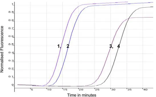
Figure 2. Comparison of the Sensitivity ofSRALAMP Under Different Conditions.Sensitivity was compared using preheated and cold template forT. b. rhodesienseisolate LVH 56 (1 and 3) and 058 (2 and 4), respectively, and as monitored using the Rotor-Gene 3000 thermocycler. Preheating of the template accelerates the detection of a positive reaction by approximately 12 min.
doi:10.1371/journal.pntd.0000147.g002
Figure 3. Visual Appearance of PostassaySRALAMP Reactions from Isolates after the Addition of 1ml of 1/10 Dilution of SYBR Green I.Positive samples produce a green colour almost immediately (tubes 1, 2, and 3 containing 100 pg ofT. b. rhodesienseDNA) while negatives remain orange (tube 4 and NC, negative control).
highlights another advantage of LAMP technique: the results of amplification can visually be observed through addition of a DNA intercalating dye (Figure 3), eliminating the need for gel electrophoresis and greatly reducing the time taken for result analysis.
When pure trypanosome DNA was used, the detection limit of the
SRALAMP test without loop primers was an equivalent of 1,000 trypanosomes/ml. This limit was improved to an equivalent of one trypanosome/ml with the inclusion of loop primers. Increased sensitivity and reduction in LAMP reaction time with the addition of loop primers is well documented [20] and has been demonstrated in detection of Mycobacterium [11], periodontal pathogens [12], and
Plasmodium falciparum malaria [10]. Loop primers accelerate the LAMP reaction by hybridising to the stem-loop region, initiating further DNA amplification [20]. When different templates were used, heat-treated buffy coat from mice blood performed better than the supernatant obtained after boiling blood samples. The higher sensitivity recorded could be the effect of concentrating the parasites in the buffy coat through centrifugation; therefore, buffy coat seems a superior template forSRALAMP test.
The robustness of the LAMP test is demonstrated by the ability to amplify target DNA from various templates without the expensive and time-consuming process of DNA purification. We observed no inhibitory effects in using 2–5ml of supernatant in a 25ml reaction or an increase in sensitivity beyond 2ml, indicating that this volume was the optimal for our samples. The possibility of using heat-processed samples without compromising sensitivity eliminates the need for DNA extraction and further shortens the LAMP reaction. Other studies have shown superior tolerance of LAMP tests for biological substances [9,13] and heat processed blood has been used successfully in detection Malaria [10]. The method of template preparation for use in LAMP tests, however, needs to be further developed.
The potential usefulness of SRA LAMP is confirmed by its ability to detect T. b. rhodesiense directly from parasitaemic and apparently aparasitaemic clinical samples (human blood and CSF). The human blood (JE2 and JE3) and CSF samples JE8–JE10 used in the present study were negative by microscopy at the time of sampling. Parasites were demonstrated only following inoculation
of the samples in mice. When the samples were tested, they were positive bySRALAMP assay while only JE4, JE9, and JE10 were positive using TBR PCR (Table 1) [7]. Detection of aparasitaemic samples demonstrates one of the practical values ofSRALAMP in sleeping sickness diagnosis-time-consuming parasite multiplication assays in mice are unnecessary, and early diagnosis increases the chances of cure after treatment.
In the present study, amplification of the target sequence was confirmed by restriction enzyme digestion using RsaI, melting curve analysis, and sequence analysis. It is important to distinguish
T b. rhodesienseandT. b. gambiensesince the two parasitic infections have different treatments. In recent years theT. b. rhodesienseregion in Southern Uganda has been expanding towards the T. b. gambiensefocus as a result of livestock movement [21,22]. There is therefore a need to continue development of rapid and sensitive techniques to differentiate the two parasites and to compliment the available PCR tests, and to this end theSRA LAMP assay has shown great potential for this application.
The LAMP test should theoretically not amplify nontarget sequences, since the specificity is enhanced by using a set of six primers. However there is a high risk of amplicon contamination since the tubes have to be opened to add the dye. Analysis of any false positive reactions through sequencing and restriction enzyme analysis would easily distinguish between false positive and contamination. To reduce the chances of contamination, similar protocols to those followed for PCR are required. However, the great potential for LAMP is that reactions can be performed and results read without opening tubes [23]. On this end, more work is needed to develop such a closed reaction system for diagnosing sleeping sickness.
This study has shown that the SRA LAMP assay could be developed into an assay forT. b. rhodesiensethat is simple to use at point of care. The detection of the equivalent of one trypanosome/ ml in the buffy coat (with the possibility of reducing this further to 0.1 trypanosomes/ml) compares well with the normal parasitae-mia in humans. Since DNA amplification and reading of results require minimum equipment, the technique has great potential for use in the HAT-endemic countries as back-up test for other HAT tests currently in use.
Figure 4. Melting Curves forT. b. rhodesiense SRALAMP Product as Monitored in Rotor-Gene 3000.The curves were obtained after LAMP amplification for 35 min and detected on the FAM channel using 1uC steps, and a hold of 30 sec, at each step from 60 to 96uC. All isolates had a melting temperature (Tm) of,87.5uC indicating similar sequences, and hence similar amplicon.
Acknowledgments
The authors acknowledge the provision of samples from Wendy Gibson (University of Bristol, UK), Johnson Ouma (Trypanosomiasis Research Centre, KARI, Kenya), Philippe Bu¨scher (Institute of Tropical Medicine, Belgium), Enock Matovu (Makerere University, Uganda), and Stafford Kibona (National Institute of Medical Research, Tabora, Tanzania).
Author Contributions
Conceived and designed the experiments: AT ZN. Performed the experiments: ZN TA AM JE. Analyzed the data: AT ZN JN TA AM JE. Contributed reagents/materials/analysis tools: AT JN AM. Wrote the paper: ZN JN.
References
1. De Greef C, Imberechts H, Matthyssons G, Van Meirvenne N, Hamers R (1989) A gene only expressed in serum resistant variants ofTrypanosoma brucei rhodesiense. Mol Biochem Parasitol 36: 169–176.
2. De Greef C, Hamers R (1994) The serum resistance associated (SRA) gene of Trypanosoma brucei rhodesienseencodes a VSG-like protein. Mol Biochem Parasitol 68: 277–284.
3. Gibson W, Backhouse T, Griffiths A (2002) The human serum resistance associated gene is ubiquitous and conserved inTrypanosoma brucei rhodesiense throughout East Africa. Infect Genet Evol 25: 1–8.
4. Welburn SC, Picozzi K, Fevre EM, Coleman PG, Odiit M, et al. (2001) Identification of human-infective trypanosomes in animal reservoir of sleeping sickness in Uganda by means of serum-resistance-associated (SRA) gene. Lancet 358: 2017–2019.
5. Njiru ZK, Ndung’u K, Matete G, Ndung’u JM, Gibson WC (2004) Detection of Trypanosoma brucei rhodesiensein animals from sleeping sickness foci in East Africa using the serum resistance associated (SRA) gene. Acta Tropica 90: 249–254. 6. Truc P, Mathieu-Daude´ F, Tibayrec M (1991) Multilocus isoenzyme
identification of Trypanosoma bruceistocks isolated in central Africa: evidence for an animal reservoir of sleeping sickness in Congo. Acta Tropica 49: 127–135. 7. Masiga DK, Smyth AJ, Hayes P, Bromidge TJ, Gibson WC (1992) Sensitive detection of trypanosomes in tsetse flies by DNA amplification. Int J Parasitol 22: 909–918.
8. Notomi T, Okayama H, Masubuchi H, Yonekawa T, Watanabe, et al. (2000) Loop-mediated isothermal amplification of DNA. Nucleic Acids Res 28(12): E63.
9. Kaneko H, Kawana T, Fukushima E, Suzutani T (2007) Tolerance of loop-mediated isothermal amplification to a culture medium and biological substances. J Biochem Biophys Methods 70: 499–501.
10. Poon LL, Wong BW, Ma EH, Chan KH, Chow LM, et al. (2006) Sensitive and inexpensive molecular test forfalciparummalaria: detectingPlasmodium falciparum DNA directly from heat-treated blood by loop-mediated isothermal amplifica-tion. Clin Chem 52: 303–306.
11. Iwamoto T, Sonobe T, Hayashi K (2003) Loop-mediated amplification for direct detection ofMycobacterium tuberculosis,M. aviumandM. intracellulare in sputum samples. J Clin Microbiol 41: 2616–2622.
12. Yoshida A, Nagashima S, Ansai T, Tachibana M, Kato H, et al. (2005) Loop-mediated Isothermal Amplification method for rapid detection of the
periondontopathic bacteria Porphyromona gingivalis, Tannerella forsythia and Treponema denticola. J Clin Microbiol 43: 2418–2424.
13. Yamada Y, Itoh M, Yoshida M (2006) Sensitive and rapid diagnosis of human parvovirus B19 infection by Loop-Mediated Isothermal Amplification. Br J Dermatol 155: 50–55.
14. Ikadai H, Tanaka H, Shibahara N, Matsuu A, Uechi M, Itoh N, Oshiro S, Kudo N, Igarashi I, Oyamada T (2004) Molecular evidence of infections with Babesia gibsoniparasites in Japan and evaluation of the diagnostic potential of a loop-mediated isothermal amplification method. J Clinic Microbiol 42: 2465–2469.
15. Kuboki N, Inoue N, Sakurai T, Di Cello F, Grab DJ, Suzuki H, Sugimoto C, Igarashi I (2003) Loop-mediated isothermal amplification for detection of African trypanosomes. J Clin Microbiol 41: 5517–5524.
16. Thekisoe OM, Kuboki N, Nambota A, Fujisaki K, Sugimoto C, Igarashi I, Yasuda J, Inoue N (2007) Species-specific loop-mediated isothermal amplifica-tion (LAMP) for diagnosis of trypanosomosis. Acta Tropica. 102: 182–189. 17. Radwanska M, Claes F, Magez S, Magnus E, Perez-Morga D, et al. (2002)
Novel primer sequences for polymerase chain reaction-based detection of Trypanosoma brucei gambiense. Am J Trop Med Hyg 67: 289–295.
18. Cobb B, Clarkson JM (1994) A simple procedure for optimising the polymerase chain reaction (PCR) using modified Taguchi methods. Nucleic Acids Res 22: 3301–3805.
19. Monis PT, Giglio S, Saint CP (2005) Comparison of SYTO9 and SYBR Green I for real-time polymerase chain and investigation of the effect of dye concentration on amplification and DNA melting curve analysis. Anal Biochem 30: 24–34.
20. Nagamine K, Hase T, Notomi T (2002) Accelerated reaction by loop-mediated isothermal amplification using loop primers. Mol Cell Probes 16: 223–229. 21. Fevre EM, Coleman PG, Odiit MD, Magona J, Welburn SC, et al. (2001) The
origins of a new sleeping sickness outbreak (caused by Trypanosoma brucei infection) in eastern Uganda. Lancet. 358: 625–628.