Mediated
N
-Oxide Reduction of Cyadox as Revealed by
Docking and Mutagenesis Studies
Chigang Chen., Guyue Cheng., Haihong Hao, Menghong Dai, Xu Wang, Lingli Huang, Zhenli Liu,
Zonghui Yuan*
National Reference Laboratory of Veterinary Drug Residues (HZAU) and MOA Key Laboratory for the Detection of Veterinary Drug Residues in Foods, Huazhong Agricultural University, Wuhan, Hubei, China
Abstract
Xanthine oxidoreductase (XOR) is a cytoplasmic molybdenum-containing oxidoreductase, catalyzing both endogenous purines and exogenous compounds. It is suggested that XOR in porcine hepatocytes catalyzes theN-oxide reduction of quinoxaline 1,4-di-N-oxides (QdNOs). To elucidate the molecular mechanism underlying this metabolism, the cDNA of porcine XOR was cloned and heterologously expressed in Spodoptera frugiperdainsect cells. The bovine XOR, showing sequence identity of 91% to porcine XOR, was employed as template for homology modeling. By docking cyadox, a representative compound of QdNOs, into porcine XOR model, eight amino acid residues, Gly47, Asn352, Ser360, Arg427, Asp430, Asp431, Ser1227 and Lys1230, were located at distances of less than 4A˚ to cyadox. Site-directed mutagenesis was performed to analyze their catalytic functions. Compared with wild type porcine XOR, G47A, S360P, D431A, S1227A, and K1230A displayed altered kinetic parameters in cyadox reduction, similarly to that in xanthine oxidation, indicating these mutations influenced electron-donating process of xanthine before subsequent electron transfer to cyadox to fulfill theN -oxide reduction. Differently, R427E and D430H, both located in the 424–434 loop, exhibited a much lower Km and a decreased Vmaxrespectively in cyadox reduction. Arg427 may be related to the substrate binding of porcine XOR to cyadox, and Asp430 is suggested to be involved in the transfer of electron to cyadox. This study initially reveals the possible catalytic mechanism of porcine XOR in cyadox metabolism, providing with novel insights into the structure-function relationship of XOR in the reduction of exogenous di-N-oxides.
Citation:Chen C, Cheng G, Hao H, Dai M, Wang X, et al. (2013) Mechanism of Porcine Liver Xanthine Oxidoreductase MediatedN-Oxide Reduction of Cyadox as Revealed by Docking and Mutagenesis Studies. PLoS ONE 8(9): e73912. doi:10.1371/journal.pone.0073912
Editor:Paul Taylor, University of Edinburgh, United Kingdom
ReceivedApril 22, 2013;AcceptedJuly 24, 2013;PublishedSeptember 9, 2013
Copyright:ß2013 Chen et al. This is an open-access article distributed under the terms of the Creative Commons Attribution License, which permits
unrestricted use, distribution, and reproduction in any medium, provided the original author and source are credited.
Funding:This work was supported by the National Basic Research Program of China (grant no. 2009CB118800), the National High Technology Research and Development Program of China (grant no. 2011AA10A214), the National Natural Science Foundation of China (grant no. 31272614) and the Fundamental Research Funds for the Central Universities (program no. 2013QC002). The funders had no role in study design, data collection and analysis, decision to publish, or preparation of the manuscript. No additional external funding was received for this study.
Competing Interests:The authors have declared that no competing interests exist. * E-mail: yuan5802@mail.hzau.edu.cn
.These authors contributed equally to this work.
Background
Xanthine oxidoreductase (XOR) is a molybdo-flavoenzyme, existing as two forms, xanthine dehydrogenase (XDH; EC 1.17.1. 4) and xanthine oxidase (XO; EC 1.17.3.2) [1]. XOR plays an important role in the catabolism of purines in mammalians. This enzyme catalyzes the oxidation of hypoxanthine to xanthine and further catalyzes the oxidation of xanthine to uric acid. XOR is synthesized as its prevalent XDH form, and can be converted to XO form, either reversibly by disulfide formation or irreversibly by proteolytic cleavage [2]. XOR is involved in many physiolog-ical and pathologphysiolog-ical processes. For example, XOR functions as a source of reactive oxygen species (ROS), which are pathogenic agents in ischemia-reperfusion injury [3]. XOR also plays critical roles in the metabolism of exogenous compounds as one of the most important drug-metabolizing enzymes in addition to cytochrome P450 enzymes. In the presence of an adequate electron donor, XOR can mediate the reduction of various
compounds, such as nitrobezoic acid, 1-nitropyrene, 2-nitrofluor-ene, 4-nitroquinolineN-oxide, nitrofurazone and nicotinamideN -oxide [1].
and XO is the terminal electron acceptor to FAD, as XDH strongly prefers nicotinamide adenine dinucleotide (NAD+
) over molecular oxygen (O2), while XO can only use O2[9]. Recently, the XDH crystallized in complex with NAD+has been solved [2]. Although the conformation change of 423–433 loop (bovine numbering) near the flavin binding site is important for the conversion from XDH to XO [10], conformational change in this loop region was not observed in response to NAD+
binding [2]. Quinoxaline-1,4-di-N-oxides (QdNOs) have shown manifold biological properties, including antibacterial, antiviral, anticancer, antifungal, and insecticidal activities [11]. Cyadox, a QdNO derivative, is a novel antimicrobial and growth-promoting agent used in feed. It was reported that cyadox exhibited less toxicity, greater growth-promoting effect and higher antimicrobial activity, compared with those of other QdNO growth promoters [12–15]. Bothin vivo andin vitro studies showed that cyadox wasN-oxide reductively metabolized in rat, chicken and swine [16–19]. Recent study demonstrated that cyadox could be enzymatically reduced to cyadox-4-monoxide and cyadox-1-monoxide, catalyzed by alde-hyde oxidase and XOR in the cytosol, and by cytochrome b5 reductase in the microsomes [20].In vitrostudy demonstrated that QdNOs caused hypoxia-selective DNA cleavage by redox-activation under xanthine/XOR system [21]. Till now, there is no study about the catalytic mechanism of XOR in the reduction of exogenous compounds of di-N-oxides. Since cyadox is mainly used in swine, and swine is one of the most important food producing animals as well as a useful model for medical research [22], it is necessary to investigate the catalytic mechanism of porcine XOR in the reductive metabolism of cyadox.
Molecular docking is a method which predicts the preferred orientation of one molecule to a second when bound to each other to form a stable complex [23]. Site-directed mutagenesis of a key amino acid residue may result in property changes of the protein, affecting its function correspondingly. By combined use of these two approaches, several structure-function relationships of XOR have been explained. For example, inhibitors were docked into the active site of the bovine milk XDH to study the structure-activity relationship between the inhibitory features and the different docking energies [24]. By mutating Cys43, Cys51 (ligands to the Fe/S II) and Cys115 (ligand to Fe/S I) of rat XOR, the sequence of the intramolecular electron transfer was identified occurring as follows: Mo-ptRFe/S IRFe/S IIRFAD [25]. By site-directed mutagenesis, Trp335 and Phe336 were demonstrated to be important residues for converting rat XOR to highly superox-ide-productive XO [26].
In this study, cyadox was chosen as the substrate to investigate the catalytic mechanism of porcine XOR in the reduction of an exogenous electron acceptor. Given that the porcine XOR gene is not available in GenBank database, the cDNA of this gene was cloned and heterologously expressed in Spodoptera frugiperda (Sf9) insect cells. The homology modeling of porcine XOR was conducted using the reported crystal structure of bovine milk XOR as template. Molecular docking between porcine XOR as the receptor and cyadox as the ligand was performed to discover the possible key amino acid residues responsible for the binding or catalyzing of cyadox by XOR. These residues were then mutated by site-directed mutagenesis technology. The enzymatic activities of the wild type porcine XOR and its mutants for the physiological substrate, xanthine, and the exogenous substrate, cyadox, were analyzed. It was demonstrated that Arg427 and Asp430 respec-tively determined the substrate binding and electron transfer process during the reduction of cyadox. This study not only gives insights into the metabolic process of cyadox in swine that is associated with the pharmacological and toxicological activities of
this compound, but also enlarges the knowledge about the structure-function relationship of XOR in the metabolism of exogenous compounds.
Materials and Methods
Ethics Statement
Animal procedure was carried out in strict accordance with the recommendations in the guidelines of the Committee on the Care and Use of Laboratory Animals of China. The protocol was approved by the Ethical Committee of the Faculty of Veterinary Medicine (Huazhong Agricultural University).
Chemicals
Cyadox (C12H9N5O3, 99.8%) and cyadox-1-monoxide (C12H9N5O2, 99.5%) were synthesized by Institute of Veterinary Pharmaceutical of Huazhong Agricultural University (Wuhan, China). Xanthine and uric acid were purchased from Sigma (Saint Louis, USA). The restriction enzymes were purchased from Takara (Dalian, China). All other chemicals and reagents commercially available were of the highest analytical grade.
Animal, cells and plasmids
4 month old male healthy swine (Danish Landrace6Yorkshire 6 Duroc crossbreed) was purchased from the China Swine Breeding and Test Center (Wuhan, China). The swine was fed with a basal diet [18] containing no antimicrobial agents/ compounds and was acclimatized for 1 week prior to starting the experiment. The swine had access to waterad libitum. The environmental temperature of the rooms was controlled at approximately 20uC.
Esherichia coliDH 5a, E. coliDH10Bac and pMD 18-T vector were purchased from Takara (Dalian, China).Spodoptera frugiperda
(Sf9) cells and baculovirus transfer vector pFastBac HTb were purchased from BD Gentest (New Jersey, USA).
DNA manipulation, plasmid construction and mutagenesis
After 12 hours fast, the swine was exsanguinated by jugular artery bleeding. Swine liver was obtained and stored at270uC until use. Total RNA was isolated from swine liver cells using TRIzolH Reagents (Invitrogen) following the manufacturer’s instructions. The concentration and purity of RNA were estimated based on the absorbances at 260 and 280 nm. PCR primers were designed according to the conservative DNA sequences of XORs from rat [27], mouse [28], human [29], bovine milk [30] and cat [31] (Table 1).
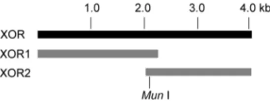
To construct the full-length cDNA of porcine XOR, two cDNA fragments, XOR1 (containing nucleotide 1–2211) and XOR2 (containing nucleotide 2082–4005) (Figure 1) were obtained by reverse transcription of total RNA extracted from swine hepato-cytes, using the pair of primers of XOR1 and XOR2, respectively. The two cDNA fragments were then inserted into the pMD 18-T vector to form the plasmids of XOR1 and pMD18-T-XOR2 respectively, and the two plasmids were digested withMun
I andSalI followed by ligating the two digested products, XOR19
primers. Then the product was digested withSalI andKpnI, and inserted in frame intoSalI -KpnI sites of pFastBac HTb vector to form the recombinant plasmid of pFastBac HTb-XOR with an N-terminal 6-His fusion. The mutants were constructed by one step site-directed mutagenesis as previously described [33]. All the constructed plasmids were sequenced and confirmed by GenScript (Nanjing, China).
Expression of the porcine XOR and its mutants
The recombinant vector of pFastBac HTb-XOR and the empty vector of pFastBac HTb were respectively transformed intoE. coli
DH10Bac cells containing the baculovirus genome, and positive clones were selected as white colonies, using 5-bromo-4-chloro-3-indolyl-b-D-galactopyranoside (X-gal) as substrate. The recombi-nant baculovirus DNA was isolated from DH10Bac white colonies and then transfected into Sf9 cells by Insect GeneJuiceH Transfection Reagent (Merck, Darmstadt, Germany). The medi-um containing viruses was collected three to four days post transfection and preserved as virus stock.
For each batch of enzyme preparation, Sf9 cells were cultured to 26107cells/liter at 27uC in flasks in Grace’s insect medium containing 10% fetal bovine serum (FBS) (Invitrogen, Carlsbad, CA, USA), 10mM riboflavin, 10mM guanosine, 80mM FeSO4 and 0.5 mM Na2MoO4. Then the cells were infected with recombinant virus at concentration six times higher than that
under expression conditions. The cells were harvested and homogenized as previously described [32], and the Sf9 cytosolic proteins containing the recombinant enzymes were obtained following the procedural instruction of Membrane and Cytosol Protein Extraction Kit (Beyotime, Jiangsu, China).
SDS-PAGE and protein quantification
The SDS-PAGE was carried out with glycine-Tris buffer system [34]. The gel was stained with Coomassie brilliant blue G-250. The amount of cytosolic proteins was determined according to the Bradford method [35] with bovine serum albumin (BSA) as standard. The amounts of target proteins (XOR) were estimated by comparing band intensities with BSA on the gel using GeneTools gel imaging software (Syngene, Los Altos, CA, USA).
Immunoblot analysis
Proteins separated by SDS-PAGE were electrophoretically transferred onto a nitrocellulose membrane. The blotted mem-brane was blocked with skim milk. The memmem-brane was then incubated with the monoclonal 6-His antibody (Beyotime) diluted 1:2000 in TBS containing 0.05% (v/v) Tween-20 (TBST) at room temperature for 1 h. After washing three times with TBST, the blotted membrane was treated with horseradish peroxidase (HRP)-conjugated goat anti-mouse IgG secondary antibody (Beyotime) diluted 1:500 in TBST at room temperature for 1 h, followed by washing three times with TBST. The immunoreactive proteins were identified using 3,39-Diaminobenzidine Tertrahy-drochloride Horseradish Peroxidase Color Development Kit (Beyotime).
Homology model and protein-ligand interaction study For homology structure modeling, protein sequence alignment was performed by Clustal X software [36]. A three dimensional (3D) homology model of porcine XOR was constructed by Protein Modeling module of Discovery Studio 2.1 (DS 2.1) software (Accelrys, San Diego, USA) available from http://www.accelrys. com/. To choose a template structure appropriate for modeling, the protein sequence of porcine XOR was screened against PDB structure database (http://www.rcsb.org/pdb/home/home.do). From a series of templates, the 3D structure of bovine milk XOR (PDB ID: 1FIQ), which shows the highest sequence identity (91%) to that of porcine XOR, was selected as the modeling template. The protein model was generated, refined and validated as previously described [37].
For protein-ligand interaction study, the cyadox structure was generated with ChemDraw software (CambridgeSoft, Waltham, MA, USA), and optimized using ‘‘Prepare Ligands’’ in the DS 2.1 for docking. Cyadox as the ligand molecule was docked into the refined protein model of porcine XOR using LibDock and Flexible Docking tool of DS 2.1, which employs a genetic algorithm and allows full ligand flexibility and partial protein flexibility. The most suitable docking mode with highest score and lowest energy conformation was finally selected. The binding sites Table 1.Primers for porcine XOR cDNA amplification and
site-directed mutagenesis.
Primer Sequence (from 59to 39)
XOR1 Fwd: GACCCCCAACCTGTTGACAATG Rev: GTACAACTCGCCTGAAACAACATTA XOR2 Fwd: TCCAGCCATCATCACAATTGAGGAT Rev: CTGCACCTCTCACACATTCACGTTC XORa Fwd: GCAGTCGAC
ATGACAGCAGATGAGT Rev: GGGGTACCTCAGACCCTCAGAGAC G47Ab
Fwd: CTGTGGAGAAGGGGCCTGCGGAGCATGCA Rev: TGCATGCTCCGCAGGCCCCTTCTCCACAG N352Ab
Fwd: GTCCATCGGAGGGGCCATCATCACCGCCA Rev: TGGCGGTGATGATGGCCCCTCCGATGGAC S360Pb Fwd: CCGCCAGCCCCATCGCCGACCTCAACCCTGT
Rev: ACAGGGTTGAGGTCGGCGATGGGGCTGGCGG R427Eb
Fwd: CAAGCAGGCCTCCGAGAGAGAAGATGACA Rev: TGTCATCTTCTCTCTCGGAGGCCTGCTTG D430Hb
Fwd: CTCCCGGAGAGAACATGACATAGCGAAGG Rev: CCTTCGCTATGTCATGTTCTCTCCGGGAG D431Ab
Fwd: CCCGGAGAGAAGATGCCATAGCGAAGGTGAC Rev:GTCACCTTCGCTATGGCATCTTCTCTCCGGG S1227Ab
Fwd: CACCCGCGGCCCCGCTACCTACAAGATCC Rev: GGATCTTGTAGGTAGCGGGGCCGCGGGTG K1230Ab
Fwd: CCCCAGCACCTACGCTATCCCTGCCTTCG Rev: CGAAGGCAGGGATAGCGTAGGTGCTGGGG
aPrimers for wild type porcine XOR cDNA amplification. The restriction sites of SalI andKpnI are underlined in the forward and reward primer, respectively. The start and stop codons are in bold.
bPrimers for site-directed mutagenesis. The mutated sites are underlined in bold and italic.
doi:10.1371/journal.pone.0073912.t001
Figure 1. Cloning of porcine XOR cDNA.
were defined as amino acid residues in distances less than 4 A˚ to cyadox.
Enzymatic activity assay
The Sf9 cytosolic proteins containing the recombinant enzymes were used as the enzyme sources, and the enzymatic activities were measured using cyadox and xanthine as substrate. Cytosolic proteins of Sf9 cells transfected with recombinant baculovirus DNA containing the empty vector of pFastBac HTb were defined as XOR-free enzyme source and were used as negative control. For oxidation of xanthine, the reaction mixture contained 0.1mg/ml porcine XOR or its
mutants, and 50,1000mM xanthine in 50 mM Tris-HCl buffer
(pH 7.4) in a final volume of 200ml. The reaction was initiated by incubation at 37uC for 20 min. For reduction of cyadox, 25,400mM of cyadox were added in the reaction mixture
containing 0.1mg/ml porcine XOR or its mutants and 100mM of xanthine in 50 mM Tris-HCl buffer (pH 7.4) in a final volume of 200ml, and then the mixtures were incubated at 37uC
for 60 min and were kept in anaerobic condition (99.999% N2). The reactions were stopped by boiling for 12 min followed by centrifugation at 12,000 g and 4uC for 12 min. The superna-tants were then subjected to high-performance liquid chroma-tography (HPLC) for analysis.
HPLC was performed using Waters 2695 HPLC System equipped with the Waters 2687 Duall absorbance detector. To separate cyadox and its reduced metabolite, cyadox-1-monoxide, 40mL of sample was injected into Agilent EclipseTM XDB-C18 column (250 mm64.6 mm, 5mm). The mobile phase consisted of solvent A (0.5% formic acid) and solvent B (acetonitrile). The gradient elution program was set as follows: 0–5 min, 15% solvent B; 5–15 min, 15% to 45% solvent B; 15–15.1 min, 45% to 15% solvent B; 15.1–20 min, 15% solvent B. The wavelength was set at 305 nm. For the separation of xanthine and its metabolite, uric acid, the isocratic elution program was as follows: 5% solvent B. The wavelength was set at 265 nm. Quantitative analysis of cyadox-1-monoxide or uric acid was carried out according to the peak area.
Kinetic parameters for the biotransformation of xanthine and cyadox were calculated by fitting data into the Michaelis-Menten equation, v = Vmax[S]/(Km+[S]), transformed as double recipro-cal equation, 1/v = 1/Vmax+Km/Vmax[S]. The intrinsic clearance (CLint) is the proportionality constant between rate of metabolism (v) and the drug substrate concentration at the enzyme site ([S]). When Km..[S], CLint= v/[S] = Vmax/Km.
Statistical analysis
Descriptive statistical parameters such as mean and standard deviation were calculated. Statistical analysis of the data was performed using Microsoft Excel 2003.
Results
Cloning, sequencing and homology analysis of porcine XOR
After cloning and sequencing, the sequence of porcine liver XOR cDNA was submitted to GenBank (accession number: JN896312.1). The cDNA consists of 4005 bp nucleotides, translated as 1334 aa peptide. The entire protein sequence of porcine XOR was aligned with those of reported mammalian XORs by Clustal X [36] (Figure S1). All these mammalian XORs showed similar length and conserved amino acid sequences (80% or more overall identity). The identities of the protein sequence of porcine (Sus scrofa) XOR (protein ID:
AEW10559) with those of bovine (Bos taurus) (NP_776397) [30], cat (Felis catus) (NP_001009217) [31], human (Homo sapiens) (NP_000370) [29], mouse (Mus musculus) (NP_035853) [28] and rat (Rattus norvegicus) (NP_058850) [27] were 91%, 87%, 89%, 85% and 86%, respectively. In porcine XOR, Glu804 and Arg882 (swine numbering) in the active sites of Mo-pt center, and loop 424–434 (swine numbering) involved in the conversion from XDH to XO are universally conserved when compared with XORs from other mammals (Figure S1).
Homology model of porcine XOR and docking of cyadox The X-ray crystal structure of bovine milk XOR (PDB ID: 1FIQ) was chosen as template since it exhibited highest identity (91%) of protein sequence to that of porcine XOR. Among the five models generated by the MODELER program in DS 2.1 software, the model with the lowest probability density function (PDF) total energy and discrete optimized potential energy (DOPE) score was selected and further refined by energy minimization (Figure 2A). The quality of the refined model was assessed by PROCHECK Ramachandran plot, which evaluates the stereochemical quality of a protein structure by analyzing residue-by-residue geometry and overall structural geometry [38]. The refined model demonstrated that 91.5% of the residues were in the most favored regions, 5.7% in additional allowed regions, and 2.8% in generously allowed regions. The refined model yielded Verify Score of 616.831, which was well within the range of high quality. Overall, the refined homology model of porcine XOR monomer is all well within the acceptable range.
As shown in Figure 2A, the porcine XOR monomer model, consisting of three domains, iron-sulfur center domain (residues 3– 165), FAD domain (residues 227–532), and Mo-pt domain (residues 592–1333) from N to C terminus, was similar to the structures of other XORs reported to date. The Mo-pt cofactor, the two iron-sulfur centers (Fe/S I and Fe/S II) and the FAD cofactor formed an electron transfer chain, and the distances were less than 15 A˚ between adjacent cofactors. A long narrow channel existed between Mo-pt domain and the other two domains, and this channel was suggested to be involved in the substrate entering and binding [39]. The model of porcine XOR docked with cyadox demonstrated that the ligand was in a deep hydrophobic pocket, surrounded by the side chains of Gly47, Asn352, Ser360, Arg427, Asp430, Asp431, Ser1227 and Lys1230 (Figure 2B). All these amino acid residues were located in flexible loops and appeared in distances of less than 4A˚ to cyadox. These residues might be closely related to the catalytic process of porcine XOR in the metabolism of cyadox, and were investigated by the following mutagenesis study.
Expression of porcine XOR and its mutants in Sf9 insect cells
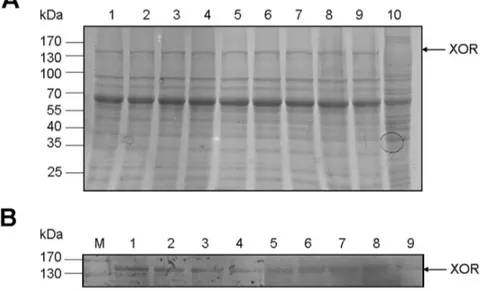
Porcine XOR was expressed in Sf9 insect cells and approximate 2 mg of XOR was harvested per 107 cells. Compared with the cytosolic proteins form cells harboring the empty vector (Figure 3A, lane 10), the porcine XOR, appearing as a 150 kDa peptide, was expressed in the cytosol of Sf9 insect cells (Figure 3, lane 9). This molecular weight of 150 kDa is in consistence with the fact that porcine XOR contains 1334 aa with a molecular mass of 146974.9 Da.
Ser1227 and Lys1230 were replaced with non-polar residue alanine. The expressions of these eight mutants were confirmed by SDS-PAGE and immunoblot analysis (Figure 3, lane 1–8).
The enzymatic activity of porcine XOR in the metabolism of xanthine and cyadox
As shown in Figure S2B, nearly no uric acid was generated when xanthine was incubated with Sf9 cell cytosol free of porcine XOR. When xanthine was incubated with the cytosol containing recombinant porcine XOR, the uric acid was generated (Figure S2C), demonstrating that the heterogeneously expressed porcine XOR was of biological activity. In the absence of xanthine, cyadox could not be metabolized to monoxide when incubated with the cytosol containing porcine XOR (Figure S3C), while in the presence of xanthine, the cyadox-1-monoxide was produced (Figure S3D), indicating that cyadox required an electron donated by xanthine to be reduced to produce cyadox-1-monoxide. The cytosol without recombinant porcine XOR could not metabolize cyadox (Figure S3B).
Enzyme kinetic analysis of recombinant porcine XOR and its mutants
First, the kinetic parameters of porcine XOR and eight mutants were compared, in the oxidation of their physiological substrate, xanthine. As shown in Table 2, the CLint value of D431A (392.31 mL/mg XOR/min) was higher than that of the wild type XOR (356.85 mL/mg XOR/min). S360P (44.05 mL/mg XOR/ min), S1227A (77.54 mL/mg XOR/min), and K1230A (102.63 mL/mg XOR/min) exhibited much lower CLintvalues, and R427E showed a slight lower CLintvalues compared with the wild type. The Km values of S360P (64.28mM), S1227A (50.80mM) and K1230A (39.25mM) were much higher than that of wild type (21.69mM), indicating these mutants had much
weaker substrate affinity.
Using cyadox as the terminal electron acceptor, the kinetic parameters of porcine XOR and its mutants were analyzed based on the yield of cyadox-1-monoxide (Table 3). Although cyadox could be metabolized by porcine XOR to yield both cyadox-1-monoxide and cyadox-4-cyadox-1-monoxide, it could also be non-enzymatically reduced to cyadox-4-monoxide mediated by heme groups of catalase in the cytosol [20]. Furthermore, as shown in Figure S3, cyadox-1-monoxide was the main product of cyadox catalyzed by porcine XOR. Therefore, only the production of cyadox-1-monoxide was monitored here to gain the kinetic parameters. In consistence with the changes of kinetic parameters in the oxidation of xanthine, the CLintvalues of S360P (8.3 mL/ mg XOR/min), S1227A (5.5 mL/mg XOR/min), K1230A (5.4 mL/mg XOR/min) were much lower than that of the wild type (23.17 mL/mg XOR/min) because of the lower Vmaxand higher Km values. Besides, D431A (24.77 mL/mg XOR/min) showed higher CLint value, and G47A exhibited only a slight higher Vmax, compared with the wild type. In contrast, the CLint value of R427E (68.67 mL/mg XOR/min) was much higher than that of the wild type, and the Km(20.86mM) was much lower than that of wild type (58.20mM), demonstrating R427E had a strong substrate affinity. Meanwhile, D430H exhibited both lower Vmax and CLintvalues than those of the wild type.
Discussion
The present study has demonstrated that the porcine XOR was expressed in Sf9 cells with XDH/XO activity. The enzyme possesses common redox active cofactors, forming an electron transfer pathway terminated by a flavin cofactor. When cyadox, an exogenous electron acceptor, was docked into porcine XOR, eight amino acid residues, which are conservative among the XORs from other mammalian species, were screened out as the potential cyadox-binding sites. The side chain of cyadox oriented toward Gly47 located in iron-sulfur center domain, Arg427 in the FAD domain, and Ser1227 and Lys1230 in the Mo-pt domain. Meanwhile, the benzene ring of quinoxaline heterocycle faced toward Asn352 and Ser360 in the FAD domain, and the pyrazine ring was located near Asp430 and Asp431 in the FAD domain. It has been shown that one electron reduction of cyadox occurs at one of the two NRO groups on the pyrazine ring [11]. However, our docking result demonstrated that the pyrazine ring of cyadox faced at FAD center, and this was different form the studies about the reduction of nicotinamideN-oxide and nitrite by XOR which both suggested that the reactions involved the abstraction of an oxygen atom and occurred at the molybdenum centre [40,41]. This is probably due to the influence of the large side chain of cyadox upon binding to the enzyme.
In order to obtain the enzyme kinetic parameters of porcine XOR in the reduction of cyadox, the production of one of the Figure 2. Structural overview (A) and the binding pocket (B) of
porcine XOR docked with cyadox.(A) From N to C terminus, iron-sulfur centers domain (purple), FAD domain (dark blue), and molybdopterin (Mo-pt) domain (green) are presented in the 3D structure of porcine XOR model, and the Mo-pt cofactor (yellow), the two iron-sulfur centers (Fe/S I and Fe/S II, red), and the FAD cofactor (bright green) are also included. Cyadox (pink) is docked into the porcine XOR model. (B) Side chains of eight key amino acid residues, Gly47 (blue, in iron-sulfur centers domain), Asn352, Ser360, Arg427, Asp430 and Asp431 (green, in FAD domain), Ser1227 and Lys1230 (yellow, in Mo-pt domain), were screened in a distance of less than 4A˚ with cyadox by molecular docking. The Mo-pt cofactor (red), the Fe/S I and Fe/S II (grey), the FAD cofactor (purple), cyadox (dark red) and xanthine (blue and grey) are also included.
reductive metabolites, cyadox-1-monoxide, was monitored, as to exclude the influence of the fact that cyadox could also be reduced to cyadox-4-monoxide by non-enzymatic reduction mediated by heme groups of catalase in the cytosol [20]. Since oxygen could oxidize cyadox-1-monoxide back to its dioxide form [21], the reaction was kept under anaerobic condition. G47A, S360P, D431A, S1227A, and K1230A displayed differential enzyme kinetic parameters compared with those of the wide type (Table 3). However, these changes were similar to those in the oxidation of xanthine (Table 2), indicating that these mutations affect the metabolism of xanthine which serves as the electron donor, resulting in impairment of the subsequent electron transfer that is critical for the reduction of cyadox as the electron acceptor. Among these residues, S360P, S1227A, K1230A exhibited
significantly lower CLintvalues for xanthine because of the lower Vmax and higher Km values. Since Ser1227 and Lys1230 are located in the Mo-pt domain, which contains the binding and active sites of XOR for xanthine, the mutations of these residues may affect the binding and catalyzing of xanthine by XOR. Since Ser360 is located at theN-terminal ofa10 helix (Figure S1), and proline is a rigid amino acid which can destroy the formation of a-helix, this mutation presumably results in the whole structural change of porcine XOR.
Different from the alterations of kinetic parameters in the oxidation of xanthine (Table 2), mutation of Arg427 to glutamic acid displayed a higher CLintand a much lower Kmvalues than those of the wild type in the reduction of cyadox (Table 3), suggesting that R427E had a strong substrate binding affinity for cyadox. Arg427 is located in the 424–434 loop (swine numbering, equivalent to 423–433 loop in bovine XOR, Figure S1), the orientation of which is important for the transition from XDH to XO [10]. The two forms of this enzyme prefer different ultimate substrate (NAD+
for XDH and O2for XO) for electron transfer, and possess different redox potentials for the flavin semiquinone/ hydroquinone pair (Esq/hq) [42,43]. It is reported that Arg426 (bovine numbering, equal to Arg427 in porcine XOR) is one of the key residues that differentiate the Esq/hq for XDH and XO significantly [2]. We suppose that the charge change of R427E may influence the substrate specificity and binding of the enzyme. More mutants need to be made to elucidate the exact role of Arg427 in the reduction of cyadox. Since the mutagenic activity of QdNOs is mainly caused by the existence ofN-oxide groups in QdNO structures [11], the greater CLintof R427E for cyadox is of favorable applications in the detoxification of cyadox in animal feed.
Meanwhile, D430H exhibited a lower CLintdue to the lower Vmax values, indicating a deficiency in reduction of cyadox (Table 3). Asp430 is also located in the 424–434 loop. In order to activate an electron transfer from Fe/S II to flavin, the enzyme needs an upshift of Esq/hqto make electron transfer from Fe/S II to the flavin isoenergetic [44]. Recent study reported that no large Figure 3. SDS-PAGE (A) and western blot (B) analysis of porcine XOR and its mutants expressed in Sf9 insect cells.(A) Lanes 1–10 were loaded with cytosolic proteins of Sf9 insect cells expressing porcine XOR mutants of G47A, N352A, S360P, R427E, D430H, D431A, S1227A, K1230A, wild type XOR and empty vector control, respectively. (B) Lanes 1–9 were loaded with proteins as described above. Monoclonal 6-His antibody was used as primary antibody to immunoblot the expressed XOR proteins as described in materials and methods. Arrows indicate the position of XOR. M, protein molecular mass standard.
doi:10.1371/journal.pone.0073912.g003
Table 2.Enzyme kinetic parameters of porcine XOR and its mutants in the oxidation of xanthine.
Enzymes Vmax
(mmol/min/mg XOR) Km(mM)
CLint
(mL/mg XOR/min)
Wild type 7.7460.03 21.6962.27 356.8568.13 G47A 7.9160.01** 24.1361.71 328.66622.86 N352A 7.6960.03 22.7060.65 339.0868.13 S360P 2.8360.03** 64.2861.00** 44.0560.27** R427E 7.7560.04 23.2460.90 333.3365.12* D430H 7.4760.03** 21.3060.59 350.9968.71 D431A 8.5260.05** 21.7260.74 392.31610.88* S1227A 3.9460.06** 50.8063.04** 77.5461.70** K1230A 4.0260.03** 39.2561.73** 102.6363.72**
Note: The values are expressed as means6standard deviations of the results of three independent experiments.
**(p,0.01) and *(p,0.05) indicate statistically significant difference in enzyme kinetic parameters between the wild type and each mutant.
conformational change was observed in response to NAD+binding at bovine XDH. Instead, the positive charge of the NAD+
ring, deprotonation of Asp429 (equivalent to Asp430 in porcine XOR), and capping of the bulk surface of the flavin by the NAD+ molecule all contributed to altering Esq/hqupon NAD+
binding to XDH [2]. Based on this data, we speculate that D430H of porcine XOR harms the deprotonation of Asp430 and influence the Esq/hq alteration which is needed for the electron transfer to the terminal electron acceptor, cyadox. In addition, the Km of K1230A was nearly three times to that of wild type in the reduction of cyadox, while only two-fold upshift of Kmwas detected in the oxidation of xanthine. It was reported that although Lys1228 (bovine numbering, equal to Lys1230 in porcine XOR) did not belong to 423–433 loop (bovine numbering), it still significantly influenced the Esq/hqfor both XDH and XO [2].
In conclusion, our work for the first time cloned the cDNA of XOR from porcine liver and obtained functionally active recombinant XOR expressed in Sf9 insect cells. Using molecular docking combined with site-directed mutagenesis techniques, it is demonstrated that Arg427 is related to the substrate binding of porcine XOR to cyadox. Asp430 probably plays a role in the electron transfer to cyadox. Besides, Gly47, Ser360, Asp431, Ser1227 and Lys1230 participate in the electron-donating process of xanthine, whose obstruction hinders the fulfillment ofN-oxide reduction of cyadox. Although molecular docking is a predictive method, it still verifies the result of mutation and kinetic analysis of porcine XOR for cyadox catabolism in our study. To get a better understanding of the catalytic mechanism underlying the reductive activation of QdNOs by porcine XOR, further study will be devoted into the crystal structure analysis of porcine XOR in complex with QdNO substrate.
Supporting Information
Figure S1 Amino acid sequence alignment of porcine XOR with other five mammalian XORs.Totally conserved
residues of XORs in all 6 mammalian species are shown by asterisks (*), partially conserved residues are shown by dots (.or:). The second structures were predicted by PSIPRED V3.0 software (http://bioinf.cs.ucl.ac.uk/psipred/). Thea-helices are shown in underlined letters, and the b-strands in boldface letters. The universally conserved Glu and Arg in the active sites of Mo-pt center are marked with filled circles (
N
). The eight key amino acid residues, Gly47, Asn352, Ser360, Arg427, Asp430, Asp431, Ser1227 and Lys1230 (swine numbering), generated by molecular docking, are indicated as numbered underlined letters in bold. (DOC)Figure S2 HPLC chromatograms of the metabolite of xathine catalyzed by recombinant porcine XOR. (A) Uric standard. (B) Xanthine incubated with Sf9 cell cytosol free of recombinant porcine XOR at 37uC for 20 min. (C) Xanthine incubated with Sf9 cell cytosol containing recombinant porcine XOR at 37uC for 20 min.
(PPT)
Figure S3 HPLC chromatograms of the metabolite of cyadox catalyzed by recombinant porcine XOR. (A) Cyadox-1-monox-ide standard. (B) Cyadox incubated with Sf9 cell cytosol free of recombinant porcine XOR with xanthine at 37uC for 60 min. (C) Cyadox incubated with Sf9 cell cytosol containing recombinant procine XOR in the absence of xanthine at 37uC for 60 min. (D) Cyadox incubated with Sf9 cell cytosol containing recombinant porcine XOR in the presence of xanthine at 37uC for 60 min.
(PPT)
Author Contributions
Conceived and designed the experiments: CC GC ZY. Performed the experiments: CC GC. Analyzed the data: CC GC HH. Contributed reagents/materials/analysis tools: MD XW LH ZL ZY. Wrote the paper: CC GC.
References
1. Kitamura S, Sugihara K, Ohta S (2006) Drug-metabolizing ability of molybdenum hydroxylases. Drug Metab Pharmacokinet 21: 83– 98.
2. Ishikita H, Eger BT, Okamoto K, Nishino T, Pai EF (2012) Protein conformational gating of enzymatic activity in xanthine oxidoreductase. J Am Chem Soc 134: 999–1009.
3. Zimmerman BJ, Granger DN (1994) Mechanisms of reperfusion injury. Am J Med Sci 307: 284–292.
4. Beedham C (1985) Molybdenum hydroxylases as drug-metabolizing enzymes. Drug Metab Rev 16: 119–156.
5. Pauff JM, Cao H, Hille R (2009) Substrate orientation and catalysis at the molybdenum site in xanthine oxidase: Crystal structures in complex with xanthine and lumazine. J Biol Chem 284: 8760–8767.
6. Cao H, Pauff JM, Hille R (2010) Substrate orientation and catalytic specificity in the action of xanthine oxidase: the sequential hydroxylation of hypoxanthine to uric acid. J Biol Chem 285: 28044–28053.
Table 3.Enzyme kinetic parameters of porcine XOR and its mutants in the reduction of cyadox.
Enzymes Vmax(mmol/min/mg XOR) Km(mM) CLint(mL/mg XOR/min)
Wild type 1.3460.05 58.2065.76 23.1761.48
G47A 1.4160.03* 65.6864.42 21.4561.05
N352A 1.2960.18 60.10612.59 21.6861.68
S360P 0.7860.04** 93.5368.15** 8.3360.36**
R427E 1.4360.05 20.8660.90** 68.6765.34**
D430H 0.9860.03** 64.6863.52 15.2260.49**
D431A 1.5260.01** 61.2960.53* 24.7760.25*
S1227A 0.8160.05** 148.20614.21** 5.4660.18**
K1230A 0.8860.08** 162.89615.50** 5.4060.10**
Note: The values are expressed as means6standard deviations of the results of three independent experiments.
7. Pauff JM, Zhang J, Bell CE, Hille R (2008) Substrate orientation in xanthine oxidase: crystal structure of enzyme in reaction with 2-hydroxy-6-methylpurine. J Biol Chem 283: 4818–4824.
8. Matsumoto K, Okamoto K, Ashizawa N, Nishino T (2011) FYX-051: a novel and potent hybrid-type inhibitor of xanthine oxidoreductase. J Pharmacol Exp Ther 336: 95–103.
9. Nishino T, Okamoto K, Eger BT, Pai EF, Nishino T (2008) Mammalian xanthine oxidoreductase - mechanism of transition from xanthine dehydroge-nase to xanthine oxidase. Febs J 275: 3278–3289.
10. Enroth C, Eger BT, Okamoto K, Nishino T, Nishino T, et al. (2000) Crystal structures of bovine milk xanthine dehydrogenase and xanthine oxidase: structure-based mechanism of conversion. Proc Natl Acad Sci U S A 97: 10723–10728.
11. Carta A, Corona P, Loriga M (2005) Quinoxaline 1,4-dioxide: a versatile scaffold endowed with manifold activities. Curr Med Chem 12: 2259–2272. 12. Fang G, He Q, Zhou S, Wang D, Zhang Y, et al. (2006) Subchronic oral toxicity
study with cyadox in Wistar rats. Food Chem Toxicol 44: 36–41.
13. He Q, Fang G, Wang Y, Wei Z, Wang D, et al. (2006) Experimental evaluation of cyadox phototoxicity to Balb/c mouse skin. Photodermatol Photoimmunol Photomed 22: 100–104.
14. Wang X, He QH, Wang YL, Ihsan A, Huang LL, et al. (2011) A chronic toxicity study of cyadox in Wistar rats. Regul Toxicol Pharmacol 59: 324–333. 15. Nabuurs MJ, van der Molen EJ, de Graaf GJ, Jager LP (1990) Clinical signs and
performance of pigs treated with different doses of carbadox, cyadox and olaquindox. Zentralbl Veterinarmed A 37: 68–76.
16. Sestakova I, Kopanica M (1988) Determination of cyadox and its metabolites in plasma by adsorptive voltammetry. Talanta 35: 816–818.
17. Liu Z, Huang L, Dai M, Chen D, Tao Y, et al. (2009) Metabolism of cyadox in rat, chicken and pig liver microsomes and identification of metabolites by accurate mass measurements using electrospray ionization hybrid ion trap/ time-of-flight mass spectrometry. Rapid Commun Mass Spectrom 23: 2026– 2034.
18. Xu N, Huang L, Liu Z, Pan Y, Wang X, et al. (2011) Metabolism of cyadox by the intestinal mucosa microsomes and gut flora of swine, and identification of metabolites by high-performance liquid chromatography combined with ion trap/time-of-flight mass spectrometry. Rapid Commun Mass Spectrom 25: 2333–2344.
19. Wu H, Li L, Shen J, Wang Y, Liu K, et al. (2012) In vitro metabolism of cyadox in rat, chicken and swine using ultra-performance liquid chromatography quadrupole time-of-flight mass spectrometry. J Pharm Biomed Anal 67–68: 175– 185.
20. Zheng M, Jiang J, Wang J, Tang X, Ouyang M, et al. (2011) The mechanism of enzymatic and non-enzymatic N-oxide reductive metabolism of cyadox in pig liver. Xenobiotica 41: 964–971.
21. Ganley B, Chowdhury G, Bhansali J, Daniels JS, Gates KS (2001) Redox-activated, hypoxia-selective DNA cleavage by quinoxaline 1,4-di-N-oxide. Bioorg Med Chem 9: 2395–2401.
22. Ekser B, Rigotti P, Gridelli B, Cooper DK (2009) Xenotransplantation of solid organs in the pig-to-primate model. Transpl Immunol 21: 87–92.
23. Lengauer T, Rarey M (1996) Computational methods for biomolecular docking. Curr Opin Struct Biol 6: 402–406.
24. Ali HI, Fujita T, Akaho E, Nagamatsu T (2010) A comparative study of AutoDock and PMF scoring performances, and SAR of 2-substituted pyrazolotriazolopyrimidines and 4-substituted pyrazolopyrimidines as potent xanthine oxidase inhibitors. J Comput Aided Mol Des 24: 57–75.
25. Iwasaki T, Okamoto K, Nishino T, Mizushima J, Hori H (2000) Sequence motif-specific assignment of two [2Fe-2S] clusters in rat xanthine oxidoreductase studied by site-directed mutagenesis. J Biochem 127: 771–778.
26. Asai R, Nishino T, Matsumura T, Okamoto K, Igarashi K, et al. (2007) Two mutations convert mammalian xanthine oxidoreductase to highly superoxide-productive xanthine oxidase. J Biochem 141: 525–534.
27. Amaya Y, Yamazaki K, Sato M, Noda K, Nishino T, et al. (1990) Proteolytic conversion of xanthine dehydrogenase from the NAD-dependent type to the O2-dependent type. Amino acid sequence of rat liver xanthine dehydrogenase and identification of the cleavage sites of the enzyme protein during irreversible conversion by trypsin. J Biol Chem 265: 14170–14175.
28. Terao M, Cazzaniga G, Ghezzi P, Bianchi M, Falciani F, et al. (1992) Molecular cloning of a cDNA coding for mouse liver xanthine dehydrogenase. Regulation of its transcript by interferons in vivo. Biochem J 283 (Pt 3): 863–870. 29. Ichida K, Amaya Y, Noda K, Minoshima S, Hosoya T, et al. (1993) Cloning of
the cDNA encoding human xanthine dehydrogenase (oxidase): structural analysis of the protein and chromosomal location of the gene. Gene 133: 279–284.
30. Berglund L, Rasmussen JT, Andersen MD, Rasmussen MS, Petersen TE (1996) Purification of the bovine xanthine oxidoreductase from milk fat globule membranes and cloning of complementary deoxyribonucleic acid. J Dairy Sci 79: 198–204.
31. Tsuchida S, Yamada R, Ikemoto S, Tagawa M (2001) Molecular cloning of a cDNA coding for feline liver xanthine dehydrogenase. J Vet Med Sci 63: 353– 355.
32. Nishino T, Amaya Y, Kawamoto S, Kashima Y, Okamoto K, et al. (2002) Purification and characterization of multiple forms of rat liver xanthine oxidoreductase expressed in baculovirus-insect cell system. J Biochem 132: 597–606.
33. Zheng L, Baumann U, Reymond JL (2004) An efficient one-step site-directed and site-saturation mutagenesis protocol. Nucleic Acids Res 32: e115. 34. King J, Laemmli UK (1971) Polypeptides of the tail fibres of bacteriophage T4.
J Mol Biol 62: 465–477.
35. Bradford MM (1976) A rapid and sensitive method for the quantitation of microgram quantities of protein utilizing the principle of protein-dye binding. Anal Biochem 72: 248–254.
36. Chenna R, Sugawara H, Koike T, Lopez R, Gibson TJ, et al. (2003) Multiple sequence alignment with the Clustal series of programs. Nucleic Acids Res 31: 3497–3500.
37. Kanth BK, Liou K, Sohng JK (2010) Homology modeling, binding site identification and docking in flavone hydroxylase CYP105P2 in Streptomyces peucetius ATCC 27952. Comput Biol Chem 34: 226–231.
38. Laskowski RA, Rullmannn JA, MacArthur MW, Kaptein R, Thornton JM (1996) AQUA and PROCHECK-NMR: programs for checking the quality of protein structures solved by NMR. J Biomol NMR 8: 477–486.
39. Okamoto K, Eger BT, Nishino T, Kondo S, Pai EF, et al. (2003) An extremely potent inhibitor of xanthine oxidoreductase. Crystal structure of the enzyme-inhibitor complex and mechanism of inhibition. J Biol Chem 278: 1848–1855. 40. Godber BL, Doel JJ, Sapkota GP, Blake DR, Stevens CR, et al. (2000) Reduction of nitrite to nitric oxide catalyzed by xanthine oxidoreductase. J Biol Chem 275: 7757–7763.
41. Murray KN, Watson JG, Chaykin S (1966) Catalysis of the direct transfer of oxygen from nicotinamide N-oxide to xanthine by xanthine oxidase. J Biol Chem 241: 4798–4801.
42. Hunt J, Massey V, Dunham WR, Sands RH (1993) Redox potentials of milk xanthine dehydrogenase. Room temperature measurement of the FAD and 2Fe/2S center potentials. J Biol Chem 268: 18685–18691.
43. Barber MJ, Siegel LM (1982) Oxidation-reduction potentials of molybdenum, flavin, and iron-sulfur centers in milk xanthine oxidase: variation with pH. Biochemistry 21: 1638–1647.