O R I G I N A L P A P E R
NADH oxidase activity of rat and human liver xanthine
oxidoreductase: potential role in superoxide production
Luisa MaiaÆRui O. DuarteÆ Ana Ponces-FreireÆ
Jose´ J. G. MouraÆLurdes Mira
Received: 4 January 2007 / Accepted: 12 March 2007 / Published online: 18 April 2007
SBIC 2007
Abstract To characterise the NADH oxidase activity of both xanthine dehydrogenase (XD) and xanthine oxidase (XO) forms of rat liver xanthine oxidoreductase (XOR) and to evaluate the potential role of this mammalian enzyme as an O2•– source, kinetics and electron paramagnetic reso-nance (EPR) spectroscopic studies were performed. A steady-state kinetics study of XD showed that it catalyses NADH oxidation, leading to the formation of one O2•– molecule and half a H2O2molecule per NADH molecule, at rates 3 times those observed for XO (29.2 ± 1.6 and 9.38 ± 0.31 min–1, respectively). EPR spectra of NADH-reduced XD and XO were qualitatively similar, but they were quantitatively quite different. While NADH efficiently reduced XD, only a great excess of NADH reduced XO. In
agreement with reductive titration data, the XD specificity constant for NADH (8.73 ± 1.36lM–1 min–1) was found to
be higher than that of the XO specificity constant (1.07 ± 0.09lM–1min–1). It was confirmed that, for the
reducing substrate xanthine, rat liver XD is also a better O2•– source than XO. These data show that the dehydrogenase form of liver XOR is, thus, intrinsically more efficient at generating O2•–than the oxidase form, independently of the reducing substrate. Most importantly, for comparative pur-poses, human liver XO activity towards NADH oxidation was also studied, and the kinetics parameters obtained were found to be very similar to those of the XO form of rat liver XOR, foreseeing potential applications of rat liver XOR as a model of the human liver enzyme.
Keywords Xanthine oxidoreductaseXanthine oxidase Xanthine dehydrogenaseRat liver Reactive oxygen species NADH
Abbreviations
AFR Activity-to-flavin ratio AO Aldehyde oxidase
EPR Electron paramagnetic resonance FAD Flavin adenine dinucleotide ROS Reactive oxygen species Sim Simulated
XD Xanthine dehydrogenase XO Xanthine oxidase XOR Xanthine oxidoreductase
Introduction
Xanthine oxidoreductase (XOR), until recently also re-ferred to as xanthine oxidase (XO), has been the subject of L. MaiaA. Ponces-FreireL. Mira
Centro de Quı´mica e Bioquı´mica,
Faculdade de Cieˆncias da Universidade de Lisboa, 1749-016 Lisbon, Portugal
L. MaiaA. Ponces-FreireL. Mira (&) Departamento de Quı´mica e Bioquı´mica, Faculdade de Cieˆncias,
Universidade de Lisboa, 1749-016 Lisbon, Portugal e-mail: lurdes.mira@fc.ul.pt
R. O. DuarteJ. J. G. Moura REQUIMTE/CQFB, Departamento de Quı´mica, Faculdade de Cieˆncias e Tecnologia da Universidade Nova de Lisboa, 2829-516 Caparica, Portugal
L. Mira
many mechanistic, structural and biophysical reviews [1–4]. Mammalian XOR is the key enzyme in the catabo-lism of purines, oxidising hypoxanthine to xanthine and xanthine to the terminal catabolite urate. XOR is a complex homodimer, containing one molybdopterin, one flavin adenine dinucleotide (FAD) and two different [2Fe–2S] centres (named Fe/S I and Fe/S II) per 145-kDa subunit [1]. The mammalian XOR is synthesised as an NAD+ -depen-dent dehydrogenase (throughout the text referred to as xanthine dehydrogenase, XD) but, although it transfers the electrons preferentially to NAD+, it can also catalyse electron transfer to O2. XD can, however, be readily con-verted to a ‘‘strict’’ oxidase form (named XO), either reversibly, through oxidation of the cysteine residues 535 and 992, or irreversibly, by proteolysis [5,6]. The cysteine oxidation (or proteolysis) causes a conformational change in the vicinity of the FAD, the site at which O2and NAD+ react, increasing the midpoint potential of the flavin moiety and blocking the access of NAD+ to FAD, but without disturbing the interactions between O2and FAD [7].
The reduction of O2, catalysed by both enzyme forms, yields O2•– and H2O2, with the stoichiometry of these reaction products being differentially affected by the form and the source of the enzyme and by the reactant concen-trations [8, 9]. Therefore, XOR can act as a source of reactive oxygen species (ROS) and it is this capacity of XOR that makes the enzyme the focus of interest in many recent biochemical and clinical studies. Normal processes of metabolism continuously form ROS, and many of them may have useful physiological functions, such as in host defence against invading pathogens, and as mediators of signal transduction [10, 11]. Their overproduction, how-ever, can play a major role in several pathological condi-tions [12–16]. Moreover, it was recently shown that XOR can catalyse the reduction of nitrates and nitrites, giving rise to both•NO [17,18] and peroxynitrite (ONOO–) [19]. This fact further amplifies the physiological and patho-logical significance of XOR. Unlike NO synthase, XOR can produce•NO under anoxic conditions and, thus, pro-mote the •NO-dependent vasodilatation in ischaemic tis-sues [20]. On the other hand, ONOO–, a powerful oxidant and destructive agent, which results from the diffusion-controlled reaction between •NO and O2•– [21], can be produced solely by the action of XOR [19]. The XOR-derived ONOO–has been proposed as a bactericidal agent in milk and the digestive tract [19].
XOR has a broad specificity for reducing substrates. In addition to the well-known oxidation of hypoxanthine and xanthine, XOR also catalyses the oxidation of a wide variety of aldehydes and substituted pyridines, purines, pteridines and related compounds, including NADH [22]. The reaction catalysed by XOR can be separated into a reductive half-reaction and an oxidative half-reaction. The
reducing substrates are oxidised at the molybdenum site, and the reduction mechanism is thought to be the same for both XD and XO [1,7]. The electrons thus transferred from the substrate to the enzyme are rapidly distributed throughout the other centres, by intramolecular electron transfer, according to their redox potentials. In the oxida-tive half-reaction, electrons are transferred from the FAD centre to NAD+or O2[1,23]. NADH, although a reducing substrate, is an important exception, since electron transfer is achieved through the FAD centre [1].
Materials and methods
Adult male Sprague–Dawley rats (3–4 months old) were obtained from the Instituto de Investigac¸a˜o Cientı´fica Bento da Rocha Cabral (Lisbon, Portugal).
All the reagents were of the highest quality available and were used as supplied. Horse heart ferricytochromec,
p-dimethylaminocinnamaldehyde, dithiothreitol, NADH, NAD+, xanthine, bovine milk XO and bovine erythrocytes superoxide dismutase were from Sigma Chemical Co. (Madrid, Spain). All the other reagents were from Merck (Darmstadt, Germany). NADH, NAD+ and xanthine con-centrations were determined spectrophotometrically using
e= 6,220 M–1cm–1 at 340 nm, e= 18,000 M–1cm–1 at
260 nm and e= 9,300 M–1cm–1 at 277 nm, respectively.
Activity assays and spectra were recorded using a PC-linked UV2-100 Unicam spectrophotometer with a temperature controlled cell unit.
Enzyme assays
XO activity was measured using 20lM xanthine in
50 mM phosphate buffer pH 7.8, at 298 K, in air-equili-brated solution, with the production of urate being moni-tored at 295 nm (e= 9,500 M–1cm–1). XD activity was
measured using the same assay mixture as described for XO, plus 85lM NAD+and monitoring NADH production
at 340 nm (e= 6,220 M–1 cm–1). One unit of catalytic
activity is defined as the amount of enzyme required to catalyse the oxidation of 1lmol min–1of substrate, under
our experimental conditions. The dehydrogenase-to-oxi-dase ratio of XOR, as defined by Waud and Rajagopalan [29], was determined as the ratio of aerobic formation of urate, measured at 295 nm, in the presence of NAD+to that in the absence of NAD+. AO activity was assayed by fol-lowing the oxidation of 25lM p
-dimethylaminocinnam-aldehyde at 398 nm (e= 30,500 M–1cm–1) in 50 mM
phosphate buffer pH 7.8, at 298 K, in air-equilibrated solution.
Purification of the XO form of XOR and AO from rat liver
XOR, in its reversible XO form, and AO were purified from rat liver as previously described by Maia and Mira [30]. Briefly, the rat liver homogenate was fractionated by heat denaturation and by ammonium sulfate precipitation to give a crude extract containing both enzymes. This ex-tract was chromatographed on a hydroxyapatite column (Bio-Rad, CA, USA) that completely separated AO from XO. Further purification of XO forms by anion-exchange chromatography on a Q-Sepharose Fast Flow column (Pharmacia Biotech, Uppsala, Sweden) resulted in a highly
purified (about 1,200-fold) preparation, with a specific activity of 3.5–3.7 U mg–1. AO was purified about 1,000-fold, with a specific activity of 3.4–3.6 U mg–1, by affinity chromatography on benzamidine-Sepharose 6B (Pharmacia Biotech, Uppsala, Sweden). Both purified enzymes dis-played the characteristic absorption spectra of highly purified enzymes, with absorbance ratios Abs280/Abs450 between 5.3 and 5.8. To evaluate the presence of inactive forms of the enzyme (desulfo and demolybdo), the activity-to-flavin ratio (AFR) of the XO form was calculated, dividing the absorbance change per minute at 295 nm by the absorbance at 450 nm of the enzyme used in the assay (200 was taken to be 100% active [31]). The final XO concentration was determined spectrophotometrically using e= 71,500 M–1cm–1 at 450 nm for the oxidised
enzyme [32], and was corrected for the presence of inactive molecules. The XO batches thus obtained did not have xanthine:NAD+ oxidoreductase activity.
Preparation of the dehydrogenase form of XOR from rat liver
XOR in its XD form was obtained through reversible reduction of oxidised XO sulfhydryl groups. Purified XO was incubated with 5 mM dithiothreitol, for 1–2 h, at 303 K, and then passed through a small G-25 column (Pharmacia Biotech, Uppsala, Sweden) equilibrated in 100 mM tris(hydroxymethyl)aminomethane (Tris)–HCl buffer, pH 7.8. Dithiothreitol treatment resulted in an 80– 85% decrease in the XO activity (xanthine:O2 oxidore-ductase activity) and a dehydrogenase-to-oxidase ratio of 5–6 was achieved, as previously described [30]. Consid-ering the intrinsic xanthine:O2 oxidoreductase activity of the XD form [31,33,34], the XD batches thus prepared are highly purified, with less than 5% irreversible XO con-tamination.
Preparation of the deflavo-XD form of XOR
THe deflavo-XD form of XOR was prepared as described by Branzoli and Massey [35].
Preparation of XO from butter milk
However, the resulting enzyme sample displayed a com-paratively low Abs280/Abs450of 9.5.
Steady-state kinetics of NADH oxidation by XOR The initial rates of NADH oxidation by both XD and XO forms of XOR were measured following the decrease in absorbance at 340 nm (e= 6,220 M–1cm–1). The enzymes
were incubated with varying concentrations of NADH (2.5–100lM) in air-equilibrated 50 mM Tris–HCl buffer,
with 1 mM EDTA, pH 7.8. Each initial rate determination was performed with three different enzyme batches, in quintuplicate, except for bovine milk XO, for which only one enzyme lot was assayed. The apparent kinetics parameters were estimated by the direct linear method of Eisenthal and Cornish-Bowden [36]. The values obtained using the double-reciprocal plot agreed within a maximum error of 7%. The specificity constants were calculated as
kNADH =kcatapp/Kmapp.
Steady-state kinetics of O2•–formation by XOR
O2•– formation during NADH oxidation by both XD and XO forms of XOR was estimated as the superoxide dismutase inhibitable reduction of cytochromec[37]. The reaction mixtures contained 50 mM Tris–HCl buffer, with 1 mM EDTA, pH 7.8, 100lM ferricytochrome c and
varying concentrations of NADH (2.5–100lM). The
ini-tial rates of O2•– formation (determined in quintuplicate and with three different enzyme batches) were evaluated by the increase in absorbance at 550 nm (De= 21,000 M–1
cm–1) in the absence and in the presence of 2,500 U cm–3 superoxide dismutase. The apparent kinetics parameters were estimated by the direct linear method of Eisenthal and Cornish-Bowden [36]. The values obtained using the double-reciprocal plot agreed within a maximum error of 3%. The specificity constants were calculated as
kNADH =kcatapp/Kmapp.
One-electron reduction flux
The percentage of oxygen one-electron reduction was calculated as half of the ratio of the O2•–production rate to the NADH oxidation rate, with the initial rates being cal-culated from the kinetics parameters determined.
Reductive titration of rat liver XOR and the deflavo-XD form
Reductive titrations of both the XD and XO forms of rat liver XOR with NADH were followed by EPR spectros-copy at 20, 40, 60 and 100 K. The enzyme samples were reduced anaerobically with NADH for 5 min in 50 mM
Tris–HCl buffer pH 7.8 at 293 K before being frozen. Enzymes were also reduced with dithionite, for 30 min. The samples were cooled with an Oxford Instruments ESR900 liquid-helium cryostat, fitted with a temperature controller. X-band EPR spectra were recorded using a Bruker EMX 6/1 spectrometer, equipped with a dual-mode ER4116DM cavity. The experimental conditions main-tained an amplitude and a frequency of modulation of 0.405 mT and 100 kHz, respectively, 0.0635-mW micro-wave power and a signal gain of 2.00·105(with NADH)
or 2.00·104(with dithionite). EPR spectra are presented
as the first derivative of absorption, in arbitrary units, and
g1,g2andg3factors stand for the low-field, medium-field and high-field lines.
The FADH•, Mo(V), Fe/S I and Fe/S II EPR signals were simulated individually with the SimFonia program from Bruker and, subsequently, added together in different proportions, until a good agreement between the simulated and the experimental spectrum was observed. Spin quan-tifications of the EPR signals were determined by double integration of the first derivative of the spectra relative to the spectrum of Cu2+-EDTA (3 mM), recorded under nonsaturating conditions.
Results
Steady-state kinetics of NADH oxidation catalysed by rat liver XOR
NADH oxidation, catalysed by both XD and XO forms of rat liver XOR, was studied by following the disappearance of NADH spectrophotometrically at 340 nm, in air-equil-ibrated buffer at 298 K. The initial rates, measured at dif-ferent NADH concentrations, are shown in Fig.1. No substrate inhibition was observed, and the experimental data fitted well to the Michaelis–Menten equation, giving the apparent kinetics parameters presented in Table1. NADH is a significantly better substrate for XD, with a higherkcatappand a lowerKmapp, than XO. Similar assays were performed in the presence of 50, 200 and 550lM
allopu-rinol (a XOR inhibitor, which acts at the molybdenum centre) and with enzyme batches with low AFR (73 and 96). Reaction profiles, identical to those of noninhibited and 100% active enzymes, were obtained in all cases. NADH oxidation was not observed in the presence of deflavo-XD, confirming that NADH oxidation by XD oc-curs at its flavin centre.
Steady-state kinetics of NADH oxidation catalysed by bovine milk XO
For comparative purposes, NADH oxidation catalysed by bovine milk XO was also studied and the initial rates for different NADH concentrations were determined (Fig.1). To emphasise the differences in the kinetics between the rat liver and the bovine milk enzymes, the rat liver XO and XD data were fitted to hyperbolic curves, while the bovine milk XO data presented were fitted to a straight line. As can be observed, although the bovine milk XO showed no saturation kinetics, the rat liver XO and XD forms are
better catalysts for NADH oxidation than the bovine milk XO, in particular at lower and physiologically more important NADH concentrations.
Steady-state kinetics of NADH oxidation catalysed by human liver XO
NADH oxidation catalysed by human liver XO was studied with an enzyme batch presenting a purification degree lower than the ones recovered from rat liver. The apparent values of Km(7.5lM) andV (5.3lM min–1) were
deter-mined by fitting the data to the Michaelis–Menten equation
0 10 20
-100 -50 0 50
Kmapp (µM)
V
p
p
a
(
µ
)
ni
m/
M
B
rat liver XD
0 10 20
-100 -50 0 50
Kmapp (µM)
V
p
p
a
(
µ
)
ni
m/
M
C
rat liver XO
0 2 4 6 8 10
0 25 50 75 100
[NADH] (µM)
v(
µ
oit
a
di
x
o
H
D
A
N
f
o
)
ni
m/
M
n
rat liver XO rat liver XD
bovine milk XO A
Fig. 1 NADH oxidation catalysed by both xanthine dehydrogenase
(XD) and xanthine oxidase (XO) forms of rat liver xanthine oxidoreductase (XOR) and by bovine milk XO. Initial rates of NADH oxidation, catalysed by rat liver XD (0.275lM) and XO (0.750lM) forms and by bovine milk XO (0.750lM), were measured spectrophotometrically at 340 nm, in air-equilibrated
tris(hydroxymethyl)aminimethane (Tris)–HCl 50 mM buffer pH 7.8. aThe values represented for the rat liver XD and XO were from a representative experiment and thehyperbolic curveswere generated with the apparent kinetics parameters given in Table1and estimated by the direct linear plots shown inbandc
Table 1 Kinetics parameters (catalytic, Michaelis–Menten and specificity constants) for NADH oxidation and O2
•–formation
Enzyme kcatapp(min–1) Kmapp(lM) kNADH(lM–1min–1)
For NADH oxidation
XD (rat liver) 29.2 ± 1.6 3.34 ± 0.34 8.73 ± 1.36
XO (bovine milk) Deviation from saturation kinetics
XO (rat liver) 9.38 ± 0.31 8.73 ± 0.44 1.07 ± 0.09
For O2•–formation
XD (rat liver) 27.4 ± 1.0 3.27 ± 0.16 8.40 ± 0.73
XO (rat liver) 9.20 ± 0.18 8.40 ± 0.26 1.10 ± 0.06
AO (rat liver) 2.63 ± 0.30 5.13 ± 0.38 0.512 ± 0.096
The values shown are means (± standard deviation) of three independent experiments performed with three different enzyme batches
(Fig.2). Akcatapp ‡13 min–1 was estimated, taking into ac-count an XO concentration lower than 0.40lM
(e450nm= 71,500 M–1 cm–1) [32]. The human and rat liver
XO specificity constants for NADH are, therefore, of the same magnitude: 1.73 and 1.07lM–1min–1, respectively.
Steady-state kinetics of O2•–formation during NADH oxidation catalysed by rat liver XOR
During NADH oxidation catalysed by both XD and XO forms of rat liver XOR, O2is reduced to O2•–and H2O2. The O2•–formation, estimated as the superoxide dismutase inhibitable reduction of cytochromec, followed saturation kinetics with no evidence of substrate inhibition (for 2.5– 100lM NADH). The initial rates of O2•–generation were
fitted to a Michaelis–Menten equation and the calculated apparent kinetics parameters are shown in Table1. Simi-larly, it was observed that when NADH oxidation was followed, the XD specificity constant for NADH was higher (8.40 ± 0.73lM–1min–1) than that obtained for XO
(1.10 ± 0.06lM–1min–1). Accordingly, these results show
that the XD form is intrinsically more efficient at gener-ating O2•–than the XO form of XOR. It is important to note that the values of the XO and XD kinetics constants for NADH, determined via O2•– production, were close to those obtained via NADH oxidation. These results indicate that, when the reductive substrate is NADH, the fraction of O2•–/H2O2 formed does not depend on its concentration.
This became evident when the percentages of oxygen one-electron reductions were calculated and found to be approximately constant for any NADH concentration (Fig.3). These results must be emphasised, since they contradict those obtained for the bovine milk XO during xanthine oxidation, in which the percentage of O2•– gen-erated decreases (25–35%) as the concentration of xanthine increases [8,9].
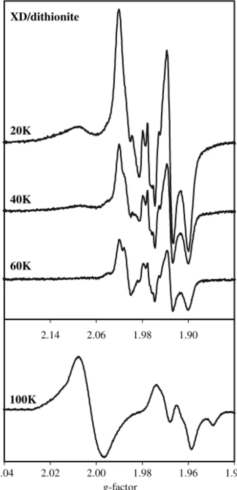
EPR spectral properties of reduced rat liver XOR
Since each of the redox centres of XOR is paramagnetic in one of its oxidation states, the reduction profiles of FADH•, Mo(V) and reduced Fe/S centres may be studied by EPR spectroscopy [38]. To characterise the EPR signals of re-dox centres of XD and XO forms of rat liver XOR, the purified enzymes were reduced with an excess of dithionite for 30 min, and the X-band EPR spectra were acquired at different temperatures (20–100 K). The EPR spectra of the dithionite-reduced XD form (Fig.4) and XO form (not shown) were qualitatively similar and showed the signals from FADH•, two Fe/S and Mo(V) centres. These signals were simulated individually at 20 and 100 K (Fig.5) and the EPR parameters found are presented in Tables 2and3. The signals obtained are closely related to those observed and described for bovine milk XO and for avian liver XD [39–45]. Both active and desulfo enzymes are reduced simultaneously by dithionite and, accordingly, the Mo(V)
0 1 2 3 4 5
0 25 50 75 100
[NADH] (µM)
v
(µ
oit
a
di
x
o
H
D
A
N
f
o
)
ni
m/
Mn
human liver XO
0 10
-100 -50 0 50
Kmapp (µM)
V
p
p
a
(µ
)
ni
m/
M
Fig. 2 NADH oxidation catalysed by human liver XO. Initial rates of
NADH oxidation, catalysed by human liver XO, were measured spectrophotometrically at 340 nm, in air-equilibrated Tris–HCl 50 mM buffer pH 7.8. Thehyperbolic curvewas generated with the parameters given in Table1. The apparent kinetics parameters were estimated by the direct linear plot shown in theinset
0 25 50 75 100
0 25 50 75 100
[initial NADH] (µM)
)
%
(
n
oit
c
u
de
r
n
ort
ce
le
-e
n
o
ne
g
y
x
O
XO
XD
Fig. 3 Percentage of one-electron oxygen reduction, catalysed by
both XD and XO forms of rat liver XOR during NADH oxidation. The percentage of one-electron oxygen reduction was calculated to be half of the ratio of the O2
•–formation rate to the NADH oxidation
signal was simulated as a mixture of slow and rapid type 1. The isotropic splitting (D= 1.2 mT) of the Mo(V) signals, observed at temperatures of or below 60 K (with or without the Fe/S II reduced), results from the magnetic interaction between the Mo(V) and the Fe/S I centre, as observed by Lowe et al. [46] with bovine milk XO. Also, no interactions were detected between Mo(V) and Fe/S II or between FAD and Mo(V).
Both XD and XO forms of rat liver XOR were also reduced with increasing quantities of NADH. The EPR spectra at 20 and 100 K of NADH-reduced XD (Fig.6) and XO (not shown) were qualitatively similar to the dithionite-reduced spectra. The EPR signals from NADH-reduced enzymes were simulated individually and the EPR parameters determined are shown in Tables2 and3. The simulation of the Mo(V) signal, as a mixture of slow and rapid type-1 signals, was consistent with the known
simultaneous reduction by NADH of active and desulfo enzymes, via the flavin (with subsequent reduction of Mo(V) [47]. In addition, the Mo(V) signals were also found to split (D= 1.2 mT) in the presence of the Fe/S I centre (at temperatures below 60 K). The spectra were, however, quantitatively quite different, with the extent of the XD reduction being higher than that of XO. Figure7 summa-rises the results of the reductive titration experiments.
The spectra of deflavo-XD in the presence of NADH display no EPR signals (with enzyme-to-NADH molar ratios of 1:500 or less), indicating that the NADH reduction of XD occurs via FAD. In the presence of dithionite, the deflavo enzyme was reduced and the resulting EPR spectra were similar to those of dithionite-reduced XD and XO (Fig.8), showing the Fe/S centres and Mo(V) signals, but without the flavin signal.
Discussion
NADH oxidase activity of XOR has long been recognised [48–50], but only recently was its potential as a source of ROS realised. Most interest was mainly focused on free-radical metabolism, mediated through hypoxanthine/xan-thine oxidation by XO, in biochemical mechanisms asso-ciated with a wide variety of human diseases. This ‘‘new’’ ROS-generating pathway, of XOR and NADH, must then be reevaluated in those clinical conditions where an in-crease in NADH is expected, such as in diseases where hypoxic and reperfusion cycles exist, in ethanol hepato-toxicity and in diabetes [12,14,51–54].
To characterise this potential mammalian liver source of ROS, NADH oxidase activity was studied using both XD and XO forms of rat liver XOR. In addition to the fact that mammalian liver enzymes have been poorly studied, other reasons justify the further investigation of liver XOR. The specific activity of human XOR is high in liver and when liver damage occurs, there is an increase in the levels of circulating enzyme, which can bind to vascular endothe-lium, causing injury [14]. For the diseases referred to above, it seemed more meaningful to carry out studies with liver enzymes than with milk enzymes. Rat liver enzymes, for instance, are kinetically quite different from the human milk enzymes, which show little XOR activity [24], thus hindering comparative studies between xanthine and NADH (physiological reducing substrates) as a ROS source. In addition, a direct comparison between XD and XO forms from the same source can be made. Furthermore, the recently reported XOR-catalysed production of•NO, in the presence of nitrite, with either NADH or xanthine as reducing substrates under anaerobic conditions, undoubt-edly contributed to further stimulate interest in the physi-ological and pathphysi-ological roles of the enzyme. XOR, unlike 1.90
1.98 2.06 2.14 XD/dithionite
40K
60K 20K
1.94 1.96 1.98 2.00 2.02 2.04
g-factor 100K
Fig. 4 Electron paramagnetic resonance (EPR) spectra of the
1.90 1.98
2.06 2.14 2.22
g-factor
XD/dithionite, 20K
sim. Mo (V)-slow sim. FADH. sim. Fe/S II sum sim.
sim. Mo(V)-rapid sim. Fe/S I experim.
1.94 1.96
1.98 2.00 2.02
g-factor
XD/dithionite, 100K
sim. Mo(V)-slow sim. FADH. sum sim.
sim. Mo(V)-rapid experim.
Fig. 5 Simulation of the EPR
spectra of the dithionite-reduced XD form of rat liver XOR at 20 and 100 K. Each EPR signal of dithionite-reduced XD, at 20 and 100 K, was simulated individually with the parameters given in Tables2and3. The experimental spectra were recorded with the experimental conditions indicated in Fig.3
Table 2 Electron paramagnetic resonance (EPR) signal parameters of the Fe/S II, Fe/S I and FADH•from NADH and dithionite-reduced rat
liver XD and XO (at 20 K)
Signal Reducing substrate Enzyme g1[line width (mT)] g2[line width (mT)] g3[line width (mT)]
Fe/S II NADH XD or XO 2.0945 (3.00) 1.9910 (2.50) 1.8895 (2.25)
Dithionite XD 2.0945 (4.00) 1.9910 (2.50) 1.8910 (2.50)
Dithionite XO 2.1120 (5.50) 1.9910 (4.50) 1.8910 (4.00)
Fe/S I NADH XD or XO 2.0210 (1.90) 1.9315 (1.57) 1.8990 (2.375)
Dithionite XD 2.0210 (1.90) 1.9320 (1.57) 1.8995 (2.37)
Dithionite XO 2.0215 (1.90) 1.9327 (1.57) 1.8987 (2.37)
Signal Reducing substrate Enzyme g[line width (mT)]
FADH•
NADH XD or XO 2.0055 (3.60)
Dithionite XD 2.0053 (4.80)
Dithionite XO 2.0060 (1.95)
FADHreduced flavin adenine dinucleotide,gline width
Table 3 EPR signals parameters of the Mo(V) from NADH and dithionite-reduced rat liver XD and XO (at 20 K)
Signal Reducing substrate
g1[line width (mT)]
g2[line width (mT)]
g3[line width (mT)]
A1H(mT) A2H(mT) A3H(mT) D1,2,3(mT)
Rapid NADH 1.9980
(0.65)
1.9723
(0.45)
1.9672
(0.35)
1.000
0.500
1.500 1.532 1.200
Dithionite 1.9980
(0.65)
1.9723
(0.45)
1.9672
(0.35)
1.000
0.500
1.500
0.300
1.532
0.250
1.200
Slow NADH 1.9711
(0.40)
1.9664
(0.30)
1.9557
(0.60)
1.450 1.600 1.400 1.200
Dithionite 1.9719
(0.60)
1.9668
(0.50)
1.9566
(0.60)
1.450
0.160
1.600
0.160
1.600
0.160
•
NO synthase, can be an important source of •NO in ischaemia, thus promoting the•NO-induced vasodilatation [17,18].
EPR spectra of NADH-reduced XD and XO were quite useful in order to define the spectral parameters of the EPR signals detected for reduced iron–sulfur centres and Mo(V)
species. The data make quite clear, as discussed below, that NADH efficiently reduces XD but a large excess of NADH is needed to efficiently reduce XO.
The kinetics studies carried out to compare the NADH oxidation catalysed by the rat liver XD and XO forms of rat liver XOR demonstrated that both enzymes do catalyse NADH oxidation with O2•– generation. It is, however, significant that the XD form is a better catalyst for NADH oxidation than is the XO form, having a higher specificity constant, whether determined by NADH consumption (8.73 ± 1.36 and 1.07 ± 0.09lM–1min–1, respectively) or
by O2•– formation (8.40 ± 0.73 and 1.10 ± 0.06 lM–1
min–1, respectively).
The percentage of one-electron oxygen reduction ca-talysed by rat liver XOR was found to be constant as the NADH concentration increased by 50%, indicating that one molecule of O2•– is formed per NADH molecule. These results are clearly different from those described for xan-thine oxidation by XO, where the fraction of O2•–formed decreases as xanthine concentration increases [8,9,56,57]. Therefore, the chemical equation for the NADH oxidation reaction can be written as
2NADHþ3O2!2NADþþ2O2þH2O2: ð1Þ In addition, it must be emphasised that the kinetics parameters obtained for human liver XO towards NADH oxidation were found to be very similar to those of XO forms of rat liver XOR. Considering the important physi-ological and pathphysi-ological roles of human XOR, these
re-1.90 1.98 2.06
2.14 2.22
g-factor
XD/NADH, 20K
XD/NADH 1/4
XD/NADH 1/2 XD/NADH 1/6 XD/NADH 1/14
1.94 1.96
1.98 2.00
2.02
g-factor
XD/NADH, 100K
XD/NADH 1/4
XD/NADH 1/2 XD/NADH 1/6 XD/NADH 1/14
Fig. 6 EPR spectra of the XD
form of rat liver XOR reduced with increasing quantities of NADH at 20 and 100 K. The enzyme (40 nmol) was reduced anaerobically, for about 5 min, with NADH, at the XD-to-NADH ratios indicated, and the EPR spectra were recorded at 20 and 100 K. Other experimental conditions were as follows: microwave frequency, 9.65 GHz; microwave power, 0.0635 mW; modulation amplitude, 4.05 G; receiver gain, 2.00·105
Fig. 7 Anaerobic titration of both XD and XO forms of rat liver
sults enable us to use rat liver XOR as a first approach in studies whose results are intended to be extrapolated to the human enzyme.
In agreement with the kinetics parameters obtained for both enzyme forms, much higher intensity EPR spectra were observed with NADH-reduced XD than with XO. While FAD removal completely hampers enzyme reduc-tion by NADH (no EPR signals were observed) and abol-ishes NADH oxidase activity, the kinetics reaction profiles obtained with inactive (desulfo and demolybdo) and allo-purinol-inhibited enzymes were identical to those of native enzymes. These results show that the NADH reduction of rat liver XOR occurs via FAD and that alterations at the molybdenum centre do not affect the NADH oxidation kinetics.
With NADH as reducing substrate, the XD form is also more efficient than XO in catalysing the reduction of nitrite to•NO [17]. This fact is particularly relevant in vivo, since the XD form is the predominant form in vivo.
Some studies with xanthine as a reducing substrate were carried out (data not shown), and demonstrated that rat liver XD is a better O2•– source than XO, as previously described [1, 33, 55]. The dehydrogenase form of liver XOR is thus intrinsically more efficient at generating O2•– than the oxidase form, independently of the reducing substrate. These are remarkable results, in light of the common association of ROS-mediated damage with oxi-dase enzymes. Therefore, XO is not essential for XOR-catalysed generation of ROS, overcoming the increasingly questioned conversion of XD to XO.
Many pathological conditions arise from free-radical oxidation of DNA, proteins and lipids, and the products of lipid peroxidation; in particular, aldehydes, directly or indirectly, affect many functions integral to cellular homeostasis [58]. Taking into account the ability of XOR to catalyse O2•–/H2O2generation, it is expected that XOR may induce the peroxidation of lipids. In fact, we have shown that rat liver XOR can promote the oxidation of lipids during NADH:O2and xanthine:O2turnover [59]. In addition, higher peroxidation extents were observed in the presence of NADH, when compared with equimolar con-centrations of xanthine, and the rat liver XD form was found to be the most efficient at promoting this oxidative process. Compared with XOR, AO seems to play a minor role in NADH-dependent liposomal lipid damage, which is in agreement with the comparatively low values of the kinetics parameters found for O2•–formation.
Moreover, a new XOR-dependent route for the genera-tion of peroxynitrite, a strong inflammatory oxidant, was described [19]. Both •NO and O2•– can be produced by XOR, but the efficiency of ONOO–production depends on the presence or absence of superoxide dismutase and on the rate of O2•–generation. Our results have shown that the XD form of rat liver XOR is more effective at generating O2•– than is the XO form. Therefore, following the same rea-soning as Godberg et al. [19], in the presence of oxygen, the XD form will have fewer electrons available for the reduction of nitrite than will XO, and it will generate less ONOO–. This condition is more consistent with the likely role of the predominant in vivo XD form in physiological function.
On the whole, we conclude that XOR can be a source of both ROS and reactive nitrogen species, which can induce destructive and useful effects, but the physiological and pathological implications of this complex enzyme are still not completely clarified.
It is worth noting that our results, obtained with rat liver XOR and bovine milk XO, show that enzymes from dif-ferent sources have difdif-ferent kinetics properties. Never-theless, the kinetics parameters of human liver XO for NADH oxidation were, indeed, very similar to those of rat liver XO and we believe that these results foresee potential 1.90 1.82
1.98 2.06 2.14 2.22
Deflavo-XD/dithionite
30K 10K
1.94 1.96
1.98 2.00
g-factor
100K
Fig. 8 EPR spectra of the dithionite-reduced deflavo-XD form of rat
applications of rat liver enzyme XOR as a model for the human liver enzymes.
References
1. Hille R, Nishino T (1995) FASEB J 9:995–1003 2. Hille R (2005) Arch Biochem Biophys 433:107–116
3. Brondino CD, Romao MJ, Moura I, Moura JJG (2006) Curr Opin Chem Biol 10:109–114
4. Hille R (2006) Eur J Inorg Chem 1913–1926
5. Amaya Y, Yamazaki K, Sato M, Noda K, Nishino T, Nishino T (1990) J Biol Chem 265:14170–14175
6. Nishino T, Nishino T (1997) J Biol Chem 272:29859–29864 7. Enroth C, Eger BT, Okamoto K, Nishino T, Nishino T, Pai E
(2000) Proc Natl Acad Sci USA 97:10723–10728
8. Rubbo H, Radi R, Prodanov E (1991) Biochem Biophys Acta 1074:386–391
9. Hausladen A, Fridovich I (1993) Arch Biochem Biophys 304:479–482
10. Suzuki YJ, Forman HJ, Sevanian A (1997) Free Radic Biol Med 22:269–285
11. Babior MB (2000) Am J Med 109:33–44
12. Nishino T, Nakanishi S, Okamoto K, Mizushima J, Hori H, Iwasaki T, Nishino T, Ichimori K, Nakazawa H (1997) Biochem Soc Trans 25:783–786
13. Wright RM, Repine JE (1997) Biochem Soc Trans 25:799–804 14. Harrison R (2002) Free Radic Biol Med 33:774–797
15. Mira L, Maia L, Barreira L, Manso CF (1995) Arch Biochem Biophys 318:53–58
16. Sahinoglu T, Stevens CR, Bhatt B, Blake DR (1996) Methods 9:628–634
17. Godberg BLJ, Doel JJ, Sapkota GP, Blake DR, Stevens CR, Eisenthal R, Harrison R (2000) J Biol Chem 275:7757–7763 18. Li H, Samouilov A, Liu X, Zweier JL (2001) J Biol Chem
276:24482–24489
19. Godberg BLJ, Doel JJ, Durgan J, Eisenthal R, Harrison R (2000) FEBS Lett 475:93–96
20. Palmer R, Ferrige A, Moncada S (1987) Nature 327:524–526 21. Beckman JS, Beckman TW, Chen J, Marshall PA, Freeman BA
(1991) Proc Natl Acad Sci USA 87:1620–1624
22. Krenitsky TA, Neil SM, Elion GM, Hitchings GH (1972) Arch Biochem Biophys 150:585–599
23. Komai H, Massey V, Palmer G (1969) J Biol Chem 244:1692– 1700
24. Sanders SA, Eisenthal R, Harrison R (1997) Eur J Biochem 245:541–548
25. Williamson JR (1966) J Biol Chem 241:5026–5036 26. Lieber CS (1988) N Engl J Med 319:1639–1650
27. Lieber CS, Savollainem M (1984) Alcohol Clin Exp Res 8:409– 423
28. Hille R (1996) Chem Rev 96:2757–2816
29. Waud WR, Rajagopalan KV (1976) Arch Biochem Biophys 172:354–364
30. Maia L, Mira L (2002) Arch Biochem Biophys 400:48–53 31. Saito T, Nishino T (1989) J Biol Chem 264:10015–10022 32. Johson JL, Waud WR, Cohen HJ, Rajagopalan KV (1974) J Biol
Chem 249:5056–5061
33. Nishino T, Nishino T, Schopfer LM, Massey V (1989) J Biol Chem 264:2518–2527
34. Harris CM, Massey V (1997) J Biol Chem 272:8370–8379 35. Branzoli U, Massey V (1974) J Biol Chem 249:4339–4345 36. Cornish-Bowden A (1995) Fundamentals of enzyme kinetics.
Portland, London
37. Fridovich I (1986) In: Greenwald RA (ed) Handbook of methods for oxygen radical research. CRC, Boca Raton
38. Hille R, Massey V (1985) In: Spiro TG (ed) Molybdenum en-zymes. Wiley, New York, pp 443–518
39. Palmer G, Massey V (1969) J Biol Chem 244:2614–2620 40. Bray RC, Vanngard T (1969) Biochem J 114:725–734
41. Hille R, Hagen WR, Dunham WR (1985) J Biol Chem 260:10569–10575
42. Bray RC, Barber MJ, Lowe DJ (1978) Biochem J 171:653–658 43. Gutteridge S, Tanner SJ, Bray RC (1978) Biochem J 175:887–
897
44. Barber MJ, Bray RC, Lowe DJ, Coughlan MP (1976) Biochem J 153:297–307
45. Barber MJ, Coughlan MP, Kanda M, Rajagopalan KV (1980) Arch Biochem Biophys 201:468–475
46. Lowe DJ, Lynden-Bell RM, Bray RC (1972) Biochem J 130:239– 249
47. Swann JC, Bray RC (1972) Eur J Biochem 26:407–415 48. Murray KN, Chaykin S (1966) J Biol Chem 241:3468–3473 49. Landon EJ, Myles M (1967) Biochem Biophys Acta 143:429–431 50. Massey V, Brumby PE, Komai H, Palmer G (1969) J Biol Chem
244:1682–1691
51. Harrison R (1997) Biochem Soc Trans 25:786–791
52. Wright RM, McManaman JL, Repine JE (1999) Free Radic Biol Med 26:348–354
53. Kato S, Kawase T, Alderman J, Inatomi N, Lieber C (1990) Gastroenterology 98:203–210
54. Chung SSM, Ho ECM, Lam KSL, Chung SK (2003) J Am Soc Nephrol 14:S233–S236
55. Hunt J, Massey V (1992) J Biol Chem 267:21479–21485 56. Fridovich I (1970) J Biol Chem 245:4053–4057
57. Porras AG, Olson JS, Palmer G (1981) J Biol Chem 256:9096– 9103


![Table 3 EPR signals parameters of the Mo(V) from NADH and dithionite-reduced rat liver XD and XO (at 20 K) Signal Reducing substrate g 1 [line width(mT)] g 2 [line width(mT)] g 3 [line width(mT)] A 1 H (mT) A 2 H (mT) A 3 H (mT) D 1,2,3 (mT) Rapid NADH 1.9](https://thumb-eu.123doks.com/thumbv2/123dok_br/16472399.731754/8.892.76.819.862.1065/table-signals-parameters-dithionite-reduced-signal-reducing-substrate.webp)


