PERITONITIS DUE TO TRANSVERSE COLON PERFORATION: A RARE
PRESENTATION
Vandana B. Giriradder1, Veerendraswamy S. M2, Sagar K3, Ravikanth J4
HOW TO CITE THIS ARTICLE:
Vandana B. Giriradder, Veerendraswamy S. M, Sagar K, Ravikanth J. “Peritonitis Due to Transverse Colon Perforation: A Rare Presentation”. Journal of Evidence based Medicine and Healthcare; Volume 2, Issue 35, August 31, 2015; Page: 5538-5546, DOI: 10.18410/jebmh/2015/767
ABSTRACT: Abdominal tuberculosis is defined as infection of the peritoneum, hollow or solid abdominal organs with Mycobacterium tuberculi. TB can affect any part of the gastrointestinal (GI) tract including anus, peritoneum and hepatobiliary system. The peritoneum and the ileocaecal region are the most likely sites of infection. The clinical manifestations of abdominal tuberculosis are nonspecific and mimic various GI disorders and cause delay in diagnosis and management. This pathology has several complications, including free intestinal perforation.
KEYWORDS: Transverse colon, Perforation, Abdominal tuberculosis, Peritonitis.
INTRODUCTION: Tuberculosis is endemic in India, and involvement of the gastrointestinal tract is frequently encountered. Abdominal tuberculosis (TB) is the sixth commonest extra-pulmonary TB form after lymphatic, genitourinary, bone and joint, miliary and meningeal tuberculosis. TB may involve any site of the gastrointestinal tract, but the commonest site of involvement is ileocecal region.[1] On the other hand, isolated extra-caecal colonic involvement of tuberculosis is
uncommon. While perforation is a serious and uncommon complication of abdominal TB and the reported incidence of free perforation in patients with tubercular enteritis varies from 0-11%, perforation due to tubercular colitis is virtually unknown.[2]
The authors present a case of colonic TB which was manifested by intestinal perforation.
CASE REPORT: 28 years old female patient XYZ presented to casualty. With chief complaints of pain abdomen since 1 month, insidious in onset, diffuse, gradually progressive, spasmodic in nature, severe since 3 days, no aggravating and relieving factors. Also complains distension of abdomen since 3 days, insidious in onset, gradually progressive.
Complains of fever since 2 weeks insidious in onset, moderate degree, intermittent, not associated with chills and rigors. History of not passing stools since 2 days.
No history of vomiting, no history of burning micturition. History of irregular menstrual cycle. Patient was nulliparous with married life of 9 years. No history of similar complains in the past, no history of diabetes, hypertension and tuberculosis.
On examination patient was conscious, oriented, vitals stable, anemic, not jaundiced, no lymphadenopathy. Per abdomen examination revealed distended abdomen, diffuse tenderness with guarding and rigidity, free fluid present, bowel sounds enhanced. Per rectal examination showed roomy rectum, no faecal impaction, normal faecal staining present. Respiratory system, Cardiovascular system and Central nervous system examination found normal.
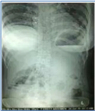
function tests, Serum electrolytes, Liver function tests, S.amylase & S.lipase were within normal limits. Erect x-ray abdomen showed air under diaphragm (Figure 1).
Outside USG scan revealed Mild spleenomegaly (? Infectious), Right ovarian 3.3x2.7cm and left ovarian 3.1x2.8 cm haemorrhagic cysts, Very minimal ascites and mild prominent bowel loops (correlate with erect abdomen X-ray to rule out hollow viscus perforation/ obstruction.)
Under the provisional diagnosis of Perforative Peritonitis patient was resuscitated and subjected to emergency Exploratory Laparotomy on 24/1/15 which revealed phlegminous mass with omental adhesions. Large mass around 10x10 cms arising from distal part of Transverse colon to the extent of splenic flexure with multiple perforations. (Figure 2) Perforation posteriorly formed an abscess in lesser sac of stomach with accumulation of frank pus. Multiple mesenteric lymph nodes enlarged, firm in consistency. Planned to go ahead with Left Hemicolectomy.
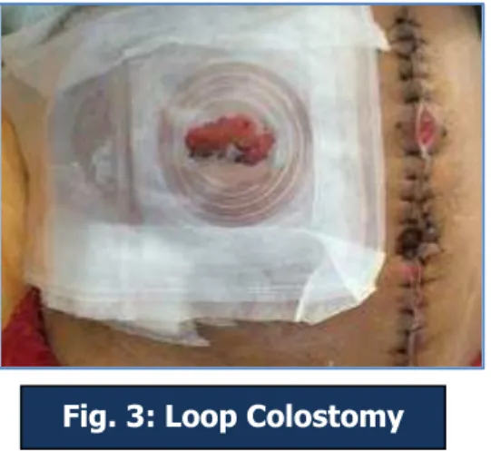
Intra operatively mass was separated from omental adhesions then lesser sac accessed. Pus samples were taken and sent for culture sensitivity. Left Hemicolectomy and Transverse loop colostomy (Figure 3) followed by Hartmann’s procedure done. Post-operatively patient was on ventilator support for 2 days. Treated with appropriate antibiotics, analgesics and supportive
Fig. 1: Air under the right dome of diaphragm
stitch abscess let it out. Abdominal drains removed on post-operative day 6. Patient had fever for 1 day on post-operative day 7.
Culture sensitivity of peritoneal abscess sample: Klebsiella species isolated which was treated empirically by spectrum antibiotics.
Macroscopic specimen showed constriction at a distance of 6cm from one resected margin with an ulcerated lesion measuring 7x4cm. Another constricted segment noted measuring 4x2.5cm at 5cm distance from other resected end. 9 lymph nodes were isolated largest measuring 1x1cm.
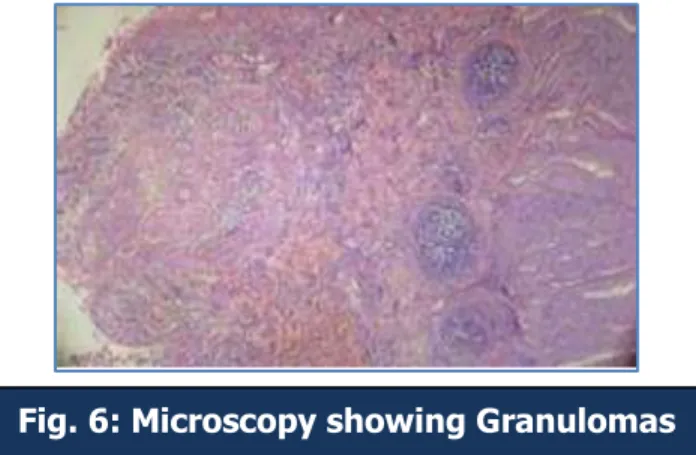
Microscopic examination revealed focally ulcerated mucosa and submucosa showing several confluent epitheloid cell granulomas. Granulomas showed central area of necrosis
bordered by epitheloid cells, langhan’s type of giant cells and lymphocytes. These granulomas
were seen infiltrating up to the serosal surface. Lymphnodes showing confluent granulomas having central area of necrosis bordered by epitheloid cells, langhan’s type of giant cells and lymphocytes.
Fig. 3: Loop Colostomy
CATEGORY –I ATT was started on 06/02/15 on advice of RNTCP. Patient is currently doing well after follow up of 5 months.
DISCUSSION: Tuberculosis is a chronic granulomatous disease caused by aerobic bacteria Mycobacterium tuberculosis. It remains the worldwide problem despite the discovery of the causative organism for more than a century ago. Pulmonary tuberculosis is the most common form and it primarily involves the lung but any part of the body can be involved by the disease.[3,4] Abdominal tuberculosis (TB) constitutes a major public health problem in developing
countries and associated with significant morbidity and mortality.[5,6] The incidence of abdominal
TB is increasing in western and developed countries due to immigration from developing countries, aging populations, increasing incidence of human immunodeficiency virus (HIV) infection, and misdiagnosis with ineffectual treatment.[7,8] For these reasons, abdominal TB
represents an interesting challenge for surgeons in developed countries as well.
Tuberculosis (TB) can involve any part of the gastrointestinal tract from mouth to anus, the peritoneum and the pancreatobiliary system. It can have a varied presentation, frequently mimicking other common and rare diseases.[9] The clinician must look for tuberculosis, and
confirm or exclude this treatable malady in any patient who presents with gastrointestinal disease.
Abdominal TB is a disease that predominantly affects young adults. Two-thirds of all cases involve patients between 21 and 40 years of age. There is no difference in the incidence rate between male and female subjects, although some studies suggest a slightly increased female predisposition.[10]
The postulated mechanisms by which the tubercule bacilli reach the gastrointestinal tract are: (i) hematogenous spread from the primary lung focus in childhood, with later reactivation; (ii) ingestion of bacilli in sputum from active pulmonary focus; (iii) direct spread from adjacent organs; and (iv) and through lymph channels from infected nodes.[10]
The earlier belief that most cases are due to reactivation of quiescent foci is being challenged with a recent study using DNA fingerprinting showing that 40 per cent cases are due
Clinical presentation can be acute, chronic, or both acute and chronic. In the majority of cases, constitutional symptoms are present, including fever (40-70%), pain (80-95%), diarrhea (11-20%), constipation, alternating constipation and diarrhea, weight loss (40-90%), anorexia, malaise, ascites, abdominal distension, night sweating, and hematochezia.[7,10,13] Despite a
popular misconception that intestinal complications are often linked to pulmonary TB, only 15- 20% of the patients with gastrointestinal tubercular complications have concomitantly active pulmonary TB. Thirty to 50% of patients with abdominal TB have a normal chest film.[13]
Colonic tuberculosis can present in several forms. Segmental tuberculosis of transverse colon is a rare malady.[14,15,16,17] The most common involvement is in the form of segmental ulcers
and colitis, inflammatory strictures and hypertrophic lesions resembling polyps or masses.[18,19]
The hypertrophic form causes a diagnostic dilemma at the time of surgery because it mimics colonic carcinoma,[19] a problem which was encountered in the present case.
Histopathologic findings are very informative in diagnosis of intestinal TB. Characteristic histological features are submucosal and serosal caseating granuloma. Generally, it is recommended to interpret histopathologic findings in conjunction with culture or PCR results in order to enhance diagnostic accuracy.[20, 21]
There are several complications involving intestinal TB, including bowel obstruction (31.7%), intestinal perforation (4.9%), enterocutaneous fistula (2.4%), and small bowel volvulus resulting from mesenteric lymphadenitis (2.4%).[22] Different studies typically denote different
percentages for these complications.[23]
It accounts for 1-10% of abdominal TB cases and has a poor prognosis, with a mortality rate higher than 30%.[24, 25, 13]
Even if a patient presents with all the appropriate signs and symptoms, there is little conclusive evidence that alludes to intestinal TB perforation. The percentage of intestinal perforation due to abdominal TB is very low, even in countries with high TB incidence. In 2006, Jhobta et al.,[26] reported that 3.9% of peritonitis cases in a series of 504 consecutive intestinal
perforations resulted from TB-induced intestinal perforation. Another article, considering a series of 204 consecutive patients, reported only 4 cases (1.9%) of intestinal perforation resulting from TB.[27] It should be pointed out that incidence rates have decreased significantly in the past 20
years. In 1986, Kapoor et al.,[28] reported a series of 6 cases with an incidence of TB-induced
perforation of 13.3%. In those years, the percentages of TB-induced intestinal perforations ranged from 7.5% to 12.2%.[26, 28, 29]
Tubercular perforations are usually ileal and are associated with distal strictures. In a study by Nagi et al., [30] only 10.8% of the 684 cases with abdominal TB had TB affecting the
colon. Wig et al., previously described 10 such cases encountered over a period of 7 years.[31]
Patients with free perforation of a tuberculous colon are extremely ill, and immediate surgical intervention is essential after a brief period of intensive resuscitation. The principles of restoration of bowel continuity remain the same as those following colonic resection for any other disease. Anti-tubercular drugs remain the mainstay of treatment after histopathological confirmation of the diagnosis.
The diagnosis of intestinal perforation is not difficult in most cases. For the surgeon, the difficulty arises when trying to reach an intra-operative diagnosis and when subsequently planning appropriate treatment for the patient. Surgical treatment of tuberculous perforations is rather controversial. Although pharmacological treatment remains the central pillar of abdominal TB, emergency surgery is often required for its acute complications[32] particularly for
perforations. There is very little scientific evidence with which to base an argument for the best way to treat these complications of abdominal TB, and the noticeable lack of literature in this field certainly does not help.
The bulk of this topic’s background is derived from a series of case reports and case series published in a 40-year period. In all, 119 cases have been described, with various means of treatment. In many of the reported cases, the surgical treatment and the outcomes were not adequately described. However, direct closure of the perforation with or without bypass is generally associated with poorer results.[33] Resection and anastomosis is therefore
recommended,[34] especially if combined with postoperative anti-tubercular therapy.[35,36,37]
However, regardless of the surgical procedure, the mortality rate is relatively high, ranging from 30%[33,34] to 60%.[13] This high rate of mortality is principally due to the poor clinical perioperative
conditions of patients undergoing surgery. In addition to already being a highly debilitating
pathology, the peritonitis resulting from the perforation overwhelms the patient’s capacity to bear
surgical stress. In all the reviewed literature, very few reports are found that resembled our case. We believe that our course of action in this case was a safe and responsible way to approach the problem.
In the presence of extensive adhesions, no attempt should be made to locate the perforation, as injury to the adherent intestinal loops is likely and focal fistula may result in the postoperative period. [28] In this case, it is possible to place drains and immediately begin an
ex-juvantibus anti-tubercular therapy once the surgical intervention has been completed.
REFERENCES:
1. Sarrami AH, Sharifi M, Ahsan M, Afsharmoghaddam N. Multiple intestinal perforations as a primary manifestation of abdominal tuberculosis in a HIV-infected patient. JSCR. 2010 10:7 2. Narendra Mohan Gupta, Tashi Motup, Kusum Joshi. Isolated Colonic Tuberculous
Perforation as a Rare Cause of peritonitis: Report of a Case. Jpn J Surg (1999), 29: 273-275 3. Park K, editor. Park’s textbook of Preventive & Social Medicine. 19th ed. India: M/S
Banarasidas Bhanot; 2007. p. 768.
4. Kapoor VK. Abdominal tuberculosis: The Indian contribution. Indian J Gastroenterol.
1998;17:141–47.
5. Global tuberculosis control 2012. [Internet]: World Health Organization. (Online) http://www.who.int/tb/publications/global_report/en/index.html Cited 2011 April 11.
6. Wadhwa N, Agarwal S, Mishra K. Reappraisal of abdominal tuberculosis. J Indian Med
Assoc. 2004;102:31–32
7. Tanrikulu AC, Aldemir M, Gurkan F, Suner A, Dagli CE, Ece A. Clinical review of tuberculous peritonitis in 39 patients in Diyarbakir, Turkey. J Gastroenterol Hepatol 2005; 20:906-9. 8. Lingenfelser T, Zak J, Marks IN, Steyn E, Halkett J, Price SK. Abdominal tuberculosis: still a
potentially lethal disease. Am J Gastroenterol 1993; 88:744-50.
9. Peda Veerraju E. Abdominal tuberculosis. In: Satya Sri S, editor. Textbook of pulmonary and extrapulmonary tuberculosis. 3rd ed. New Delhi: Interprint; 1998 p. 250-2.
10.M.P. Sharma, Vikram Bhatia. Abdominal Tuberculosis. Indian J Med Res 120, October 2004, pp 305-315
11.Wig KL, Chitkara NK, Gupta SP, Kishore K, Manchanda RL. Ileoceacal tuberculosis with particular reference to isolation of Mycobacterium tuberculosis. Am Rev Respir Dis 1961; 84: 169-78.
12.Vij JC, Malhotra V, Choudhary V, Jain NK, Prasaed G, Choudhary A, et al. A clinicopathological study of abdominal tuberculosis. Indian J Tuberc1992; 39: 213-20. 13.Clarke DL, Thomson SR, Bissetty T, Madiba TE, Buccimazza I, Anderson F. A single surgical
unit’s experience with abdominal tuberculosis in the HIV/AIDS era. World J Surg 2007; 31:1087-98.
14.Chawla S, Mukherjee P, Ber K: Segmental tuberculosis of the colon (a report of 10 cases). Clin Radiol 22: 104- 109, 1971
15.Crohn BB, Yarnis H: Primary Ileocaecal Tuberculosis. NY State Med 40: 158-166, 1940 16.Kolawole TM, Lewis EA: A radiologic study of tuberculosis of the abdomen (gastrointestinal
tract) AJR123:348-358, 1975
17.Werbeloff L, Novis BH, Banks S, Marks IN: The radiology of tuberculosis of the gastrointestinal tract. Br Radiol 46: 329-336, 1973
18.Singh V, Kumar P, Kamal J, Prakash V, Vaiphei K, Singh K. Clinicocolonoscopic profile of colonic tuberculosis. Am J Gastroenterol 1996; 91: 565-568.
19.Devanesan JD, Sable RA, Pitchumoni CS, Lev R, Zapiach L. Segmental tuberculosis of the colon mimicking carcinoma. Arch Surg 1980; 115: 90-91
21.Jin XJ, Kim JM, Kim HK, Kim L, Choi SJ, Park IS et al. Histopathology and TB-PCR kit analysis in differentiating the diagnosis of intestinal tuberculosis and Crohn's disease. World J Gastroenterol 2010; 16(20):2496-2503
22.Akinoğlu A, Bilgin I. Tuberculous enteritis and peritonitis. Can J Surg 1988; 31: 55-8. 23.Marks IN. Abdominal tuberculosis. Ballieres Clin Med Comunicable Dis 1988; 3: 329-48. 24.Bhansali SK. Abdominal tuberculosis. Experiences with 300 cases. Am J Gastroenterol 1977;
67: 324-37.
25.Segal I, Tim LO, Mirwis J, Hamilton DG, Mannell A. Pitfalls in the diagnosis of gastrointestinal tuberculosis. Am J Gastroenterol 1981; 75: 30-5.
26.Jhobta RS, Attri AK, Kaushik R, Sharma R, Jhobta A. Spectrum of perforation peritonitis in India-review of 504 consecutive cases. World J Emerg Surg 2006; 1: 26.
27.Khanna AK, Misra MK. Typhoid perforation of the gut. Postgrad Med J 1984; 60: 523-5. 28.Kapoor VK, Kriplani AK, Chattopadhyay TK, Sharma LK. Tuberculous perforations of the
small intestine. Ind J Tub 1986; 33: 188-9.
29.Kakar A, Aranya RC, Nair SK. Acute perforation of small intestine due to tuberculosis. Aust N Z J Surg 1983; 53: 381-3.
30.Nagi B, Kochhar R, Bhasin DK et al. Colorectal tuberculosis. Eur Radiol2003; 13: 1907– 1912.
31.J D Wig, A K Malik, A Chaudhary, N M Gupta. Free perforation of tuberculous ulcers of the small bowel. Indian J Gastroenterol 1985; Vol 4 No 4: 259-261
32.Tan KK, Chen K, Sim R. The spectrum of abdominal tuberculosis in a developed country: a
single institution’s experience over 7 years. J Gastrointest Surg 2009; 13: 142-7.
33.Bhansali SK, Desai AN, Dhaboowala CB. Tuberculous perforation of the small intestine. A clinical analysis of 19 cases. J Assoc Physicians India 1968; 16: 351-5.
34.Aston NO, de Costa AM. Tuberculous perforation of the small bowel. Postgrad Med J 1985; 61: 251-2.
35.Salvati V, Fumo F, D’Armiento FP, Fumo M, Cerrone C. Primary intestinal tuberculosis. Minerva Chir 1996; 51:567-71.
36.Shah S, Thomas V, Mathan M, Chacko A, Chandy G, Ramakrishna BS, et al. Colonoscopic study of 50 patients with colonic tuberculosis. Gut 1992; 33:347-51.
4. Post Graduate, Department of General Surgery, SSIMS & RC, Davangere.
NAME ADDRESS EMAIL ID OF THE CORRESPONDING AUTHOR: Dr. Vandana B. Giriradder, No. 122, Sarayu Girls Hostel, Gnanshankar Campus,
NH-4 Bypass, Davangere-577005. E-mail: [email protected]
Date of Submission: 19/08/2015. Date of Peer Review: 20/08/2015. Date of Acceptance: 24/08/2015. Date of Publishing: 31/08/2015. AUTHORS:
1. Vandana B. Giriradder 2. Veerendraswamy S. M. 3. Sagar K.
4. Ravikanth J.
PARTICULARS OF CONTRIBUTORS: 1. Post Graduate, Department of General
Surgery, SSIMS & RC, Davangere. 2. Professor & HOD, Department of
General Surgery, SSIMS & RC, Davangere.