Different Donor Cell Culture Methods Can
Influence the Developmental Ability of
Cloned Sheep Embryos
LiBing Ma1☯*, XiYu Liu1☯, FengMei Wang2☯, XiaoYing He1☯, Shan Chen1, WenDa Li1
1School of Mathematics, Physics and Biological Engineering, Inner Mongolia University of Science & Technology, Baotou, Inner Mongolia, China,2Baotou Light Industry Vocational Technical College, Baotou, Inner Mongolia, China
☯These authors contributed equally to this work. *[email protected]
Abstract
It was proposed that arresting nuclear donor cells in G0/G1 phase facilitates the develop-ment of embryos that are derived from somatic cell nuclear transfer (SCNT). Full confluency or serum starvation is commonly used to arrestin vitrocultured somatic cells in G0/G1
phase. However, it is controversial as to whether these two methods have the same effi-ciency in arresting somatic cells in G0/G1 phase. Moreover, it is unclear whether the cloned embryos have comparable developmental ability after somatic cells are subjected to one of these methods and then used as nuclear donors in SCNT. In the present study,in vitro
cul-tured sheep skin fibroblasts were divided into four groups: (1) culcul-tured to 70–80%
con-fluency (control group), (2) cultured to full concon-fluency, (3) starved in low serum medium for 4 d, or (4) cultured to full confluency and then further starved for 4 d. Flow cytometry was used to assay the percentage of fibroblasts in G0/G1 phase, and cell counting was used to assay the viability of the fibroblasts. Then, real-time reverse transcription PCR was used to deter-mine the levels of expression of several cell cycle-related genes. Subsequently, the four groups of fibroblasts were separately used as nuclear donors in SCNT, and the develop-mental ability and the quality of the cloned embryos were compared. The results showed that the percentage of fibroblasts in G0/G1 phase, the viability of fibroblasts, and the expres-sion levels of cell cycle-related genes was different among the four groups of fibroblasts. Moreover, the quality of the cloned embryos was comparable after these four groups of fibroblasts were separately used as nuclear donors in SCNT. However, cloned embryos derived from fibroblasts that were cultured to full confluency combined with serum starvation had the highest developmental ability. The results of the present study indicate that there are synergistic effects of full confluency and serum starvation on arresting fibroblasts in G0/ G1 phase, and the short-term treatment of nuclear donor cells with these two methods could improve the efficiency of SCNT.
OPEN ACCESS
Citation:Ma L, Liu X, Wang F, He X, Chen S, Li W (2015) Different Donor Cell Culture Methods Can Influence the Developmental Ability of Cloned Sheep Embryos. PLoS ONE 10(8): e0135344. doi:10.1371/ journal.pone.0135344
Editor:Xiuchun (Cindy) Tian, USA, UNITED STATES
Received:December 1, 2014
Accepted:July 21, 2015
Published:August 20, 2015
Copyright:© 2015 Ma et al. This is an open access article distributed under the terms of theCreative Commons Attribution License, which permits unrestricted use, distribution, and reproduction in any medium, provided the original author and source are credited.
Data Availability Statement:All relevant data are within the paper.
Funding:This work was funded by National Natural Science Foundation of China, grant number 31160245; National Natural Science Foundation of China (http://www.nsfc.gov.cn/), to Ma LiBing; and Natural Science Foundation of Inner Mongolia Autonomous Region of China, grant number 2012MS0503 (http://www.nmkjt.gov.cn/), to He XiaoYing.
Introduction
In the technology of somatic cell nuclear transfer (SCNT), a differentiated somatic nucleus is transferred into the cytoplasm of a mature, enucleated oocyte; the cytoplasm of the oocyte has the ability to reprogram the somatic nucleus into a totipotent state. Therefore, SCNT-derived embryos of high quality can develop to term. However, the efficiency of SCNT technology is low, and it can be influenced by many factors, such as the quality of the mature oocytes [1], the
type, passage number and cell cycle phase of the donor cells [2–4], and the procedure used for
SCNT [5].
In early studies, somatic cells arrested in G0/G1 phase were recommended as the ideal nuclear donors in SCNT because they facilitated coordination in the cell cycle of the somatic
nuclei and the cytoplasm of oocytes [6,7]. Moreover, during SCNT, if the nuclear genetic
material was totally removed from an oocyte, and a somatic cell in G0/G1 phase was injected (or fused) into this enucleated oocyte, after this reconstructed embryo was activated, and a cer-tain protein synthesis inhibitor was used to prevent the exclusion of genetic material, this SCNT-derived embryo would have the correct number of chromosomes (diploid) [8]. When
somatic cells are culturedin vitro, the pool of cells exists in different phases of the cell cycle.
Therefore, two methods, serum starvation or culture to full confluency, have been used to
arrest somatic cells in G0/G1 phase, so that they can be used as nuclear donors in SCNT [7,9–
11]. However, there is some controversy as to whether these two methods can efficiently arrest
somatic cells in G0/G1 phase [12–15]. For example, full confluency and serum starvation had
the same efficiency in arresting cattle granulosa cells and fibroblasts and canine dermal
fibro-blasts in G0/G1 phase [12,13]. However, full confluency was more efficient than serum
starva-tion in arresting goat dermal fibroblasts in G0/G1 phase [15]. In contrast to full confluency, serum starvation for 3 days could more efficiently arrest cat skin fibroblasts in G0/G1 phase [16]. We considered the possibility that somatic cells derived from different species may have a different response to full confluency or serum starvation. Additionally, if somatic cells are sub-jected to one of these two methods and then used as nuclear donors in SCNT, it is unclear whether the resulting cloned embryos are comparable in developmental ability and quality.
In the present study,in vitrocultured sheep skin fibroblasts were differentially cultured to
70–80% confluency (with or without further starvation in low serum medium for 4 d) or full
confluency (with or without further starvation in low serum medium for 4 d), and flow cytom-etry was used to assay the percentage of fibroblasts from each method that was in G0/G1 phase, and cell counting was used to assay the viability of the fibroblasts. Real-time reverse transcription PCR (real-time RT PCR) was used to determine the levels of expression of several cell-cycle-related genes in the differentially cultured fibroblasts. Subsequently, the different groups of fibroblasts were used as nuclear donors, and the developmental ability and the qual-ity of the SCNT-derived embryos were compared.
Materials and Methods
Unless otherwise indicated, all chemicals were purchased from Sigma-Aldrich Corporation (St. Louis, MO, USA).
Somatic cells cultured
in vitro
and frozen in liquid nitrogen
Sheep skin fibroblasts were isolated from the ear of a Mongolian sheep (Ovis aries) (a healthy,
two-year old, virgin ewe) obtained from the Nanqiao slaughterhouse in Baotou city. The pri-mary culture of fibroblasts was performed as previously described with some modifications [17,18]. Briefly, tissues were mechanically dissociated, and explants were cultured in
supplemented with 10% (v/v) fetal bovine serum (FBS, HyClone, Logan, UT, USA), 100 IU/ml
penicillin and 100μg/ml streptomycin at 38°C in a humidified atmosphere of 5% CO2. When
the cells from the explants reached 90% confluency, they were removed with 0.25% (m/v)
tryp-sin-0.05% (m/v) ethylenediaminetetraacetic acid (EDTA) treatment, washed 2–3 times in PBS,
counted, frozen into aliquots in 10% (v/v) DMSO, 20% (v/v) FBS and 70% (v/v) DMEM, and stored in liquid nitrogen.
Somatic cell culture methods and cell cycle analysis
Thawed fibroblasts were plated in 12-well plates and cultured in normal DMEM using the
con-ditions described above. When the fibroblasts reached 70–80% confluency (control group),
every 1–2 plates of fibroblasts were subjected to one of the following culture methods: low
serum starvation (0.5% (v/v) FBS in DMEM) for 4 d or cultured to full confluency in normal DMEM with or without further starvation for 4 d. Three wells of fibroblasts from each
treat-ment (70–80% confluency, full confluency, serum starvation and full confluency with serum
starvation) were removed from the plate, washed 2–3 times in PBS, and then immobilized and
stained using the Cell Cycle Detection Kit (KeyGen, Nanjing, China) according to the
manu-facturer’s protocols. Subsequently, the cell cycle of the fibroblasts was assayed with flow
cytom-etry (BD FACSCanto II, Becton, Dickinson and Company, Franklin Lakes, NJ, USA).
All fibroblasts in one well from each culture method were harvested, washed 1–2 times in
PBS. After dyed with 0.4% (m/v) trypan blue (dissolved in PBS) for 3 min, the viability ((No. of viable cells/No. of total cells)×100%) of the fibroblasts was assayed by cell counting. For each culture method, three wells of fibroblasts were separately used as three biological replicates for cell counting; and for each biological replicate, three technical replicates were performed, and the mean±standard deviation was calculated.
Real-time RT PCR
Cell extracts were prepared according to a previously described procedure [19] with slight modifications. For each culture method, three wells of fibroblasts were separately used as three biological replicates for real-time RT PCR; and for each biological replicate, three technical rep-licates were performed. Briefly, after each culture method, one well of fibroblasts were removed
from the plate, washed 2–3 times, and harvested by centrifugation. After the supernatant was
discarded, 98μl of ice-cold Cells-to-cDNA II Cell Lysis Buffer (Ambion, USA) were added to
each microcentrifuge tube. The mixture was incubated at 75°C for 15 min, and then placed on
ice. After 2μl of RNase-free DNase I (2 U/μl, Ambion, USA) were added, the mixture was
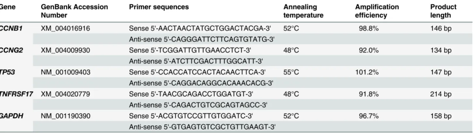
incu-bated at 37°C for 15 min to degrade the genomic DNA, and the remaining RNA was used as the template for real-time RT PCR. The primers for real-time RT PCR were designed according
to the mRNA sequences ofCCNB1(cyclin B1),CCNG2(cyclin G2),TP53(tumor protein p53),
TNFRSF17(tumor necrosis factor receptor superfamily member 17) andGAPDH
(glyceralde-hyde-3-phosphate dehydrogenase) using Primer Premier 5.0 software and are shown in Table 1. Real-time RT PCR was performed using the One Step SYBR PrimeScript PLUS RT-PCR Kit (TaKaRa Biotech. (Dalian) Co., Ltd., Dalian, China) according to the
manufactur-er’s protocol. The real-time RT PCR mixture consisted of 2μl total RNA, 1μl forward primer
(10μM), 1μl reverse primer (10μM), 0.5μl PrimeScript PLUS RTase Mix, 1.5μl TaKaRa Ex
Taq HS Mix, 12.5μl 2×One Step SYBR RT-PCR Buffer, and 6.5μl RNase Free dH2O in a total
volume of 25μl. Real-time RT PCR was performed by holding the reactions at a specific
tem-perature (45°C forCCNG2andTNFRSF17; 50°C forTP53; 48°C forCCNB1andGAPDH) for
45 min, a 95°C incubation for 2 min to inactivate the PrimeScript PLUS RTase, and 40 cycles
and extension at 72°C for 30 s using a real-time thermal cycler (Smart Cycler II, Cepheid, Sun-nyvale, CA, USA). Next, melt curves were generated by slowly heating (0.2°C/s) the PCR prod-ucts from 56°C to 95°C. The real-time RT PCR prodprod-ucts were further confirmed by
electrophoresis.GAPDHwas used as a reference gene, and the expression of the
cell-cycle-related genes (CCNB1,CCNG2,TNFRSF17andTP53) from fibroblasts in each group was
nor-malized to that of untreated fibroblasts (control group) using the 2-ΔΔC
Tmethod.
Somatic cell nuclear transfer
The procedure for sheep oocyte collection, maturation and the removal of cumulus cells was described previously [18]. Denuded oocytes with a polar body were incubated in H-SOF
sup-plemented with 7.5μg/ml cytochalasin B, 10μg/ml Hoechst 33342 and 10% (v/v) FBS at
38.5°C for 15 min, and then mounted onto a micromanipulator (NT-88NE, Nikon-Narishige, Tokyo, Japan) equipped with epifluorescence. The first polar body and adjacent cytoplasm were removed using an aspiration micropipette. Enucleated oocytes were analyzed by exposure to UV light, and only oocytes from which all chromosomes were removed were used for SCNT. One fibroblast was injected into the perivitelline space of an enucleated sheep oocyte using an injection micropipette. The karyoplast-cytoplast couplets were equilibrated in an elec-trofusion medium (0.3 M mannitol, 0.5 mM HEPES, 1% (m/v) fatty acid-free BSA, 0.05 mM
CaCl2and 0.1 mM MgCl2) for 3 min, then transferred into a cell fusion chamber containing
the same medium for electrofusion using a fusion machine (EP-1 Voltain, CryoLogic Pty. Ltd., Melbourne, Australia). After manual alignment, the karyoplast-cytoplast couplets were
sub-jected to a double DC fusion pulse of 1.25 kV/cm for 80μs, as described previously [18]. After
electrofusion, the karyoplast-cytoplast couplets were transferred into H-SOF supplemented with 10% (v/v) FBS to complete the fusion process. The fused embryos were activated by
cul-turing them in H-SOF containing 5μM ionomycin and 10% (v/v) FBS for 5 min and were
sub-sequently cultured in SOF containing 2 mM 6-DMAP and 10% (v/v) FBS for 4 h. Activated
cloned embryos were washed 2–3 times, and then cultured at 38°C in a humidified atmosphere
of 5% CO2in SOF supplemented with 2% (v/v) essential amino acids (Gibco), 1% (v/v)
non-essential amino acids (Gibco), 8 mg/ml fatty acid-free BSA, 5% (v/v) FBS and 1 mM glutamine. The progression of embryonic development was monitored every 24 h and half of the culture
medium was refreshed every 48 h. All groups of cloned embryos were culturedin vitrofor 12
days, all blastocysts were counted during culture. The cell number in the early blastocysts
(obtained on the 8th day ofin vitroculture) was assayed to determine the quality of the cloned
Table 1. The Primers for Real-Time RT PCR.
Gene GenBank Accession Number
Primer sequences Annealing
temperature
Amplification efficiency
Product length
CCNB1 XM_004016916 Sense 5'-AACTAACTATGCTGGACTACGA-3' 52°C 98.8% 146 bp
Anti-sense 5'-CAGGGATTCTTCAGTGTATG-3'
CCNG2 XM_004009930 Sense 5'-TCGGATTGTTGAACCTCT-3' 48°C 92.0% 134 bp Anti-sense 5'-ATCTTCGACTTTGGCATT-3'
TP53 NM_001009403 Sense 5'-CCACCATCCACTACAACTTCA-3' 55°C 101.2% 147 bp
Anti-sense 5'-CAGGACAGGCACAAACACG-3'
TNFRSF17 XM_004020779 Sense 5'-TAACGCAGACCTGGATGT-3' 48°C 91.8% 214 bp Anti-sense 5'-CAGACTGTCGCAGTAGCC-3'
GAPDH NM_001190390 Sense 5'-ACGTGTCCGTTGTGGATC-3' 52°C 96.7% 158 bp
Anti-sense 5'-GTGAGTGTCGCTGTTGAAGT-3'
embryos using a previously described protocol [20]. Briefly, blastocysts were incubated for 30 min at 38°C in PBS supplemented with 1.0 mg/ml Hoechst 33342, then, placed in a drop of mounting medium under a coverslip on a glass slide, the coverslip was slightly pressed, so that all cells in the blastocyst were spread out, and the cell number was counted under an inverted microscope (Ti-U, Nikon, Tokyo, Japan) equipped for epifluorescence. Three blastocysts derived from each group of fibroblasts were assayed.
Statistical analyses
The number of fibroblasts in the different phases of cell cycle as well as the developmental rate of cloned embryos was compared for statistical significance by the chi-square analysis using SPSS software (SPSS Inc., Chicago, IL, USA). The viability of fibroblasts, the levels of expres-sion for the cell cycle-related genes and the cell number from the cloned early blastocysts were compared for statistical significance by one-way analysis of variance, and further compared
statistically by least significant difference (LSD). Differences with a p-value<0.05 were
consid-ered to be statistically significant.
Results
The cell cycle of differentially cultured fibroblasts
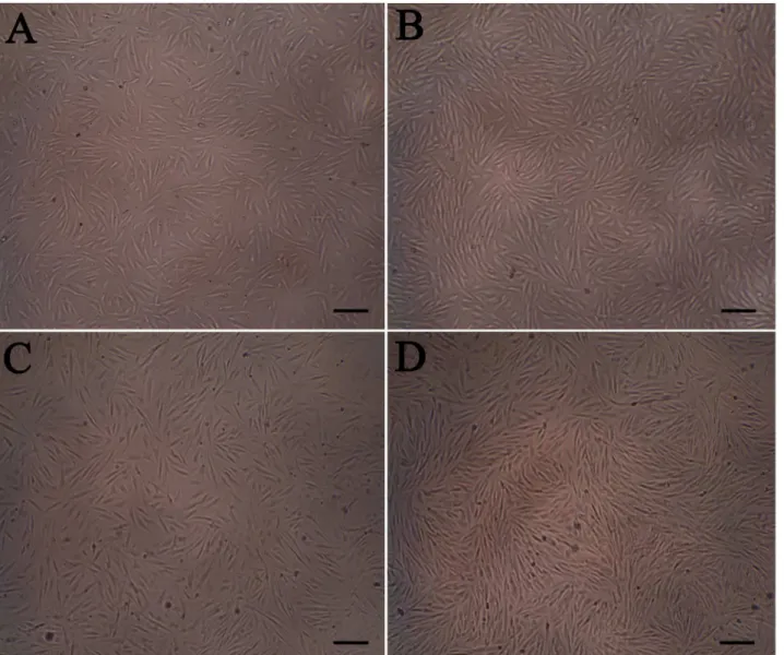
After culture with different methods, there was no apparent change in fibroblasts morphology (Fig 1). The percentages of fibroblasts in the different phases of the cell cycle were assayed by
flow cytometry, and the results are shown inFig 2andTable 2.
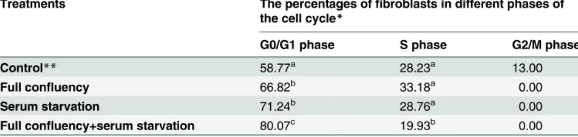
As shown inTable 2, the effect of full confluency or serum starvation on the cell cycle of
fibroblasts was similar; both culture methods significantly (p<0.05) increased the percentage
of fibroblasts in G0/G1 phase and decreased the percentages of fibroblasts in S and G2/M phases. Of the three culture methods, full confluency combined with serum starvation was the most efficient in arresting fibroblasts in G0/G1 phase, as the percentage of fibroblasts in G0/G1 phase was the highest in this group. In contrast, the percentage of fibroblasts in S phase was lowest in this group. Moreover, all of the culture methods could efficiently inhibit fibroblasts from entering into G2/M phase because none of the fibroblasts remained in G2/M phase in any of the groups.
The viability of fibroblasts in the groups of 70–80% confluency, full confluency, serum
starvation and full confluency with serum starvation were 92.47±3.07%, 90.30±2.66%, 78.87±3.40% and 76.30±3.09%, respectively. The difference in cell viability between
70–80% confluency group and full confluency group was not significant, the same result
was also obtained between serum starvation group and full confluency with serum
starva-tion group. However, the latter two groups had significantly (p<0.05) lower cell viability
than those of the former two groups.
Expression of cell cycle-related genes in differentially cultured
fibroblasts
Real-time RT PCR was used to assay the effects of the different culture methods on the
expres-sion levels of cell cycle-related genes, and the results are shown inFig 3.
After differential culture methods, the expression of cell cycle-related genes was different.
The expression ofCCNB1, a gene related to cell cycle and division, was decreased in all groups.
Compared to the control group, the levels ofCCNB1expression were 64.6±8.4%, 43.3±7.9%
CCNG2expression, another gene related to cell cycle and division, was increased in the
serum starvation and full confluency with serum starvation groups. Compared to the control
group, the levels ofCCNG2expression were 298.6±28.1% and 283.9±43.4% in these two
groups, respectively. However, in the full confluency group,CCNG2expression was
compara-ble to the control group (94.6±18.5% relative to control group).
TP53is a tumor suppressor gene, and the protein encoded by this gene can arrest a cell at
the G1/S regulation point to inhibit cell proliferation. Compared to the control group,TP53
expression increased to 162.9±17.4%, 234.7±33.3% and 324.7±32.4% in the full confluency, serum starvation and full confluency with serum starvation groups, respectively.
The protein encoded byTNFRSF17(also designated as B-cell maturation antigen (BCMA)),
a member of the TNF-receptor superfamily, is important for B cell development and autoim-mune response. Moreover, this protein can bind to various members of the TNFR-associated
factor family, such as APRIL, to promote cell growth and proliferation [21,22]. The effects of
the different culture methods onTNFRSF17expression were different. Both full confluency
Fig 1.In VitroCultured Fibroblasts Using Different Methods.A, 70–80% confluency; B, full confluency; C, serum starvation; D, full confluency with serum
starvation. Scale bar: 50μm.
and full confluency with serum starvation could decreaseTNFRSF17expression to 72.9±12.7%
and 41.4±11.1% relative to the control group. However, after serum starvation,TNFRSF17
expression remained comparable to the control group (94.4±24.5%).
The development and quality of cloned embryos derived from
differentially cultured fibroblasts
After culture using different methods, fibroblasts were used as nuclear donors for SCNT. The
cloned embryos could develop to blastocysts in 7–8 days and the blastocysts hatched in 9–10
Fig 2. The Percentages of Fibroblasts in Different Phases of the Cell Cycle was Assayed by Flow Cytometry.A, 70–80% confluency; B, full
confluency; C, serum starvation; D, full confluency with serum starvation.
doi:10.1371/journal.pone.0135344.g002
Table 2. The Percentages of Fibroblasts in Different Phases of the Cell Cycle under Different Culture Methods.
Treatments The percentages offibroblasts in different phases of the cell cycle*
G0/G1 phase S phase G2/M phase
Control** 58.77a 28.23a 13.00
Full confluency 66.82b 33.18a 0.00
Serum starvation 71.24b 28.76a 0.00
Full confluency+serum starvation 80.07c 19.93b 0.00
a, b, c
Values with different superscripts within the same column are significantly different (P<0.05).
*Data were collected from one series offlow cytometry assay, and significant differences were determined by comparing the number offibroblasts in the different phases of the cell cycle.
days (Fig 4). The developmental ability of the cloned embryos in the present study was
compa-rable with the results of our and other previous studies [18,23,24], and the results are shown
inTable 3.
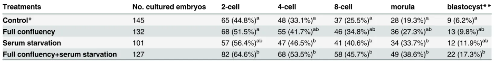
Compared with embryos derived from fibroblasts in the control group, embryos derived from fibroblasts cultured in serum starvation or full confluency with serum starvation
condi-tions had a significantly (p<0.05) higher rates of 4-cell, 8-cell and morula stages. However, the
use of full confluency fibroblasts only slightly increased the developmental ability of cloned embryos. In all developmental stages, the difference in developmental rate of the cloned
embryos was not significant (p>0.05) between the control group and the full confluency
group. Moreover, of the three culture methods, full confluency with serum starvation showed the greatest increase in the developmental ability of the cloned embryos.
The cell number in cloned early blastocysts (obtained on the 8th day ofin vitroculture)
was assayed after staining with Hoechst 33342 (Fig 4). There was no significant difference in
the cell number of cloned blastocysts derived from 70–80% confluency (105.7±7.0), full
con-fluency (96.3±8.7), serum starvation (95.7±6.5) and full concon-fluency with serum starvation (101.7±16.8), respectively. This result suggests that the different fibroblasts culture methods did not affect the quality of the resulting cloned embryos.
Discussion
Presently, two types of methods, full confluency (or contact inhibition) and serum starvation,
are commonly used to arrestin vitrocultured somatic cells in G0/G1 phase. More than half a
century ago, it was discovered that non-malignant cells would stop proliferating when they Fig 3. The Relative Expression of Cell Cycle-Related Genes was Assayed by Real-Time RT PCR.The expression of genes in each group was normalized to the control group. Data were collected from three replicates and are presented as the mean±standard deviation. Bars with different lowercase
letters are significantly different (p<0.05).
Fig 4.In VitroCultured Cloned Sheep Embryos.A, 2- and 8-cell embryos; B, early blastocyst; C, hatching blastocyst; D, early blastocyst stained with Hoechst 33342. Scale bar: 20μm.
doi:10.1371/journal.pone.0135344.g004
Table 3. The Developmental Ability of Cloned Embryos Derived from Differentially Cultured Fibroblasts.
Treatments No. cultured embryos 2-cell 4-cell 8-cell morula blastocyst**
Control* 145 65 (44.8%)a 48 (33.1%)a 37 (25.5%)a 28 (19.3%)a 9 (6.2%)a
Full confluency 132 68 (51.5%)a 55 (41.7%)ab 46 (34.8%)ab 36 (27.3%)ab 13 (9.8%)ab
Serum starvation 101 57 (56.4%)ab 47 (46.5%)b 41 (40.6%)b 34 (33.7%)b 12 (11.9%)ab Full confluency+serum starvation 127 82 (64.6%)b 68 (53.5%)b 58 (45.7%)b 49 (38.6%)b 22 (17.3%)b
a, bValues with different superscripts within the same column are signi
ficantly different (P<0.05). *Control:fibroblasts were cultured to 70–80% confluency.
**The data include all blastocysts from 12 days of culture. Data were collected from 10 series of SCNT experiments.
reached full confluence, despite the availability of extracellular nutrients and growth factors
[25]. Recently, it was proposed that full confluency could upregulate p27Kip1through the p38α
-Spry2-EGFR pathway, to arrest cell proliferation [26]. Serum starvation can also lead to cell cycle arrest in G0/G1 phase [27]. However, its mechanism may be different from that of full confluency. It was proposed that serum starvation arrested the cell cycle through the
Skp2-p27-CDK2 (or CDK4) pathway [28]. In the present study, both the percentage of fibro-blasts in G0/G1 phase and the expression levels of cell cycle-related genes were different after three types of culture methods. These results further implied that the mechanisms of arresting cell cycle by the previously described methods were different. For example, full confluency
could result in a significant decrease inTNFRSF17expression, but serum starvation had little
effect on the expression of this gene. Similar results were also obtained in other studies [29–
31]. The biological relevance of BCMA, the protein encoded byTNFRSF17, in maintaining the
viability and proliferation of Hodgkin and Reed-Sternberg (HRS) lymphoma cells has been demonstrated by Chiu et al [32]. APRIL and BAFF could deliver nonredundant signals via BCMA and TACI receptors through both autocrine and paracrine pathways. These signals
caused NF-κB activation; antiapoptotic BCL-2 and BCL-X
Lup-regulation, proapoptotic BAX
down-regulation, growth-inducing c-MYC protein up-regulation, and as a result, the survival and proliferation of HRS cells were enhanced [32]. In fact, cell cycle arrest upon confluency may be evoked by cell-cell communication, which is transduced into the cytoplasm and
nucleus through certain signaling pathways [33,34]. When cells were cultured in low serum
medium, they could not obtain sufficient growth factors for their proliferation. Subsequently, growth factor-related signaling pathways and metabolic pathways were affected, resulting in
cell cycle arrest [16,35].
There was a controversy as to whether full confluency and serum starvation had the same
efficiency in arrestingin vitrocultured cells in G0/G1 phase because similar and different
effi-ciencies have been reported [12–15]. We considered that this contradiction may be because
cells derived from different species had distinct responses to different culture methods, and the time of culture was also a pivotal factor in arrest efficiency. For example, different methods (full confluency, serum starvation and chemical inhibitors) had different levels of efficiency in G0/G1 phase arrest in fibroblasts derived from different species of cats [36]. In addition, pro-longing the time of serum starvation could more efficiently arrest goat fibroblasts in G0/G1 phase [15]. In the present study, full confluency and serum starvation had the same efficiency in arresting sheep skin fibroblasts in G0/G1 phase. Moreover, the synergistic effects of these two types of methods could further increase the percentage of fibroblasts in G0/G1 phase. Simi-lar results were also obtained in another study using domestic cat fibroblasts [14]. As discussed above, although prolonging the time of serum starvation could improve arrest efficiency, the percentage of dead cells also increased [15]. Therefore, short-term culture with a combination of these two methods may be an alternative strategy.
In present study, the blastocyst rate of cloned embryos in control group was as low as 6.2% (No. of blastocysts/No. of cultured embryos). This result was comparable with the result of a recent sheep SCNT study, in which fresh lymphocytes were used as nuclear donors, and sheep enucleated MII oocytes were used as nuclear recipients. The blastocyst rate of cloned embryos was also as low as 7.0% (25/356, No. of blastocysts/No. of cultured embryos) [23]. Therefore, it was considered that arresting donor cells in G0/G1 phase facilitated the development of cloned embryos derived from SCNT [8]. In previous studies of SCNT, serum starvation or full
con-fluency was commonly used to culture nuclear donors [7,9–11], and the combination of these
two methods for culturing nuclear donors was only reported in few studies [18,19]. In a bovine
results of the present study were in accord with this study; the blastocyst rates of cloned embryos in the groups of full confluency and serum starvation were 9.8% and 11.9% (No. of blastocysts/No. of cultured embryos), respectively. Our results were comparable with the results of a recent sheep SCNT study, in which sheep confluent cumulus cells were used as nuclear donors, and sheep enucleated MII oocytes were used as nuclear recipients; the blasto-cyst rates of cloned embryos were 7.1%-11.7% (5/70-11/94, No. of blastoblasto-cysts/No. of cultured embryos) [24]. In fact, in our a recent study, sheep skin fibroblasts were cultured to full
con-fluency with further serum starvation for 3–5 d and then used as nuclear donors, sheep
enucle-ated MII oocytes were used as nuclear recipients, the blastocyst rate of cloned embryos was 14.6% (15/103, No. of blastocysts/No. of cultured embryos) [18], this result was comparable with the result of present study (17.3%, No. of blastocysts/No. of cultured embryos). However, in that study, we did not further study the effects of different donor cell culture methods on the developmental ability of sheep cloned embryos.
In the present study, full confluency or serum starvation could efficiently arrest fibroblasts in G0/G1 phase. However, cloned embryos derived from these fibroblasts had a comparable total developmental ability with those derived from untreated fibroblasts, and only fibroblasts cultured to full confluency with serum starvation could produce cloned embryos with a signifi-cantly higher developmental ability. As discussed above, the mechanism of arresting cell cycle by full confluency or serum starvation was different. A combination of these two methods could arrest somatic cells in a more stable G0/G1 phase. After these pretreated somatic cells were transferred into the cytoplasm of enucleated oocytes, the nuclei and cytoplasm of the cloned embryos were coordinated in the cell cycle, which facilitated the development of cloned embryos. Moreover, many genes were downregulated in cells arrested in G0/G1 phase [31], which may have facilitated the reprogramming of the somatic nuclei and the development of cloned embryos. Therefore, we propose that full confluency combined with serum starvation
could more efficiently arrestin vitrocultured somatic cells in G0/G1 phase, and when these
cells were used as nuclear donors, the resulting cloned embryos would exhibit higher develop-mental ability. This finding may improve the efficiency of SCNT.
Acknowledgments
We thank the master’s students Liu Juan and Cheng Teng for their assistance during this study
and Prof. Wang JianYing for his critical comments.
Author Contributions
Conceived and designed the experiments: LM. Performed the experiments: LM XL FW XH. Analyzed the data: LM. Contributed reagents/materials/analysis tools: SC WL. Wrote the paper: LM.
References
1. Bhojwani S, Alm H, Torner H, Kanitz W, Poehland R. Selection of developmentally competent oocytes through brilliant cresyl blue stain enhances blastocyst development rate after bovine nuclear transfer. Theriogenology 2007; 67: 341–345. PMID:16999988
2. Sung LY, Gao S, Shen H, Yu H, Song Y, Smith SL, et al. Differentiated cells are more efficient than adult stem cells for cloning by somatic cell nuclear transfer. Nat Genet. 2006; 38: 1323–1328. PMID: 17013394
4. Wells DN, Laible G, Tucker FC, Miller AL, Oliver JE, Xiang T, et al. Coordination between donor cell type and cell cycle stage improves nuclear cloning efficiency in cattle. Theriogenology 2003, 59(1): 45–59. PMID:12499017
5. Moulavi F, Hosseini SM, Hajian M, Forouzanfar M, Abedi P, Ostadhosseini S, et al. Nuclear transfer technique affects mRNA abundance, developmental competence and cell fate of the reconstituted sheep oocytes. Reproduction 2013; 145: 345–355. doi:10.1530/REP-12-0318PMID:23401598
6. Campbell KH, Loi P, Otaegui PJ, Wilmut I. Cell cycle co-ordination in embryo cloning by nuclear trans-fer. Rev Reprod. 1996; 1: 40–46. PMID:9414437
7. Wilmut I, Schnieke AE, McWhir J, Kind AJ, Campbell KH. Viable offspring derived from fetal and adult mammalian cells. Nature 1997; 385: 810–813. PMID:9039911
8. Ma LB, He XY, Wang FM, Cheng T, Liu XY. Somatic cell reprogrammed by oocyte: process and barri-cade. Anim Cells and Syst. 2014; 18: 161–171.
9. Galli C, Lagutina I, Crotti G, Colleoni S, Turini P, Ponderato N, et al. Pregnancy: a cloned horse born to its dam twin. Nature 2003; 424: 635.
10. Kim MK, Jang G, Oh HJ, Yuda F, Kim HJ, Hwang WS, et al. Endangered wolves cloned from adult somatic cells. Cloning Stem Cells 2007; 9: 130–137. PMID:17386020
11. Wani NA, Wernery U, Hassan FA, Wernery R, Skidmore JA. Production of the first cloned camel by somatic cell nuclear transfer. Biol Reprod. 2010; 82: 373–379. doi:10.1095/biolreprod.109.081083
PMID:19812298
12. Hayes O, Ramos B, Rodríguez LL, Aguilar A, Badía T, Castro FO. Cell confluency is as efficient as serum starvation for inducing arrest in the G0/G1 phase of the cell cycle in granulosa and fibroblast cells of cattle. Anim Reprod Sci. 2005; 87: 181–192. PMID:15911169
13. Khammanit R, Chantakru S, Kitiyanant Y, Saikhun J. Effect of serum starvation and chemical inhibitors on cell cycle synchronization of canine dermal fibroblasts. Theriogenology 2008; 70: 27–34. doi:10. 1016/j.theriogenology.2008.02.015PMID:18423836
14. de Barros FR, Goissis MD, Caetano HV, Paula-Lopes FF, Peres MA, Assumpção ME, et al. Serum starvation and full confluency for cell cycle synchronization of domestic cat (felis catus) foetal fibro-blasts. Reprod Domest Anim. 2010; 45: 38–41. doi:10.1111/j.1439-0531.2008.01201.xPMID: 19416486
15. Dalman A, Eftekhari-Yazdi P, Valojerdi MR, Shahverdi A, Gourabi H, Janzamin E, et al. Synchronizing cell cycle of goat fibroblasts by serum starvation causes apoptosis. Reprod Domest Anim. 2010; 45: e46–53. doi:10.1111/j.1439-0531.2009.01520.xPMID:19788523
16. Golpour M, Akhavan Niaki H, Khorasani HR, Hajian A, Mehrasa R, Mostafazadeh A. Human fibroblast switches to anaerobic metabolic pathway in response to serum starvation: a mimic of warburg effect. Int J Mol Cell Med. 2014; 3: 74–80. PMID:25035856
17. Ma LB, He XY. The levels of DNA methylation of sheep cloned embryos in different development stages. Indian J Anim Res. 2014, 48: 221–226.
18. Ma LB, He XY, Wang FM, Cao JW, Cheng T. The development and expression of pluripotency genes in embryos derived from nuclear transfer and in vitro fertilization. Zygote 2014; 22: 540–548. doi:10. 1017/S0967199413000129PMID:23731893
19. Ma LB, Yang L, Zhang Y, Cao JW, Hua S, Li JX. Quantitative analysis of mitochondrial RNA in goat–
sheep cloned embryos. Mol Reprod Dev. 2008; 75: 33–39. PMID:17570506
20. Hua S, Zhang Y, Li XC, Ma LB, Cao JW, Dai JP, et al. Effects of granulosa cell mitochondria transfer on the early development of bovine embryos in vitro. Cloning Stem Cells 2007; 9: 237–246. PMID: 17579556
21. Rickert RC, Jellusova J, Miletic AV. Signaling by the tumor necrosis factor receptor superfamily in B-cell biology and disease. Immunol Rev. 2011; 244: 115–133. doi:10.1111/j.1600-065X.2011.01067.x
PMID:22017435
22. Sanchez E, Li M, Kitto A, Li J, Wang CS, Kirk DT, et al. Serum B-cell maturation antigen is elevated in multiple myeloma and correlates with disease status and survival. Br J Haematol. 2012; 158: 727–738.
doi:10.1111/j.1365-2141.2012.09241.xPMID:22804669
23. Iuso D, Czernik M, Di Egidio F, Sampino S, Zacchini F, Bochenek M, et al. Genomic stability of lyophi-lized sheep somatic cells before and after nuclear transfer. PLoS One 2013; 8: e51317. doi:10.1371/ journal.pone.0051317PMID:23308098
25. Levine EM, Becker Y, Boone CW, Eagle H. Contact inhibition, macromolecular synthesis, and polyribo-somes in cultured human diploid fibroblasts. Proc Natl Acad Sci USA. 1965; 53: 350–356. PMID: 14294068
26. Swat A, Dolado I, Rojas JM, Nebreda AR. Cell density-dependent inhibition of epidermal growth factor receptor signaling by p38alpha mitogen-activated protein kinase via Sprouty2 downregulation. Mol Cell Biol. 2009; 29: 3332–3343. doi:10.1128/MCB.01955-08PMID:19364817
27. Cooper S. Reappraisal of serum starvation, the restriction point, G0, and G1 phase arrest points. FASEB J. 2003; 17: 333–340. PMID:12631573
28. Shin JS, Hong SW, Lee SL, Kim TH, Park IC, An SK, et al. Serum starvation induces G1 arrest through suppression of Skp2-CDK2 and CDK4 in SK-OV-3 cells. Int J Oncol. 2008; 32: 435–439. PMID: 18202766
29. Gustincich S, Schneider C. Serum deprivation response gene is induced by serum starvation but not by contact inhibition. Cell Growth Differ. 1993; 4: 753–760. PMID:8241023
30. Gos M, Miloszewska J, Swoboda P, Trembacz H, Skierski J, Janik P. Cellular quiescence induced by contact inhibition or serum withdrawal in C3H10T1/2 cells. Cell Prolif. 2005; 38: 107–116. PMID: 15842254
31. Coller HA, Sang L, Roberts JM. A new description of cellular quiescence. PLoS Biol. 2006; 4: e83. PMID:16509772
32. Chiu A, Xu W, He B, Dillon SR, Gross JA, Sievers E, et al. Hodgkin lymphoma cells express TACI and BCMA receptors and generate survival and proliferation signals in response to BAFF and APRIL. Blood 2007; 109: 729–739. PMID:16960154
33. Curto M, Cole BK, Lallemand D, Liu CH, McClatchey AI. Contact-dependent inhibition of EGFR signal-ing by Nf2/Merlin. J Cell Biol. 2007; 177: 893–903. PMID:17548515
34. Kim JH, Asthagiri AR. Matrix stiffening sensitizes epithelial cells to EGF and enables the loss of contact inhibition of proliferation. J Cell Sci. 2011; 124: 1280–1287. doi:10.1242/jcs.078394PMID:21429934
35. Zimmermann O, Zwaka TP, Marx N, Torzewski M, Bucher A, Guilliard P, et al. Serum starvation and growth factor receptor expression in vascular smooth muscle cells. J Vasc Res, 2006; 43: 157–165.
PMID:16407661