Pravastatin induces cell cycle arrest and decreased production
of VEGF and bFGF in multiple myeloma cell line
P. J. J. Trojan
a, M. S. Bohatch-Junior
a, M. F. Otuki
b, F. Souza-Fonseca-Guimarães
c,
P. V. Svidnicki
d, V. Nogaroto
d, D. Fernandes
b, E. A. Krum
band G. M. Favero
a*
aLaboratório Multidisciplinar de Ciências Biológicas e da Saúde, Universidade Estadual de Ponta Grossa – UEPG,Campus de Uvaranas, Av. General Carlos Cavalcanti, 4748, CEP 84030-900, Ponta Grossa, PR, Brazil bDepartamento de Ciências Farmacêuticas, Universidade Estadual de Ponta Grossa – UEPG, Campus de Uvaranas,
Av. General Carlos Cavalcanti, 4748, CEP 84030-900, Ponta Grossa, PR, Brazil cUnit Cytokines and Inflammation, Department Infection and Epidemiology Institut Pasteur,
25-28 rue du Docteur Roux, 75015, Paris, France
dLaboratório de Citogenética e Evolução, Departamento de Biologia Molecular, Estrutural e Genética, Universidade Estadual de Ponta Grossa – UEPG, Campus de Uvaranas, Av. General Carlos Cavalcanti, 4748,
CEP 84030-900, Ponta Grossa, PR, Brazil *e-mail: gmfavero@uepg.br
Received: June 23, 2014 – Accepted: October 17, 2014 – Distributed: February 29, 2016 (With 4 Figures)
Abstract
Multiple myeloma (MM) is a B cell bone marrow neoplasia characterized by inflammation with an intense secretion of growth factors that promote tumor growth, cell survival, migration and invasion. The aim of this study was to evaluate the effects of pravastatin, a drug used to reduce cholesterol, in a MM cell line.Cell cycle and viability were determinate by Trypan Blue and Propidium Iodide. IL6, VEGF, bFGF and TGFβ were quantified by ELISA and qRT-PCR including here de HMG CoA reductase. It was observed reduction of cell viability, increase of cells in G0/G1 phase of the cell cycle and reducing the factors VEGF and bFGF without influence on 3-Methyl-Glutaryl Coenzyme A reductase expression.The results demonstrated that pravastatin induces cell cycle arrest in G0/G1 and decreased production of growth factors in Multiple Myeloma cell line.
Keywords: pravastatin, Multiple Myeloma, 3-Methyl-Glutaryl Coenzyme A reductase, vascular endothelial growth
factor, fibroblast growth factor.
Pravastatina induz parada no ciclo celular e diminuição na produção de
VEGF e bFGF em linhagem de Mieloma Multiplo
Resumo
O Mieloma Múltiplo é uma neoplasia de linfócitos B da medula óssea, caracterizada por inflamação com uma intensa secreção de fatores de crescimento que promovem o aumento do volume do tumor, sobrevivência celular, migração e invasão. O objetivo deste estudo foi avaliar os efeitos da pravastatina, uma droga usada para reduzir o colesterol, em um linhagem de MM. O ciclo celular e viabilidade foram determinadas por Trypan Blue e iodeto de propídio. IL6, VEGF, bFGF e TGF foram quantificadas por ELISA e qRT-PCR, incluindo aqui de HMG CoA redutase. Observou-se a redução da viabilidade das células, aumento de células na fase G0/G1 do ciclo celular e redução no VEGF e bFGF, sem influência na expressão da enzima 3-Metil-Glutaril Coenzima A redutase. Os resultados demonstraram que a pravastatina induz parada no ciclo celular em G0/G1 e diminuição da produção de fatores de crescimento em várias
linhas de células de Mieloma.
Palavras-chave: pravastatina, Mieloma Multiplo, 3-Metil-Glutaril Coenzima A redutase, fator de crescimento do endotélio vascular, fator de crescimento de fibroblasto.
1. Introduction
Multiple Myeloma (MM) is a neoplastic disease that
affects antibodies-secreting B cells fully differentiated,
which emerges and expands at the bone marrow (Zhan et al.,
results in bone destruction, suppress antibody production
and lead to renal failure (Ludwig et al., 2010; Raab et al., 2009). The plasma cell adhesion on bone marrow increases
the secretion of cytokines which in turn stimulate the proliferation of tumor cells contributing to their survival,
migration and induce resistance to chemotherapeutic agents (Balakumaran et al., 2010; Peng et al., 2011).
The disease evolution is, partially, possible due to
angiogenic mechanisms. Several studies have sought
the relationship between growth factors such as FGF (fibroblast growth factor) and VEGF (vascular endothelial growth factor), to prognosis of the disease and sensitive
to therapy (Greco et al., 2009). It was demonstrated that
the levels of VEGF and FGF are elevated in patients with MM and often correlated among themselves and with other angiogenic factors (Greco et al., 2009). The binding
of TGFβ (transforming growth factor β) to its receptor promotes activation of intracellular mediators, whose effects
include immunomodulation, cell cycle and angiogenesis (Ma and Ma, 2011).
Over the last decade, Statins, the most commercial drug all over the world, prescribed as cholesterol-lowering agents,
have revealed other effects, including immunomodulation, neuroprotection, improvement of bone metabolism and antitumour. The inhibition of the enzyme HMG-CoA reductase, an essential key in the biosynthesis of cholesterol
(Medina 2010; Musso et al., 2011), demonstrates to be the key point to reduce cholesterol and several compounds like
isopentenyladenine, dolichol, ubiquinone, geranilpirofosfato (GGPP), farnesylpyrophosphate (FPP) (Wong et al., 2007; Gauthaman et al., 2009). The reduction in synthesis of isoprenoids (GGPP, FPP), decreases it’s binding to cellular
proteins responsible for intracellular signaling such as
Rho, Rac and Cdc42 (Roy et al., 2011; Musso et al., 2011).
The aim of this study was to assess the effects of
Pravastatin, a HMG-CoA reductase inhibitor, over a
MM cell line, with a focus over cell cycle, inflammatory and growth factors response, cholesterol production and pleiotropic effects.
2. Methods
2.1. Cell culture
RPMI 8226 multiple myeloma cells were grown
in RPMI 1640 medium supplemented with 10% fetal
bovine serum (FBS), 100 U/mL penicillin/streptomycin and 24mM NaHCO3 at 37 °C in a humidified atmosphere containing 5% CO2. Cells were seeded at an initial density
of 4×10⁴ cells/cm2 (RPMI 8226). Upon confluence,
Pravastatin was added to the media at a concentration of 0.3-0.9 μM for different times.
2.2. Cell viability
Multiple Myeloma cells were plated at a density of 1×10⁶ cells/well. Six-well tissue culture plates (Corning,
NY) were used. Cells were treated with Pravastatin for three consecutive days (24h, 48 h, and 72h). Cell proliferation
and viability were determined by Trypan Blue exclusion method. Experiments were performed three times in triplicate. 2.3. Analysis of cell cycle by flow cytometry
Cell viability was examined by the trypan blue exclusion
method. Cell cycle analysis was performed by flow cytometry. Cells were seeded at a density of 2×10⁶ cells/cm2
(RPMI 8226). After 24h cells were treated with 0.3-0.9 μM of pravastatin for up to 72h. At the end of the experiment, cells were trypsinized (0.05% trypsin in 0.02% EDTA) and washed 3 times with PBS, fixed in 70% ethanol, and stained with propidium iodide (PI; 50 µg/10µL, final concentration), for 30 min in the dark, according to the
method described by Vindelov et al. (1983). All analyses
were done using a FACScalibur flow cytometer (Becton Dickinson, San Jose, CA). The red fluorescence of PI was collected through a 585/42-nm band – pass filter, and the fluorescence signals were measured in a linear scale of
1024 channels. For each sample, at least 10000 events
were acquired and the data were analyzed using two appropriate software (CELLQuest, Becton Dickinson, San Jose, CA; ModiFit LT 2.0). Cells in S/G2/M (proliferating)
and G0/G1 phases, and hypodiploid cells (cells under death process) were analized.
2.4. Evaluation of growth factors
IL-6, VEGF, bFGF and TGFβ were investigated after treatment of cells in culture with pravastatin in three different concentrations: 0.3 μM, 0.6 μM and 0.9 μM. The samples of supernatant were withdrawn after 24, 48 and 72 hours after the contact of cells with pravastatin. The supernatant was analyzed by ELISA (Enzyme Linked Immuno Sorbent Assay) to determine the levels of growth factors surveyed. The ELISA plates were read at the Life Lab reader MX range 2001 at 450/690 nm. VEGF and bFGF were quantified by RayBio® kit and IL-6 and TGFβ by eBioscience kit® following the methodology described by the manufacturers.
2.5. RNA extraction and quantitative RT-PCR (qRT-PCR) assays
Total RNA from the MM cultured cells was isolated
using the Illustra RNAspin Mini RNA Isolation Kit
(GE Healthcare), according to the manufacturer’s instructions. One microgram of total RNA were reverse transcripted using
the First-Strand cDNA Synthesis Kit (GE Healthcare), as
5’ CAAAAACGGGGGCTTCTT 3’, bFGF_r: 5’ AGCCAGGTAACGGTTAGCAC 3´) and sufficient
water to 25 µL. A negative control was also included
for each gene amplification assay. The 18S rDNA (18S_f: 5’ ATGCGTGCATTTATCA GA 3’; 18S_r: 5’AACTATCCCGTCTGCAAG 3’) was used as an internal control. The PCR cycling conditions were: 10 min 95 °C;
40 cycles: 15 seg 95 °C, 30 seg 60 °C, 15 seg 72 °C;
followed by a dissociation curve. Threshold cycle (Ct) was measured and a relative change in the expression level of one specific gene was presented as 2−ΔΔCt .
2.6. Statistical analysis
Data are expressed as the mean ± SEM performed three times in triplicate. The data presents normal distribution and homogeneity of variances. These conditions had been
proved by the Shapiro-Wilk and Bartlett test, respectively.
The statistical analysis was performed with Student’s t-test or one-way analysis of variance (ANOVA) with Bonferroni’s Multiple Comparison Test as indicated in the figure legends. A p value less than 0.05 was considered statistically significant.
3. Results
To verify if pravastatin affects cell cycle replication, firstly, we assess the cell viability. We observed a decreased at cells numbers after the addition of pravastatin only at higher concentrations (0.6 μM and 0.9 μM). Pravastatin at 0.3 μM was not able to affect cell viability. Interestingly, the concentration of 0.6 μM and 0.9 μM promoted cytostatic effect only after 72 hours of incubation, showing that this effect is both, concentration/time dependent (Figure 1).
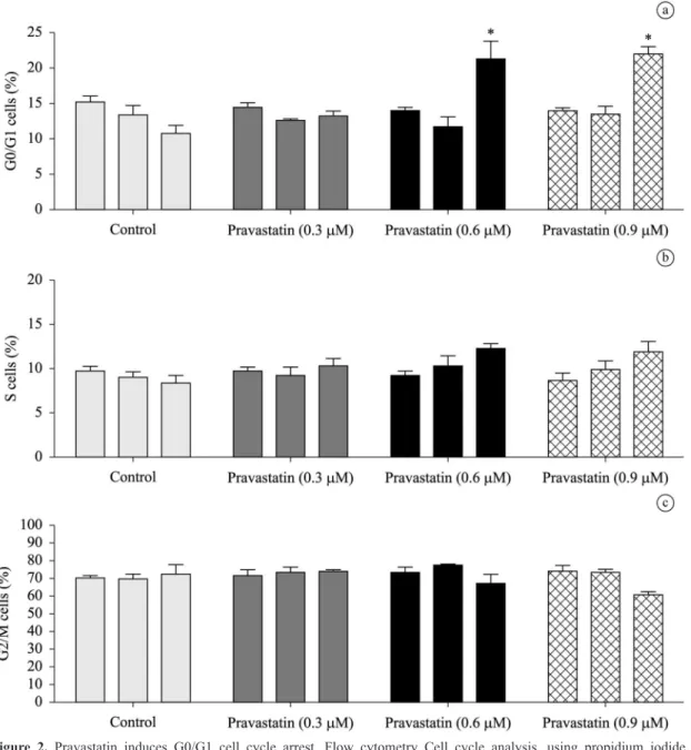
Thereafter, we observe the action of pravastatin in the cycle by the methodology of PI. The incubation of the MM cell with pravastatin (0.6 μM and 0.9 μM), result in an increase in the number of cells in G0/G1 cell cycle phase after 72 hours. The lower concentration of pravastatin (0.3 μM) was insufficient to provide cell cycle arrest in a significant quantity of cells. The Figure 2 shows the G0/G1 cell cycle arrest. It’s important to emphasize that
these cell lineage is well known to have different size and shape, and to concentrate the major amount of cells in
the G2/M, and to be able to synthesize and secrete IL-6.
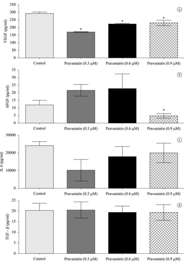
The main factors released by the cell line and that are involved at proliferation of MM cell were evaluated. These four important factors are shown at Figure 3. The three
periods, 24h, 48h and 72h were evaluated. For sake of clarity the results shown are for 48h of treatment. Pravastatin reduced VEGF and bFGF levels at supernatant of the
cells, meanwhile did not cause changes in the secretion
of IL-6 and TGFβ.
To confirm the results obtained with the ELISA we performed a qRT-PCR. In addition to the factors studied we also evaluated the expression of HMGCoA reductase gene. Pravastatin was able to increase only VEGF mRNA levels. Interestingly, the levels of HMGCoA have not changed with the use of pravastatin in the studied cells (Figure 4).
4. Discussion
The literature has reported different effects of statins in neoplastic cells, such as induction of apoptosis, decreased cell proliferation, arrest in G0/G1 cell cycle and reduction of inflammatory cytokines. These effects have been tested in different cancers such as melanoma (Favero et al., 2010), thyroid cancer (Zhong et al., 2011) osteosarcoma (Tsubaki et al., 2011), hepatocellular carcinoma (Tijeras-Raballand et al., 2010). Additionally, several studies have shown that statins
reduce cell proliferation in hematopoietic malignancies
such as chronic lymphocytic leukemia (Podhorecka et al., 2010), acute promyelocytic leukemia NB4 (Sassano et al., 2007) and MM (Otsuki et al., 2004). Wong et al. (2007)
showed that the sensitivity of cells to lovastatin is associated with a specific profile of genetic abnormalities. Another
study (Clendening et al., 2010) attributed the sensitivity
of cancer cells to statins dysregulation of the mevalonate pathway. After the evaluation of a large panel of MM cell
Wong et al. (2007) found that 50% of the studied strains are sensitive to lovastatin.
A Japanese study (Otsuki et al., 2003) observed that
about a third of the strains tested were MM pravastatin sensitive. Different strains of melanoma showed varied susceptibility to statins, the effects related to cell cycle
arrest and apoptosis (Zhong et al., 2011). This effect was observed in other cancers (Xiang et al., 2011) where
lovastatin inhibited cell proliferation in ATC strains aggressive thyroid cancer in vitro, raising the level of p27 protein, which inhibits the activity of CDK2 holding
the cells in G1 phase.
Other effects attributed to statins refer to modulation of inflammatory mediators and growth factors. Here, we showed that pravastatin reduced VEGF and bFGF levels when compared to control cells. Despite of involvement of Figure 1. Pravastatin reduces the number of viable cells
IL-6 at the progression of multiple myeloma, the mRNA and protein levels of this cytokine was not changed by pravastatin treatment. This agrees with a similar result obtained before by other study (Otsuki et al., 2003). Likewise,
despite the extensive evidence, showing that TGF-β is a key player in cell proliferation, differentiation and apoptosis, pravastatin effect on MM cells cycle is no explained by changes in this growth factor production. Nevertheless, we cannot rule out the possibility that pravastatin affect the signaling pathway of IL-6 and TGF-β.
The reduction of VEGF and FGF was observed by
others (Cho et al., 2008) who note the reduction of VEGF
in vitro and in vivo. Several studies shows (Zhong et al., 2011) that statins reduce mRNA of FGF, HGF and
TGFβ in osteosarcoma cells in culture. These growth factors are due to inhibition of GGPP (geranyl-geranyl pyrophosphate), preventing the location of Ras to the plasma membrane and subsequent activation of pathways
MEK/ERK (extracellular signal controlled by the kinase)
and PI3K/AKT (phosphatidylinositol kinase 3 / protein
kinase C).
In conclusion, the results of our work showed that
Pravastatin induces: cell cycle arrest at G0/G1 allied to a decrease in cell growth and survival in a MM cell line;
Figure 3. Pravastatin induces the loss of VEGF and FGF. Evaluation of different factors in the supernatant culture of MM
Figure 4. Pravastatin not change the expression of HMG CoA reductase. Evaluation of different mRNA by qRT PCR. The result
shown is representative of 48h of treatment performed three times in triplicate. Threshold cycle (Ct) was measured and a relative change in the expression level of one specific gene was presented as 2−ΔΔCt Each bar represents the mean with their standard
deviations. Statistical analysis was performed using Student’s t-test. * p < 0.05 compared with the control group.
decrease in VEGF and bFGF; did not change IL-6 and TGFβ. These effects should be, in part, due to pleiotropic effects and not by the 3-Methyl-Glutaryl Coenzyme A
reductase inhibition.
References
BALAKUMARAN, A., ROBEY, P.G., FEDARKO, N. and LANDGREN, O., 2010. Bone marrow microenvironment in myelomagenesis: its potential role in early diagnosis. Expert Review of Molecular Diagnostics, vol. 10, no. 4, pp. 465-480. http://dx.doi.org/10.1586/erm.10.31. PMid:20465501.
CHO, S.J., KIM, J.S., KIM, J.M., LEE, J.Y., JUNG, H.C. and SONG, I.S., 2008. Simvastatin induces apoptosis in human colon cancer cells and in tumour xenografts, and attenuates colitis-associated colon câncer in mice. International Journal of Cancer, vol. 123, no. 4, pp. 951-957. http://dx.doi.org/10.1002/ ijc.23593. PMid:18521906.
FAVERO, G.M., OTUKI, M.F., OLIVEIRA, K.A., BOHATCH JUNIOR, M.S., BORELL, P., BARROS, F.E., MARIA, D.A., FERNANDES, D. and BYDLOWSKI, S.P., 2010. Simvastatin impairs murine melanoma growth. Lipids in Health and Disease, vol. 16.
GADÓ, K., SILVA, S., PÁLÓCZI, K., DOMJÁN, G. and FALUS, A., 2001. Mouse plasmocytoma: experimental modelo of human multiple myeloma. Haematologica, vol. 86, no. 3, pp. 227-236. PMid:11255268.
GAUTHAMAN, K., MANASI, M. and BONGSO, A., 2009. Statins inhibit the growth of variant human embryonic stem cells and câncer cells in vitro but not normal human embryonic stem cells. Journal British Pharmacology, vol. 157, no. 6, pp. 962-973. http:// dx.doi.org/10.1111/j.1476-5381.2009.00241.x. PMid:19438511.
GIULIANI, N. and RIZZOLI, V., 2007. Myeloma cells and boné marrow osteoblast interactions: role in the development of osteolytic lesions in multiple myeloma. Leukemia & Lymphoma, vol. 48, no. 12, pp. 2323-2329. http://dx.doi.org/10.1080/10428190701648281. PMid:18067006.
GRECO, C., VITELLI, G., VERCILLO, G., VONA, R., GIANNARELLI, D., SPERDUTI, I., PISANI, F., CAPOLUONGO, E., PETTI, M.C. and AMEGLIO, F., 2009. Reduction of serum IGF-I levels in patients affected with monoclonal gammopathies of undetermined significance or multiple myeloma: comparision with bFGF, VEGF and k-ras gene mutation. Journal of Experimental & Clinical Cancer Research, vol. 28, no. 1, pp. 35. http://dx.doi. org/10.1186/1756-9966-28-35. PMid:19284554.
LUDWIG, H., BEKSAC, M., BLADÉ, J., BOCCADORO, M., CAVENAGH, J., CAVO, M., DIMOPOULOS, M., DRACH, J., EINSELE, H., FACON, T., GOLDSCHMIDT, H., HAROUSSEAU, J.L., HESS, U., KETTERER, N., KROPFF, M., MENDELEEVA, L., MORGAN, G., PALUMBO, A., PLESNER, T., SAN MIGUEL, J., SHPILBERG, O., SONDERGELD, P., SONNEVELD, P. and ZWEEGMAN, S., 2010. Current multiple myeloma treatment strategies with novel agents: a european perspective. The Oncologist, vol. 15, no. 1, pp. 6-25. http://dx.doi.org/10.1634/theoncologist.2009-0203. PMid:20086168.
MA, S. and MA, C.C., 2011. Recent development in pleiotropics effects of statins on cardiovascular disease trhrough regulation of transforming growth factor-beta superfamily. Cytokine & Growth Factor Reviews, vol. 22, no. 3, pp. 167-175. PMid:21700485. MEDINA, M.W., 2010. The relationship between HMGCR genetic variation alternative splicing and statin eficacy. Discovery Medicine, vol. 9, no. 49, pp. 495-499. PMid:20587337.
MUSSO, A., ZOCCHI, M.R. and POGGI, A., 2011. Relevance of the mevalonate biosynthetic pathway in the regulation of bone marrow mesenchymal stromal cell-mediated effects on T-cell proliferation and B-cell survival. Haematologica, vol. 96, no. 1, pp. 16-23. http://dx.doi.org/10.3324/haematol.2010.031633. PMid:20884711. OTSUKI, T., SAKAGUCHI, H., ETO, M., FUJII, T., HATAYAMA, T., TAKATA, A., TSUJIOKA, T., SUGIHARA, T. and HYODOH, F., 2003. IL-6 a key factor in growth inhibition of human myeloma cells induced by pravastatin, en HMG-CoA reductase inhibitor. International Journal of Oncology, vol. 23, no. 3, pp. 763-768. PMid:12888915.
OTSUKI, T., SAKAGUCHI, H., HATAYAMA, T., FUJII, T., TSUJIOKA, T., SUGIHARA, T., TAKATA, A., HYODOH, F. and ETO, M., 2004. Effects of an HMG-CoA reductase inhibitor, simvastatin, on human myeloma cells. Oncology Reports, vol. 11, no. 5, pp. 1053-1058. PMid:15069546.
PENG, H., WEN, J., LI, H., CHANG, J. and ZHOU, X., 2011. Drug inhibition profile prediction for NFκB pathway in multiple myeloma. Public library of Science, vol. 6, no. 3.
PODHORECKA, M., HALICKA, D., KLIMEK, P., KOWAL, M., CHOCHOLSKA, S. and DMOSZYNSKA, A., 2010. Simvastatin and purine analogs have a synergic effect on apoptosis of chronic lymphocytic leukemia cells. Annals of Hematology, vol. 89, no. 11, pp. 1115-1124. http://dx.doi.org/10.1007/s00277-010-0988-z. PMid:20499237.
RAAB, M.S., PODAR, K., BREITKREUTZ, I., RICHARDSON, P.G. and ANDERSON, K.C., 2009. Multiple myeloma. Lancet, vol. 374, no. 9686, pp. 324-339. http://dx.doi.org/10.1016/S0140-6736(09)60221-X. PMid:19541364.
ROY, M., KUNG, H.J. and GHOSH, P.M., 2011. Statins and prostate cancer: role of cholesterol inhibition vs. prevention of small GTP-binding proteins. American Journal of Cancer Research, vol. 1, no. 4, pp. 542-561. PMid:21984972.
SASSANO, A., KATSOULIDIS, E., ANTICO, G., ALTMAN, J.K., REDIG, A.J., MINUCCI, S., TALLMAN, M.S. and PLATANIAS, L.C., 2007. Supressive effects of statin on acute promyelocytic leukemia cells. Cancer Research, vol. 67, no. 9, pp. 4524-4532. http://dx.doi.org/10.1158/0008-5472.CAN-06-3686. PMid:17483369. TIJERAS-RABALLAND, A., HAINAUD-HAKIM, P., CONTRERES, J.O., GEST, C., LE HENAFF, C., LEVY, B.I., POCARD, M., SORIA, C. and DUPUY, E., 2010. Rosuvastatin counteracts vessel arterialisation and sinusoid capillarisation, reduces tumour growth, and prolongs survival in murine hepatocelular carcinoma. Gastroenterology Research and Practice, vol. 2010, pp. 640797. http://dx.doi.org/10.1155/2010/640797. PMid:21528105. TSUBAKI, M., YAMAZOE, Y., YANAE, M., SATOU, T., ITOH, T., KANEKO, J., KIDERA, Y., MORIYAMA, K. and NISHIDA, S., 2011. Clockade of the Ras/MEK/ERK and Ras/PI3K/AKT pathways by statins reduces the expression of bFGF, HGF and TGFβ as angiogenic factors in mouse osteossarcoma. Cytokine, vol. 54, no. 1, pp. 100-107. http://dx.doi.org/10.1016/j.cyto.2011.01.005. PMid:21292498.
VINDELOV, L.L., CHRISTENSEN, I.J. and NISSEN, N.I., 1983. A detergent-trypsin method for the preparation of nuclei for flow cytometric DNA analysis. Cytometry, vol. 3, no. 5, pp. 323-327. http://dx.doi.org/10.1002/cyto.990030503. PMid:6188586.
WONG, W.W., CLENDENING, J.W., MARTIROSYAN, A., BOUTROS, P.C., BROS, C., KHOSRAVI, F., JURISICA, I., STEWART, A.K., BERGSAGEL, P.L. and PENN, L.Z., 2007. Determinantes of sensivity to lovastatin-induced apoptosis in multiple myeloma. Molecular Cancer Therapeutics, vol. 6, no. 6, pp. 1886-1897. http://dx.doi.org/10.1158/1535-7163.MCT-06-0745. PMid:17575117.
XIANG, Y., REMILY-WOOD, E.R., OLIVEIRA, V., YARDE, D., HE, L., CHENG, J.Q., MATHEWS, L., BOUCHER, K., CUBITT, C., PEREZ, L., GAUTHIER, T.J., ESCHRICH, S.A., SHAIN, K.H., DALTON, W.S., HAZLEHURST, L. and KOOMEN, J.M., 2011. Monitoring a nuclear factor-kB signature of drug resistance in multiple myeloma. Molecular and Celular Proteomic, vol. 10, no. 11. ZHAN, F., HUANG, Y., COLLA, S., STEWART, J.P., HANAMURA, I., GUPTA, S., EPSTEIN, J., YACCOBY, S., SAWYER, J., BURINGTON, B., ANAISSIE, E., HOLLMIG, K., PINEDA-ROMAN, M., TRICOT, G., VAN RHEE, F., WALKER, R., ZANGARI, M., CROWLEY, J., BARLOGIE, B. and SHAUGHNESSY JUNIOR, J.D., 2006. The molecular classification of multiple myeloma. Blood, vol. 108, no. 6, pp. 2020-2028. http://dx.doi.org/10.1182/ blood-2005-11-013458. PMid:16728703.