Tissue Evidence of the Testosterone Role
on the Abnormal Growth and Aging
Effects Reversion in the Gerbil
(
Meriones unguiculatus
) Prostate
WELLERSON RODRIGO SCARANO,1PATRICIA SIMONE LEITE VILAMAIOR,2
ANDSEBASTIA˜ O ROBERTO TABOGA3*
1
Cell Biology Department, Biology Institute, UNICAMP, Campinas, Sa˜o Paulo, Brazil 2
Rio Preto Universitary Center, UNIRP, Biological Sciences School, Sa˜o Jose´ do Rio Preto, Sa˜o Paulo, Brazil
3
Microscopy and Microanalysis Laboratory, Department of Biology, IBILCE, Sa˜o Paulo State University, Sa˜o Jose´ do Rio Preto, Sa˜o Paulo, Brazil
ABSTRACT
Prostate differentiation during embryogenesis and its further homeo-static state maintenance during adult life depend on androgens. Abundant biological data suggest that androgens play an important role in the devel-opment of the prostate cancer and other prostatic diseases. The objective of this work was to evaluate the effects of the testosterone supplementa-tion in gerbil (a new experimental model) at different ages. Tissues from experimental animals were studied by histological and histochemistry pro-cedures, androgen receptor immunohistochemistry assay, morphometric-stereological analysis, and transmission electron microscopy (TEM). After the treatment were observed increase of prostate weight and epithelium height in all ages studied. In some adult and aged treated animals, hyper-plasic and dishyper-plasic process were observed, including prostatic intraepithe-lial neoplasias and adenocarcinomas. Increase of the thickness of the smooth muscle cell (SMC) layer was observed in pubescent and adult ani-mals and TEM revealed apparent SMC hypertrophy. An apparent increase in the frequency of blood vessels distributed by the subepithelial stroma in the treated animals was noticed. Reversion of the natural effects of aging on the prostate was observed in the aged treated animals in some acini of the gland. These data demonstrate that the gerbil prostate is susceptible to androgenic action at the studied ages and it can serve, for example, as experimental model to studies of prostate neoplasic process induction and hormonal therapy in aged animals. Anat Rec Part A, 288A:1190–1200, 2006. Ó2006 Wiley-Liss, Inc.
Key words: testosterone; prostate; stroma; epithelium; gerbil
Androgens are steroid hormones that induce the differ-entiation and maturation of the male reproductive organs and the development of the male secondary sex character-istics. Prostate differentiation during embryogenesis and its further homeostatic state maintenance during adult life depend on androgens. The normal prostatic epithelium is composed of different cells types that have varying androgen sensitivities, including androgen-independent basal stem cells, androgen-dependent luminal secretory cells, and androgen-independent but androgen-sensitive
*Correspondence to: Sebatia˜o Roberto Taboga, IBILCE, UNESP, Departamento de Biologia, Rua Cristo´va˜o Colombo, 2265, Jardim Nazareth, Sa˜o Jose´ do Rio Preto, SP, Brazil. Fax: 55-17-32212390. E-mail: [email protected]
Grant sponsor: State of Sa˜o Paulo Research Foundation (FAPESP); Grant number: 02/12942-6.
Received 25 May 2006; Accepted 9 August 2006 DOI 10.1002/ar.a.20391
Published online 9 October 2006 in Wiley InterScience (www.interscience.wiley.com).
Figures 1–3 (See overleaf.).
1191
transitional cells (Isaacs, 1999). Thus, the normal prostate is inherently heterogeneous in its sensitivity to andro-gens.
Testosterone enters the prostate cell by passive or active diffusion. Once in the cytoplasm, it will remain as testos-terone or transform to DHT by 5a-reductase and will be attached predominantly to a cytoplasmic receptor or an-drogen receptor (AR) or to a nuclear receptor (Rosner et al., 1999).
Epithelial ARs are required for expression of AR-de-pendent prostatic secretory proteins but many androgenic effects on epithelium, as regulation of proliferation of nor-mal epithelia, are elicited by paracrine factors produced by AR-positive stroma (Donjacour and Cunha, 1993). Con-versely, hormonal regulation of epithelial differentiation and functions requires direct hormonal action-mediated epithelial hormone receptors (Buchanan et al., 1998, 1999). Androgenic regulation of prostatic epithelial cells during ma-lignant transformation of prostatic epithelial cells appears to involve conversion from a paracrine to an autocrine mech-anism of androgen-stimulated growth (Gao et al., 2001).
Abundant biological data suggest that androgens play an important role in the development of the prostate can-cer. The growth and maintenance of the prostate are de-pendent on androgens, prostate cancer regresses after androgen ablation or antiandrogen therapy, and testos-terone induces prostate tumors in laboratory animals (Shirai et al., 2000; So et al., 2003; Zanetoni et al., 2005).
Current evidence indicates that serum levels of sex hor-mones carry no relations to the development of prostate cancer, and there is either no change or only a modest in-crease in prostate specific antigen (PSA) after testosterone administration (Nomura et al., 1988). The suspicion of prostate cancer is, however, an absolute contraindication for androgen therapy.
On the basis of these considerations, the aim of the pres-ent study was to investigate the effects of testosterone sup-plementation on the gerbil’s prostate at different phases of the postnatal development, trying to establish the possible model to experimental carcinogenesis. In addition, the Mongolian gerbil (Meriones unguiculatus) has been rec-ognized in some biomedical sciences, such as immunology (Nawa et al., 1994), physiology (Nolan et al., 1990), and morphology (Custo´dio et al., 2004; Pinheiro et al., 2003; Santos et al., 2003, 2006; Corradi et al., 2004). More re-cently, gerbil has also been suggested as a suitable model for studies on mammalian aging (Pegorin de Campos et al., 2006). Gerbil’s prostate has compact lobes, some-what similar to the human prostate, unlike rats and mice,
which have distinct lobes (Pinheiro et al., 2003; Go´es et al., 2006). Previous data from our laboratory has dem-onstrated that histological, histochemical, and ultrastruc-tural features of the adult gerbil’s prostate are comparable to the human prostate. Besides, we have observed that old gerbils (12 months) may spontaneously develop benign prostate hyperplasia, cancer, and other prostate disorders (Pegorin de Campos et al., 2006).
MATERIALS AND METHODS Animals and Hormone Treatments
To accomplish the work, 30 maleMeriones unguicula-tus gerbils of the following ages were used: pubescent (40 days after birth), adult (120 days after birth), and aged (12 months after birth).
For each age group, the animals were divided in two groups of five each for control and treated groups. In each case, the treated group received in alternate days subcu-taneous injections of testosterone cipionate diluted in vegetal oil (10 mg/ml) at a dose of 0.1 ml/application/animal (1 mg/application) for 21 days, while the control group re-ceived only vegetal oil (Santos et al., 2006).
After 21 days of treatment, the animals of all ages and of both groups were anesthetized lightly by CO2 inhalation and killed by cervical displacement. After this procedure, the animals were weighed and immediately decapitated to blood collecting. The ventral prostate was removed, weighed, and immediately submitted to light microscopy and ultrastructural procedures.
Animal handling and experiments were done according to the ethical guidelines of the Sa˜o Paulo State Univer-sity following the Guide for Care and Use of Laboratory Animals. The number of individuals employed in this work was justified by the large number of analytical pro-cedures employed.
Hormonal Serum Levels
Circulating plasma testosterone levels were determined by immunochemical assays. Blood was collected and the serum was separated by centrifugation and stored at 208C for subsequent hormone assay. The determination of serum levels of testosterone was performed by lumines-cence immunoassay (mouse antibodies antitestosterone; Johnson and Johnson) in automatic analyzer from Vitros-ECi-Johnson and Johnson for ultrasensitive chemilumi-nescence detection. The intra-assay and interassay varia-tion was 4.6% and 4.3%, respectively.
Fig. 1–3. Fig. 1. Histological sections from pubescent animals: H&E (a–candf); reticulin (dande). Control animals: a and d; testosterone-treated animals: b, c, e, and f. a shows the general tissue aspect. In b, the arrows point to the blood vessels (v) in the stroma, and a clear supra-nuclear area is evident (asterisk). In c occur the presence of mitotic fig-ures (fm), basal cells (bc), and prominent Golgi area (asterisk). In d and e, the arrows point to reticular fibers of the stroma. f shows the displasic secretory epithelium. l, lumen; ep, epithelium. Fig. 2. Histological sec-tions from adult animals: H&E (a–eandh); reticulin (fandg). Control ani-mals: a and f; testosterone-treated group: b, c, d, e, g, and h. a: General tissue aspect. In b, prominent supranuclear area (asterisk) of the nonal-tered epithelium. c shows high grade and low grade of the PIN. In d, ad-enocarcinoma (ad) and presence of microacini (ma) surrounded by tumorous cells are observed. e: Detail of the carcinoma showing division
Histochemistry
Ventral prostates of control and testosterone-treated groups were cut into fragments and immediately fixed by immersion for 24 hr in Karnovsky’s fixative (0.1 M So¨ren-se¨n phosphate buffer, pH 7.2, containing 5% paraformal-dehyde and 2.5% glutaralparaformal-dehyde). Fixed tissue samples were dehydrated in a graded ethanol series and embedded in glycol methacrylate resin (Leica historesin embedding kit) and part of prostate fragments was embedded in para-plast for immunohistochemical tests. Histological sections (3mm) were subjected to hematoxylin-eosin (H&E) staining for general studies, to Go¨mo¨ri’s reticulin (Go¨mo¨ri, 1937) staining for collagen and reticular fibers and to Feulgen (Mello and Vidal, 1980) staining for nuclear study. Micro-scopic analyses were performed on Zeiss-Jenaval or Olympus photomicroscopes, and the microscopic fields were digitalized using the Image-Pro Plus version 4.5 for Windows software.
Morphometric and Stereological Analysis
Using an analyzing system of images (Image Pro-Plus), H&E and Feulgen sections were analyzed. Images of 50 histological fields for each experimental group in the ages studied were analyzed, such that histological fragments of all animals were evaluated equally. The morphometric analyze was performed to evaluate epithelium height, smooth muscle cell layer thickness, and nuclear area of the secretory epithelial cells. For this comparative study, 200 measurements to each parameter were realized. Stereo-logic analyses were obtained by Weibel’s multipurpose gra-ticulate with 120 points and 60 test lines (Weibel, 1979) to compare the relative proportion (volume density) among the prostatic components (epithelium, stroma, and lumen of acinus) in the different ages in both experimental groups. The volume (or absolute volume) of each of these
compartments was determined by multiplying the volume density by the mean prostatic weight based on the deter-mination that 1 mg of fresh rat ventral tissue had a vol-ume of approximately 1 mm3 according Vilamaior et al. (2006).
Statistical Analysis
The testosterone effects on gerbil ventral prostate were evaluated by analyses of mean 6 standard deviation (SD) of several parameters such as epithelium height, smooth muscle cell thickness, nuclear perimeter, nuclear area, relative and absolute proportions of prostatic tissue compartments. Statistical analysis was performed in the Statistica 6.0 software (StatSoft). The hypothesis test Anova and Tukey’s honest significant difference (HSD) test were employed, andP0.05 was considered statisti-cally significant.
Transmission Electron Microscopy
The ventral prostates of control and treated gerbils were processed for transmission electron microscopy as described previously (De Carvalho et al., 1994), employing the fixa-tion procedure of Cotta-Pereira et al. (1976). Briefly, tissue fragments were fixed in 0.25% tannic acid plus 3% glutar-aldehyde in Millonig’s buffer, dehydrated in acetone, and embedded in Araldite resin. Ultrathin sections (50–75 nm) obtained with a diamond knife were stained by uranyl ace-tate and lead citrate. Observation and electron micro-graphs were made with a LEO-Zeiss 906 transmission electron microscope.
Immunohistochemistry (IHC)
AR (N-20; 1:100 dilution; rabbit polyclonal antibody; Santa Cruz Biotechonology, Santa Cruz, CA) was used for IHC. Immunohistochemistry staining was performed using Fig. 4–6. Fig. 4. AR immunohistochemistry. Pubescent animals:a(control) andb(treated). Fig. 5. AR
immunohistochemistry. Adult animals:a(control) andb(treated). Fig. 6. AR immunohistochemistry. Aged animals:a(control) andb(treated).
1193
the avidin-biotin complex (ABC) kit (Santa Cruz Biotech-nology). The paraplast-embedded sections (5 mm) were dewaxed and then rehydrated in graded alcohol and dis-tilled water. Antigenic recuperation was realized in citrate buffer in high temperature (1008C) for 45 min. Endogenous peroxidase activity was blocked with 0.3% hydrogen perox-ide in methanol for 45 min, followed by a quick rinse in dis-tilled water and phosphate-buffered saline (PBS). Sections were incubated with normal goat and primary antibody at 48C overnight. The slides were then incubated with the bi-otinylated antirabbit at 378C followed by peroxidase-conju-gated ABCs and diaminobenzidine (DAB). The sections were then counterstained with hematoxylin of Harris. For negative control, the primary antibody was replaced with the corresponding normal isotype serum.
RESULTS
The prostate gland in intact animals presented acini with simple cylindrical epithelium, surrounded by a fine strip of vascularized conjunctive tissue and a layer of smooth mus-cle cells (SMCs; Figs. 1a, 2a, and 3a). Among the acini, loose vascularized conjunctive tissue was observed.
The epithelial cells of the gland have evident secretory characteristics and in some places extrusion granules could be observed, typical of apocrine secretory cells (Figs. 1a and 3a). Other secretory characteristics were based on the presence of clear supranuclear areas desig-nated to be the area of the Golgi apparatus (1a, 2a and 3a). Dysplasic regions were observed in aged control ani-mals in some acini (Fig. 3a).
Ultrastructurally, the secretory glandular epithelium was formed by prismatic cells that vary from short to high, depending on the age, whereas the nucleus had ba-sal polarity and a large amount of endoplasmic reticulum (Figs. 7 and 8). Besides, it was possible to differentiate clearly between the secretor cells and the basal cells (Fig. 7). The basal cells were located at the base of the epithelium in intimate contact with the basal lamina and they were poor in endoplasmic reticulum (Fig. 7). Sometimes it was possible to identify electrondenses corpuscles, probably of ceramide bodies, between the epithelial cells (Fig. 7).
Adjacent to the epithelium and intermixed among the smooth muscle cells were observed collagen and reticular fibers (Fig. 9). However, the reticular fibers became denser at the epithelial base, adjacent to the basal mem-brane and intermixed among the SMCs (Figs. 1d, 2f, and 3f). In the aged animals, there was a stromal rearrange-ment, where accumulation of collagen fibrils was ob-served adjacent to the epithelium and among the pros-tatic acini (Figs. 3f, 10, and 11), and apparent hypertro-phy of the SMCs (Fig. 10), as can be observed in Table 1, which compares the control animals of the other ages in relation to the thickness of the SMC layer and to the rela-tive volume occupied by the stromal compartment. Besides, there was decrease in the serum testosterone levels in the aged group in relation to the pubescent and adult animals (Fig. 20).
The testosterone-treated group presented significant in-crease in the weight of the prostate, but without suffering variation in the corporal weight when compared to the con-trol groups (Table 1). Increase was observed in the height of the secretor epithelium in relation to the control group at all the ages (Figs. 1b and c, 2b, 3e, and 12, Table 1), as well as in the volume occupied by the epithelium at the
pu-bescent and adult ages. After treatment with testosterone, the nuclear area and perimeter were unaltered only in the pubescent animals, while in the adult and aged animals there were increases of the nuclear area and perimeter in the treated animals (Table 1).
The morphometric data indicated significant increase in the thickness of the SMC layer of the acini in relation to the control group at the pubescent and adult ages, while in the aged animals there was a decrease in the treated animals. In the stroma, there was no alteration in relation to the relative volume among the groups in the pubescent and adult animals, with a decrease only in the treated aged animals (Table 1). The absolute data showed significant increases in all of the appraised pa-rameters since there was increase of the prostatic weight without alteration in the body weight (Table 1).
The histopathological analysis showed that in the tes-tosterone-treated animals at all ages occurred an increase in the supranuclear area, where they concen-trate in the synthesis organelles, mainly the Golgi appa-ratus, which appears prominent and dilated after the treatment (Figs. 1b and c, 2b, 3e, 12, 13, and 15).
An enlargement of the endoplasmic reticulum cisterns was noticed, mainly in the treated adult animals, which appeared with a vesiculous aspect, in all cytoplasm of the secretory cells (Fig. 14).
Mitotic figures were evidenced at all ages after the tes-tosterone treatment (Figs. 1c, 2e, and 3c). In the pubes-cent animals, this treatment did not provoke relevant alterations, but dysplasias in some acini were observed (Fig. 1f). In the adult animals, different degrees of pros-tatic intraepithelial neoplasia (PIN) as high-grade PIN (Fig. 2c) and low-grade PIN (Fig. 2h) were observed. In general, our understanding of PIN in Mongolian gerbil prostate is based on the microscopic grading of PIN per-formed to human prostate according the architectural pattern of epithelium atypia or dysplasia and nuclear pleomorphism of epithelial cells (Taboga et al., 2003); the increase of the PIN gradation is correlated with the increase of parameters described above. Besides, some of these animals presented adenocarcinomas and presence of microacini surrounded by tumorous cells (Figs. 2d and e). In some areas of PIN, some nuclei were observed with a differentiated chromatin distribution pattern (Fig. 2h).
In the aged individuals, the treatment with testoster-one evidenced two different effects: in some acini, rever-sal of the aging process was observed, where there were noticed increase of the epithelium, decrease of fibrillar elements of the extracellular matrix, decrease in the thickness of SMC layer, and increase in the relative vol-ume of the lvol-umen (Figs. 3d and e and 18, Table 1), each resembling, phenotypically, the adult control animals (Fig. 2a); in other acini, hyperplasic processes and pres-ence of PIN were evidpres-enced (Fig. 3b, c, and h), as well as the presence of cells denominated here as basophiles of granular cytoplasmic aspect (Fig. 3i).
Figures 7–13 (See overleaf.).
1195
and density of collagen and reticular fibers were not observed under light microscopy, although in some areas rapid accumulation of collagen fibrils was observed by electronic microscopy when compared to the control group (Figs. 1e, 2g, 9, and 16).
In the aged animals, the testosterone treatment showed a pattern of stromal organization similar to that observed in untreated adult animals (Fig. 3g), differing from the pattern of the control group where abundance of compo-nents of the extracellular matrix was found (Fig. 3f).
The data also show that with the advancement of chro-nological age, the AR expression becomes less frequent, increasing the index of negative demarcation among the untreated animals (Figs. 4a, 5a, and 6a). After the treat-ment, at all of the ages, increase in the expression of the androgenic receptors was observed, mainly in the atypi-cal regions, where the epithelium showed hyperplasic and dysplasic processes (Figs. 4–6).
DISCUSSION
The histology, histochemistry, and ultrastructure of the Mongolian gerbil prostate were described by Pegorin de
Campos et al. (2006). The described aspects indicated that the prostate of this rodent seems to be a good model for experimental studies, because it is susceptible to pathological alterations similar to those found in the human prostate gland, besides being animals easy to maintain in captivity.
Androgens are required in functional activities and the normal growth of the prostate to maintain homeostasis of the organ (Cunha et al., 1986; Debes and Tindall, 2002). The functions of testosterone in the prostate are modu-lated by ARs, which control androgenic responses in the epithelium and in the stroma (Wang et al., 2001).
The increase in the weight of the organ after testosterone treatment is associated with the anabolic factor exerted by the testosterone, in which there is production of growth factors favoring prostatic hypertrophy (Thomson, 2001).
The increase of the epithelial height, along with the prominence of the Golgi area, demonstrates the probable increase in secretory activity of the epithelial cells in the treated animals in all the ages. Such association is directly related to an increase of the intracellular secre-tory machinery: rough endoplasmic reticulum (RER) and Golgi (Gross and Didio, 1987).
Fig. 7–13. Fig. 7. Ultrastructure figure. Pubescent control prostate. Detail of the epithelium (ep) and abundant endoplasmic reticulum cis-ternae (er). Presence of ceramides granules between the secretory epi-thelial cells (arrow). st, stroma; bc, basal cell; n, nuclei. Scale bar¼ 2.01mm. Fig. 8. Ultrastructure figure. Adult control prostate. Epithelial cell with secretion vesicles (arrow) and blebs (apocrine secretion). n, nuclei; st, stroma. Scale bar¼2.01mm. Fig. 9. Ultrastructure figure. Adult control prostate. Epithelium (ep)-stroma (st) transition. col, colla-gen fibers. Scale bar ¼0.72mm. Fig. 10. Ultrastructure figure. Aged
control prostate showing the stromal compartment (st). Presence of bunches of collagen fibers in subepithelial stroma (col) and smooth muscle cells. Scale bar¼2.01mm. Fig. 11. Ultrastructure figure. Aged control prostate. Subepithelial stroma with abundant collagen fibers. Scale bar¼0.56mm. Fig. 12. Ultrastructure figure. Adult treated pros-tate. A high epithelium (ep) with dilated cisternae (arrow). n, nuclei. Scale bar¼4.34mm. Fig. 13. Ultrastructure figure. Detail of the epithe-lial cell of adult treated prostate, where it is possible to observe a prom-inent Golgi (arrows). n, nuclei. Scale bar¼1.21mm.
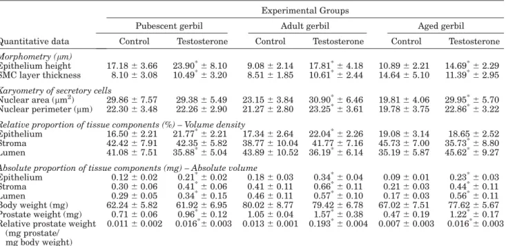
TABLE 1. Quantitative exploratory analysis from experimental animals at different ages
Quantitative data
Experimental Groups
Pubescent gerbil Adult gerbil Aged gerbil
Control Testosterone Control Testosterone Control Testosterone
Morphometry (mm)
Epithelium height 17.1863.66 23.90*68.10 9.0862.14 17.81*64.18 10.8962.21 14.69*62.29 SMC layer thickness 8.1063.08 10.49*63.20 8.5161.85 10.61*62.44 14.6465.10 11.39*62.95
Karyometry of secretory cells
Nuclear area (mm2) 29.8667.57 29.3865.49 23.1563.84 30.90*66.46 19.8164.06 29.95*65.70 Nuclear perimeter (mm) 22.3063.48 22.2662.90 21.2762.80 23.25*63.61 19.7863.75 22.86*63.22 Relative proportion of tissue components (%) – Volume density
Epithelium 16.5062.21 21.77*62.21 17.3462.64 22.04*62.26 19.0863.14 18.6562.52 Stroma 42.4267.91 42.3565.82 38.77610.04 41.7767.16 45.7367.00 35.73*68.80 Lumen 41.0867.51 35.88*65.04 43.89610.52 36.19*66.14 35.1965.87 45.62*69.27
Absolute proportion of tissue components (mg) – Absolute volume
Epithelium 0.1260.02 0.21*60.02 0.1860.03 0.34*60.04 0.0960.01 0.23*60.03 Stroma 0.3060.06 0.41*60.06 0.4160.11 0.66*60.11 0.2160.03 0.44*60.11 Lumen 0.2960.05 0.34*60.15 0.4660.11 0.57*60.10 0.1760.03 0.56*60.11 Body weight (mg) 62.2465.82 61.9266.95 80.0268.77 79.4266.78 67.0267.51 77.6265.67 Prostate weight (mg) 0.7160.06 0.96*60.12 1.0560.04 1.57*60.38 0.4760.19 1.22*60.17 Relative prostate weight
(mg prostate/ mg body weight)
0.01160.002 0.016*60.003 0.01360.001 0.193*60.004 0.00760.003 0.016*60.003
Values represent mean6SD. Statistical analysis based on the Anova and Tukey Tests. *
Figures 14–19 (See overleaf.).
1197
The testosterone participates in the process of prostate development, including the secretory processes, stimulating the synthesis of constituent substances of the sperm (Price, 1963; Aumu¨ller and Seitz, 1990; Rosai, 1996; Hayward et al., 1997; Thomson et al., 1997). Studies using castrated or aged animals with low androgenic levels show that tes-tosterone is capable of increasing the height of epithelial cells and of the secretory apparatus (Scarano et al., 2003).
Heterogeneity was observed in the expression of ARs among the acini in all age groups. This fact is associated with the existence of cellular clones more sensitive to the androgenic stimulus and its effects. This justifies the pres-ence of the acini with neoplasic lesions beside morphologi-cally normal acini. In the treated groups, there was greater density of AR-positive cells, suggesting higher susceptibil-ity to the androgenic action in these animals (Brandes, 1966). Besides, it was noticed that the cells of areas of PIN and focal hyperplasia have larger index of positive marca-tion than in normal areas, inferring that these lesions are associated to AR expression and to the androgenic incen-tive (Gao et al., 2001).
Huynh et al. (2001) suggest that, in the prostate, the production of specific growth factors such as IGF-I is de-pendent on androgens. Such factors act in the activation or inhibition of genes that control the cellular cycle, favor-ing cellular proliferation. Besides, genes that respond to androgens, through AR, are involved in the control of cel-lular division (Galbraith and Duchesne, 1997). In animals whose cells possess predisposition or alteration genetic, such interaction can aggravate the proliferative character, such as the inductor factor (Pollard and Luckert, 1986). This fact can justify the increase of the AR, mainly in hyperplasic and dysplasic regions of the epithelium.
Zanetoni et al. (2005) showed that the induction of tu-mor development is highly potentiated in the presence of testosterone in adult gerbils submitted to chemical carcino-genesis. This study points to histopathologic alterations similar to those found in our work such as PIN and adeno-carcinomas. Furthermore, the gerbils possess a high index of spontaneous histopathologic alterations during the aging process (Zanetoni and Taboga, 2001), similar to what hap-pens in humans, suggesting the existence of genetic pre-disposition that can be potentiated after hormonal supple-mentation. Induction of invasive prostate carcinomas in the rat frequently requires long-term administration of a pharmacological dose of testosterone with or without ap-plication of a chemical carcinogen (Shirai et al., 2000).
Franck-Lissbrant et al. (1998) reported that testosterone stimulates angiogenesis in the ventral prostate of mice af-ter castration, possibly from the metabolic necessity of cells after the hormonal incentive. The obtained data suggest apparent increase of the angiogenesis process in the pros-tate of treated animals, which possibly is involved in the increased energy consumption provoked by the process of cellular synthesis, since the activation of the compound AR-DNA is associated with transcriptional components and coactivators to promote gene transcription (Tsai and O’Malley, 1994).
The prominence of the smooth muscle cells, mainly in pubescent and adult animals, after treatment can be in-volved with direct anabolic processes, such as cellular hy-pertrophy and increase of contractile filaments (McArdle et al., 2003). Besides, that fact can be linked to an increase in the synthesis of elements of the extracellular matrix, on account of increase of synthesis organelles including the endoplasmic reticulum, similar to what happens in ani-mals after castration (Vilamaior et al., 2005).
In some areas, the collagen fibers appear in larger amounts in the treated pubescent and adult animals than in the group control, perhaps reflecting a discreet increase in the synthesis of that element of the extracellular ma-trix. The androgenic receptors are more abundant in the epithelium when compared to the stromal cells (Droller, 1997). Therefore, only an increase in the synthesis of the stromal cells may happen that are responsive to testoster-one, causing heterogeneity along the acini in the amount of fibrillar elements.
The decrease in the thickness of the muscular layer in treated aged animals is probably linked to alterations in the synthetic character of SMCs. With the decrease in the testosterone levels, during aging, SMCs start to develop greater synthetic activity, which justifies the increase in collagen fibers and morphologic alterations of these cells (Horsfall et al., 1994; Vilamaior et al., 2005; Pegorin de Campos et al., 2006). With the increase in the testosterone levels, after the treatment, SMCs reestablish a contractile and fusiform character, which promotes a decrease in the Fig. 20. Serum testosterone levels at the three ages studied.
Fig. 14–19. Fig. 14. Ultrastructure figure. Adult treated prostate. Secre-tory epithelial cells with dilated endomembranes of vesiculous aspect (arrows) for all cytoplasm. Presence of secretion vesicles (sv) and blebs (asterisk). n, nuclei; nu, nucleolus. Scale bar¼1.56mm. Fig. 15. Ultra-structure figure. Aged treated prostate. Detail of the secretory epithelial cell with prominent Golgi (arrows) and secretion vesicles (asterisk). n, nuclei. Scale bar¼0.56mm. Fig. 16. Ultrastructure figure. Pubescent treated prostate. Detail of the stroma showing smooth muscle cells with prominent (rer) and evident nucleolus (nu). col, collagen fibers; n, nuclei.
thickness of the muscular layer, similar to that in cas-trated animals treated with testosterone (Sugimura et al., 1986). Populations of basophilic cells were identified amid the acini epithelium, showing cytoplasmic granular aspect similar to that in neuroendocrine cells described by Capella et al. (1981).
These data demonstrate that the gerbil’s prostate is sus-ceptible to androgenic action at the studied ages, showing proliferative and dysplasic effects mainly in adult and aged animals, perhaps suggesting a possible model for the study of induced neoplasias. Besides, they show reversal of some hypertrophic effects of aging mainly on the pros-tatic stroma, which calls for future studies of this rodent in terms of finding dose-dependent responses with the objective of elucidating the reversibility of aging effects through hormonal therapy.
ACKNOWLEDGMENTS
The authors thank Mr. Luis Roberto Falleiros Ju´nior and Mrs. Rosana S. Sousa for technical assistance, as well as all other researchers at the Microscopy and Microanal-ysis Laboratory. This paper is part of the thesis presented by W.R.S. to the Institute of Biology, UNICAMP, in partial fulfillment of the requirement for a PhD degree, and was supported by grants from the Brazilian agencies National Council of Scientific and Technological Development (CNPq; fellowship to W.R.S.).
LITERATURE CITED
Aumu¨ller G, Seitz J. 1990. Protein secretion and secretory process in male accessory sex glands. Int Rev Cytol 121:127–231.
Brandes D. 1966. The fine structure and histochemistry of prostatic glands in relation to sex hormones. Int Rev Cytol 20:207–276. Buchanan DL, Kurita T, Taylor JA, Lubahn DB, Cunha GR, Cooke
PS. 1998. Role of stromal and epithelial estrogen receptors in vagi-nal epithelial proliferation, stratification, and cornification. Endo-crinology 139:4345–4352.
Buchanan DL, Setiawan T, Lubahn DB, Taylor JA, Kurita T, Cunha GR, Cooke PS. 1999. Tissue compartment-specific estrogen receptor-alpha participation in the mouse uterine epithelial secretory re-sponse. Endocrinology 140:484–491.
Capella C, Usellini L, Buffa R, Frigerio B, Solcia E. 1981. The endo-crine component of prostatic carcinomas, mixed adenocarcinoma-carcinoid tumours and non-tumour prostate: histochemical and ultrastructural identification of the endocrine cells. Histopathol-ogy 5:175–192.
Corradi LS, Go´es RM, Carvalho, HF, Taboga SR. 2004. Inhibition of 5-alpha-reductase activity induces stromal remodeling and smooth muscle de-differentiation in adult gerbil ventral prostate. Differen-tiation 72:198–208.
Cotta-Pereira G, Rodrigo FG, David-Ferreira JF. 1976. The use of tannic acid-glutaraldehyde in the study of elastic related fibers. Stain Technol 51:7–11.
Cunha GR, Donjacour AA, Sugimura Y. 1986. Stromal-epithelial interactions and heterogeneity of proliferative activity within the prostate. Biochem Cell Biol 64:608–614.
Custo´dio AM, Go´es RM, Taboga SR. 2004. Acid phosphatase activity in gerbil prostate: comparative study in male and female during postnatal development. Cell Biol Int 28:335–344.
De Carvalho HF, Lino Neto J, Taboga SR. 1994. Microfibrils: neglected components of pressure-bearing tendons. Ann Anat 176:155–159. Debes JD, Tindall DJ. 2002. The role of androgens and the androgen
receptor in prostate cancer. Cancer Lett 187:1–7.
Donjacour AA, Cunha GR. 1993. Assessment of prostatic protein secretion in tissue recombinant made of urogenital sinus mesen-chyme and urothelium from normal or androgen-insensitive mice. Endocrinology 131:2342–2350.
Droller MJ. 1997. Medical approaches in the management of pros-tatic disease. Br J Urol 79(Suppl):42–52.
Franck-Lissbrant I, Ha¨ggstro¨m S, Damber JE, Bergh A. 1998. Testos-terone stimulates angiogenesis and vascular regrowth in the ven-tral prostate in castrated rats. Endocrinology 139:451–456. Galbraith SM, Duchesne GM. 1997. Androgens and prostate cancer:
biology, pathology and hormonal therapy. Eur J Cancer 33:545–554. Gao J, Arnold JT, Isaacs JT. 2001. Conversion from a paracrine to an autocrine mechanism of androgen-stimulated growth during ma-lignant transformation of prostatic epithelial cells. Cancer Res 61: 5038–5044.
Go´es RM, Zanetoni C, Tomiosso TK, Ribeiro DL, Taboga SR. 2006. Histological response on dorsal and ventral gerbil prostatic lobes induced by different testosterone withdrawal procedures. Micron (in press).
Go¨mo¨ri G. 1937. Silver impregnation for reticulin in paraffin sections. Am J Pathol 13:993–1002.
Gross AS, Didio LJA. 1987. Comparative morphology of the prostate in adult male and female of Praomys (mastomys) natalensis stud-ies with electron microscopy. J Submicros Cytol 19:77–84. Hayward SW, Rosen MA, Cunha GR. 1997. Stromal-epithelial
interac-tions in the normal and neoplasic prostate. Br J Urol 79(Suppl 2): 18–26.
Horsfall DJ, Mayne K, Ricciardelli C, Rao M, Skinner JM, Henderson DW, Marshall VR, Tilley WD. 1994. Age-related in guinea pig pros-tatic stroma. Lab Invest 70:753–763.
Huynh H, Alpert L, Alaoui-Jamali MA, Ng CY, Chan TWM. 2001. Co-administration of finasteride and the pure anti-oestrogen ICI 182,780 act synergistically in modulating the IGF system in rat prostate. J Endocr 171:109–118.
Isaacs JT. 1999. The biology of hormone refractory prostate cancer: why does it develop? Urol Clin North Am 26:263–273.
McArdle W, Katch FI, Katch VL. 2003. Fisiologia do exercı´cio. Rio de Janeiro: Guanabara Koogan.
Mello MLS, Vidal BC. 1980. Pra´ticas de biologia celular. Campinas, Brazil: Edgard Blu¨ cher-Funcamp.
Nawa Y, Horii Y, Okada M, Arizono N. 1994. Histochemical and cyto-chemical characterization of mucosal and connective tissue mast cells of Mongolian gerbil (Meriones unguiculatus). Int Arch Allergy Immunol 104:249–254.
Nolan CC, Brown AW, Cavanagh JB. 1990. Regional variations in nerve cell resposnses to the trimethiltin intoxication in Mongolian gerbil. Acta Pathol Microbiol Immunol Scand 81:204–212. Nomura A, Heilbrun LK, Stemmermann GN, Judd HL. 1988.
Pre-diagnostic serum hormones and the risk of prostate cancer. Cancer Res 48:3515–3517.
Pegorin de Campos SGP, Zanetoni C, Goes RM, Taboga SR. 2006. Bi-ological behaviorof the gerbil ventral prostate in three phases of postnatal development. Anat Rec 288:723–733.
Pinheiro PFF, Almeida CCD, Segatelli, TM, Martinez M, Padovani CR, Martinez FE. 2003. Structure of the pelvic and penile urethra-relationship with the ducts of the sex accessory glands of the Mon-golian gerbil (Meriones unguiculatus). J Anat 202:431–444. Pollard M, Luckert PH. 1986. Production of autochthonous prostate
cancer in Lobund-Wistar rats by treatments with N-nitroso-N-methylurea and testosterone. J Natl Cancer Inst 77:583–587. Price D. 1963. Comparative aspects of development and structure in
the prostate. Natl Cancer Inst Monogr 12:1–27.
Rosai J. 1996. Male reproductive system. In: Rosai J, editor. Ackerman’s surgical pathology, vol. 1, 8th ed. St. Louis: Mosby-Year. p 1221– 1256.
Rosner W, Hryb DJ, Khan MS, Nakhla AM, Romas NA. 1999. Sex hor-mone-binding globulin mediates steroid hormone signal transduction at the plasma membrane. J Steroid Biochem Cell Biol 69:481–485. Santos FCA, Go´es RM, Carvalho HF, Taboga SR. 2003. Structure,
histochemistry and ultrastructure of the epithelium and stroma in the gerbil (Meriones unguiculatus) female prostate. Tissue Cell 35:447–457.
Santos FC, Leite RP, Custodio AM, Carvalho KB, Monteiro-Leal LH, Santos AB, Go´es RM, Carvalho HF, Taboga SR. 2006. Testosterone stimulates growth and secretory activity of the adult female prostate of the gerbil (Meriones unguiculatus). Biol Reprod 75:730–739.
1199
Scarano WR, Cordeiro RS, Go´es RM, Taboga SR. 2003. Structure and morphometry of pubertal gerbil (Meriones unguiculatus) ventral prostate after hormonal therapy. Act Microsc 12:347– 348.
Shirai T, Takahashi S, Cui L, Futakuchi M, Kato K, Tamano S, Imaida K. 2000. Experimental prostate carcinogenesis: rodent models. Mutat Res 462:219–226.
So AI, Coll AH, Gleave ME. 2003. Androgens and prostate cancer. World J Urol 21:325–337.
Sugimura Y, Cunha GR, Donjacour AA. 1986. Morphogenesis of ductal networks in the mouse prostate. Biol Reprod 34:961– 971.
Taboga SR, Santos AB, Rocha A, Vidal BC, Mello MLS. 2003. Nu-clear phenotypes and morphometry of human secretory prostatic cells: a comparative study of benign and malignant lesions in Bra-zilian patients. Caryologia 56:313–320.
Thomson AA, Foster BA, Cunha GR. 1997. Analyses of growth factor and receptor mRNA levels during development of the rat seminal vesicle and prostate. Development 124:2431–2439.
Thomson AA. 2001. Role of androgens and fibroblast growth factors in prostatic development. Reproduction 121:187–195.
Tsai MJ, O’Malley BW. 1994. Molecular mechanisms of action of ste-roid/thyroid receptor superfamily members. Annu Rev Biochem 63:451–486.
Vilamaior PSL, Taboga SR, Carvalho HF. 2005. Modulation of smooth muscle cell function: morphological evidence for a contractile to syn-thetic transition in the rat ventral prostate. Cell Biol Int 29:809–816. Vilamaior PSL, Taboga SR, Carvalho HF. 2006. Postnatal growth of the ventral prostate in Wistar rats: a stereological and morphomet-rical study. Anat Rec 288:885–892.
Wang Y, Sudilovsky D, Zhang B, Haughney PC, Rosen MS, Wu DS, Cunha TJ, Dahiya R, Cunha GR, Hayward SW. 2001. A human prostatic epithelial model of hormonal carcinogenesis. Cancer Res 61:6064–6072.
Weibel ER. 1979. Principles and methods for the morphometric study of the lung and other organs. Lab Invest 12:131–155.
Zanetoni C, Taboga SR. 2001. Age-related modifications in stromal and epithelila compartments of the male prostate of Meriones unguiculatus. Acta Microsc 3:203–204.