Research Article
Predictive Criteria to Study the Pathogenesis of
Malaria-Associated ALI/ARDS in Mice
Luana S. Ortolan,
1,2Michelle K. Sercundes,
3Renato Barboza,
4Daniela Debone,
3Oscar Murillo,
5Stefano C. F. Hagen,
6Momtchilo Russo,
1Maria Regina D’ Império Lima,
1José M. Alvarez,
1Marcos Amaku,
7Claudio R. F. Marinho,
5and Sabrina Epiphanio
1,81Departamento de Imunologia, Instituto de Ciˆencias Biom´edicas, Universidade de S˜ao Paulo, Edif´ıcio Biom´edicas IV,
Avenida Professor Lineu Prestes, No. 1730, 05508-900 S˜ao Paulo, SP, Brazil
2Departamento de Ciˆencias Biol´ogicas, Universidade Federal de S˜ao Paulo, Rua Professor Artur Riedel, No. 275,
Jardim Eldorado, 09972-270 Diadema, SP, Brazil
3Instituto de Medicina Tropical de S˜ao Paulo, Universidade de S˜ao Paulo, Avenida Dr. En´eas Carvalho de Aguiar, No. 470,
05403-000 S˜ao Paulo, SP, Brazil
4Departamento de Ciˆencias Exatas e da Terra, Universidade Federal de S˜ao Paulo, Rua Professor Artur Riedel, No. 275,
Jardim Eldorado, 09972-270 Diadema, SP, Brazil
5Departamento de Parasitologia, Instituto de Ciˆencias Biom´edicas, Universidade de S˜ao Paulo, Avenida Professor Lineu Prestes,
No. 1374, Edif´ıcio Biom´edicas II Cidade Universit´aria “Armando Salles Oliveira”, 05508-000 S˜ao Paulo, SP, Brazil
6Departamento de Cirurgia, Faculdade de Medicina Veterin´aria e Zootecnia da Universidade de S˜ao Paulo,
Avenida Professor Dr. Orlando Marques de Paiva, No. 87, Cidade Universit´aria, 05508 270 S˜ao Paulo, SP, Brazil
7Departamento de Medicina Veterin´aria Preventiva e Sa´ude Animal, Faculdade de Medicina Veterin´aria e Zootecnia da Universidade
de S˜ao Paulo, Avenida Professor Dr. Orlando Marques de Paiva, No. 87, Cidade Universit´aria, 05508 270 S˜ao Paulo, SP, Brazil
8Departamento de An´alises Cl´ınicas e Toxicol´ogicas, Faculdade de Ciˆencias Farmacˆeuticas, Universidade de S˜ao Paulo,
Avenida Professor Lineu Prestes, No. 580, Bloco 17, Cidade Universit´aria “Armando Salles Oliveira”, 05508-000 S˜ao Paulo, SP, Brazil
Correspondence should be addressed to Sabrina Epiphanio; [email protected]
Received 5 June 2014; Accepted 16 July 2014; Published 2 September 2014
Academic Editor: Mauricio Martins Rodrigues
Copyright © 2014 Luana S. Ortolan et al. his is an open access article distributed under the Creative Commons Attribution License, which permits unrestricted use, distribution, and reproduction in any medium, provided the original work is properly cited.
Malaria-associated acute lung injury/acute respiratory distress syndrome (ALI/ARDS) oten results in morbidity and mortality. Murine models to study malaria-associated ALI/ARDS have been described; we still lack a method of distinguishing which mice will develop ALI/ARDS before death. his work aimed to characterize malaria-associated ALI/ARDS in a murine model and to demonstrate the irst method to predict whether mice are sufering from ALI/ARDS before death. DBA/2 mice infected withPlasmodium bergheiANKA developing ALI/ARDS or hyperparasitemia (HP) were compared using histopathology, PaO2 measurement, pulmonary X-ray, breathing capacity, lung permeability, and serum vascular endothelial growth factor (VEGF) levels according to either the day of death or the suggested predictive criteria. We proposed a model to predict malaria-associated ALI/ARDS using breathing patterns (enhanced pause and frequency respiration) and parasitemia as predictive criteria from mice whose cause of death was known to retrospectively diagnose the sacriiced mice as likely to die of ALI/ARDS as early as 7 days ater infection. Using this method, we showed increased VEGF levels and increased lung permeability in mice predicted to die of ALI/ARDS. his proposed method for accurately identifying mice sufering from ALI/ARDS before death will enable the use of this model to study the pathogenesis of this disease.
1. Introduction
Malaria is an infectious disease with a huge impact on public health and a high mortality rate. According to the World
Health Organization, approximately 3.3 billion people were at risk of contracting malaria in 2011 [1–3]. In some indi-viduals,Plasmodiuminfection may result in severe malaria that can lead to ALI/ARDS [4, 5]. Patients infected with Volume 2014, Article ID 872464, 12 pages
P. falciparum, P. vivax, and P. knowlesi can develop ALI or ARDS with mortality rates of approximately 80% [6, 7]. Malaria-associated ALI/ARDS is thought to be due, in part, to increased alveolar permeability, parasite sequestration, and host immune response; however, the mechanisms behind it are largely unknown [4].
ALI/ARDS can occur at any time during an infection, even ater treatment with antimalarial drugs when para-sitemia has been reduced (reviewed in [4]). he development of ALI/ARDS, along with its negative outcomes, makes the prospective identiication and efective treatment of those who develop this syndrome very important. hough, there is little information on malaria-associated ALI/ARDS progres-sion, resulting in a lack of knowledge of the mechanisms of pathogenesis; therefore, the understanding of mouse models is essential.
Several reports have observed lung injury in mice infected withP. berghei(Pb) strains [8–19]. he observations have highlighted possible roles for many factors, including platelet-activating factor receptor [8], urokinase receptor [9], ICAM-1 [10, 11], CD40 [12], neutrophils [13], vascular endothelial growth factor (VEGF) [14], epithelium sodium channel activity [15], CD36-dependent parasite sequestration [16], hemozoin deposition [17], and CD8+ T lymphocytes [18], in malaria-associated lung injury.
Recently, there have been many models described that focus on the pulmonary pathology associated with malaria, including the classical C57BL/6 susceptible mouse model of cerebral malaria infection withPbANKA [16, 19]. Other models have used diferent parasite/mouse combinations that result in the mice surviving for longer periods of time (without signs of cerebral malaria), theoretically allowing the investigation of disease progression over time [14, 15, 18]. However, none of them were able to identify ALI/ARDS before death. Here, we have characterized a murine model of malaria-associated ALI/ARDS that shows similarities between humans and murine ALI/ARDS. Moreover, we proposed a method for classifying mice sufering from ALI/ARDS before the time of death as a predictive model for malaria-associated ALI/ARDS.
2. Materials and Methods
2.1. Mice and Parasites. DBA/2 male mice 6–10 weeks old
(purchased from the Department of Parasitology, University of S˜ao Paulo, Brazil) were infected with 1 ×10⁶ P. berghei
ANKA (clone 1.49 L) infected red blood cells (iRBCs), as pre-viously described [14]. Parasitemia and mortality were moni-tored daily. Parasitemia levels were analyzed using Giemsa-stained peripheral blood smears.
2.2. Anesthesia and Euthanasia. All eforts were made to
prevent undue stress or pain to the mice. Mice with signs of imminent death were euthanized to avoid sufering. Before restraint for X-rays, the mice were given ketamine (100 mg/kg) and xylazine (5 mg/kg). he mice were eutha-nized with ketamine (300 mg/kg) (Vetbrands, Brazil) and xylazine (22.5 mg/kg) (Syntec, Brazil), and consciousness was
checked by testing the pedal relex, heartbeats and breathing movements.
2.3. Histological Evaluations. Necropsy was performed in
mice dying naturally from the malaria or mice sacriiced on the 20th days ater infection (DAI) to complete the exper-iment and to avoid animal sufering. he lungs were col-lected, ixed in bufered 10% formalin and then embedded in parain, sectioned at 5�m onto slides and stained with hematoxylin-eosin (HE) and phosphotungstic acid hema-toxylin (PTAH), to emphasize ibrin, as previously described [20].
2.4. Arterial Blood Analyses and Measurements of Body
Tem-perature. Mice were placed near a heat lamp for three
minutes to increase peripheral blood low. he mice were then restrained by hand, the ventral artery of the tail was nicked with a small scalpel blade, and capillary tubes containing lith-ium-heparin (50 IU/mL) were placed underneath the cut to collect approximately 100�L of blood. he blood was imme-diately placed in an i-STAT EG 8+ cartridge and analyzed using the iSTAT System Analyzer (Abbott group). he PaO2/FiO2 was calculated assuming that the fraction of inspired O2(FiO2) was 0.21. In a subset of mice, the inguinal temperatures were assessed on day 0 and on the 5th, 7th and 9th DAI using a DT-203/60SEC digital thermometer (Becton Dickinson, Franklin Lakes, New Jersey, EUA).
2.5. X-Ray. Mice received light anesthesia on the 7th DAI and
were X-rayed for 0.066 seconds in a mA100 ine focus Bucky V mAs 6.6 (RAYtech machine KV37, USA). A trained tech-nician blinded to the infection status of the mice examined the X-rays, which were scored for signs of lung injury: 0, no change; 1, discrete and/or light opaciication; and 2, difuse opaciication. he mice were later classiied as sufering from ALI/ARDS or HP at death by the presence or absence of pleural efusion, respectively.
2.6. Determination of Respiratory Pattern. Respiratory
pat-terns (respiratory frequency (RF) and enhanced pause (Penh)) were monitored on the 5th, 7th, 9th, 15th, and 20th DAI by placing the mice in an unrestrained whole-body plethysmography chamber (WBP, Buxco Electronics, Wilm-ington, North Carolina, USA) for 10 minutes (basal level). he data were collected using Biosystems XA sotware and included the RF (breaths/minute) and variables to calculate the Penh, a theoretical variable that correlates with both pul-monary resistance and intrapleural pressure [21]. he Penh is calculated by [22]
Penh= peak expiration speed peak inspiration speed× (
expiratory time relaxation time − 1) .
(1)
2.7. Identifying ALI/ARDS in Mice before Death. To identify
7th DAI (10–12 mice per group). In the survival group, any mouse showing pleural efusion or red and congested lungs at necropsy, the cause of death was attributed to ALI/ARDS. In contrast, at necropsy, in mice without pleural efusion that died ater 13th DAI with pale lungs and high levels of parasitemia, the cause of death was attributed to HP and consequently anemia.
Individual mice sacriiced on the 7th DAI were classiied as having been likely to die of ALI/ARDS or HP, by comparing their respiratory patterns and parasitemia levels with the survival group, in which thecausa mortiswas known (Supple-mentary Figure S1 in Supple(Supple-mentary Material available online athttp://dx.doi.org/10.1155/2014/872464).
In each individual experiment, using the survival group, we established three cut-ofs using receiver operating char-acteristic (ROC) curves for the Penh, RF, and parasitemia measured on the 7th DAI, which were used as predictive criteria. he cut-ofs from this group were chosen based on the maximum sensitivity and speciicity for each parameter. he mice sacriiced on day 7 were also screened for the same parameters before sacriice, and they were grouped based on the cut-ofs from the ROC curves generated using data from the survival group. he same cut-ofs were used to retrospectively classify the sacriiced group as sufering from ALI/ARDS or HP. he mice were said to have sufered from ALI/ARDS if they were above the cut-of for at least two of the three variables. For this method to work, it was necessary that three or more animals died by ALI/ARDS in the survival group/per experiment. In all of the experiments, we calculated the sensitivity and speciicity from the survival group.
2.8. Conirming the Accuracy of the Groupings. To conirm if
and when the mice could be grouped using the respiratory pattern cut-ofs and parasitemia, conirmation experiments were performed. Two survival groups were assessed for pleural efusion and reddish lungs, which were used as the gold standards for mice dying of lung injury. hese criteria constitute a practical phenotype for assessing ALI/ARDS because they are not arbitrary and can be assessed imme-diately during the necropsy; furthermore, previous results have shown that 100% of the mice that die between 7–12 DAI with clear signs of ALI, including the presence of pulmonary edema, hemorrhages, and hypoxemia [14]. he survivors were monitored until the 20th DAI, and the cause of death was determined. At the end of the experiment, the data from one group were used to generate the ROC curves that were then used to classify the mice in the second group on the 7th DAI; these results were then compared with observa-tions of the pathology from necropsy of the second group (Supplementary Figure S2). Conirmation that the second mouse groupings were likely to be correct was performed in experiments in which the sensitivity and speciicity of the groupings using the ROC curves were calculated.
2.9. Lung Permeability and Edema. To investigate lung
per-meability, on the 7th DAI, mice were injected intravenously with 0.2 mL of 1% Evans Blue (Sigma). he mice were
sacriiced 45 minutes later, and the lungs were weighed and placed in 2 mL of formamide (Merck) for 48 hours at 37∘C. he absorbance of the formamide was then measured at
�620 nm. he amount of Evans Blue staining per gram of lung tissue was calculated from a standard curve. he sacriiced mice were classiied as sufering from ALI/ARDS by the ROC curves generated from a survival group as described above. he lung permeability of the ALI/ARDS and HP mice was expressed as fold increase in relation to that of the NI mice. To further investigate the presence of edema, in a group of survival mice, the lungs were weighed immediately ater natural death, and the wet weights were recorded and compared between ALI/ARDS and HP mice of the same age. he mice were conirmed as sufering from ALI/ARDS or HP at death by the presence or absence of pleural efusion, respectively.
2.10. VEGF in Serum. On the 7th DAI, mice were
anesthe-tized, and their serum was collected by cardiac puncture. An ELISA kit (R&D Systems) was used to quantify VEGF levels in the serum according to the manufacturer’s instructions. he VEGF level of the ALI/ARDS and HP mice was expressed as fold increase in relation to that of the NI mice. he mice were classiied as sufering from ALI/ARDS or HP by comparing the predictive criteria and VEGF levels.
2.11. Hematological Parameters Determination. Blood
sam-ples were collected in tubes containing sodium citrate as anti-coagulant. Total number of red blood cells, hemoglobin, and hematocrit were measured using V-53 reagent kit (Mindray, P.R. China) and Auto Hematology Analyzer BC-5300Vet (Mindray, Nanshan, Shenzhen, P.R. China).
2.12. Statistical Analysis. he data were analyzed by
D’Agost-ino-Pearson normality test. Nonparametric variables were compared using Mann-Whitney test. he simultaneous efects of two factors were analyzed by two-way ANOVA fol-lowing Bonferroni post-hoc test. he diferences between the groups were considered signiicant when� ≤ 0.05. Statistical analyses were performed in GraphPad Prism version 5.0, including assessments of sensitivity and speciicity. To estab-lish cut of from data, ROC curves were generated using the results of the control group in MedCalc version 8.2.1.0. he Penh, RF and, EP data were analyzed using SPSS for Microsot version 19.0 through a Pearson correlation (Penh versus RF:
−0.827,�: 0.001; Penh versus EP:−0.152,�: 0.001; RF versus EP: 0.200,�: 0.05) for further grouping. he cluster analysis was developed by case grouping using Ward’s method with a Euclidean distance analysis, which generated a dendrogram grouping each of the subjects studied and their physiologic characteristics analyzed into a particular cluster.
3. Results
3.1. Overview of Pathology at Death in PbANKA-Induced
ALI/ARDS. To characterize and discriminate the pathology
associated with ALI/ARDS or HP, DBA/2 mice were infected
followed until their deaths. Subsequently, the animals were necropsied and thecausa mortisdetermined. Survival anal-ysis revealed an average of 49.2% (25–75%) of the mice had died of ALI/ARDS between the 7th and 12th DAI, whereas the HP mice died between the 13th and 21st DAI (Figure1(a)). Although parasitemias were increased in both groups, the HP group had higher levels of parasitemia, at approximately 40– 50% on the day of death (Figure1(b)). Comparing the lung weights, we observed that the mice that died with ALI/ARDS had heavier lungs (averaging 40% more mass) than the mice that died with HP, suggesting edema (Figure1(c)).
At necropsy, noninfected (NI) mice had light pink lungs, and no liquid inside of the thoracic cavity was observed (Figure 1(d)). However, mice that died with ALI/ARDS had reddish lungs and pleural efusion (Figure 1(e)). In these mice, the histological changes where characterized by marked alveolar edema and hemorrhage, along with neu-trophil-dominant inlammatory cellular iniltration, foamy macrophages in the alveolar and interstitial sites, and destruction of the alveolar septa (Figure1(h)), as previously described [14]. In contrast to the ALI/ARDS mice, the animals that died with HP ater the 13th DAI had grayish lungs and a darkened spleen and liver but no pleural efusion (Figure1(f)) and severe anemia, with decreases in the number of erythrocytes, hemoglobin level, and hematocrit percentage (Supplementary Figure S3). hese HP mice had interstitial pneumonia with mononuclear inlammatory cells but at a later time in the infection, diagnosed on the day of death (Figure1(i)). Interestingly, in the lungs of ALI/ARDS mice we observed the presence of acellular eosinophilic membranes that adhered to the alveolar ducts and walls and hyaline membranes, a hallmark of ALI/ARDS in humans (Figures1(j) and1(k)).
3.2. Chest Radiography Shows Lung Opacity in
PbANKA-Induced ALI/ARDS. Bilateral iniltrates observed on frontal
chest radiographs are recognized as a criterion for the diag-nosis of ALI and ARDS [23]. In our work, X-ray analysis on the 7th DAI revealed lung opaciication, which was more prominent in the ALI/ARDS group than in the HP group (Figures2(a)and2(b)). NI mice had no changes in the lungs.
3.3. PbANKA-Induced ALI/ARDS Is Associated with
Hypox-emia and Decreased Body Temperature. Hypoxemia is not a
direct assessment of damageper sebut is oten a manifes-tation of injury [24]. In humans, the hypoxemia was deined by he American-European Consensus Conference as PaO2/FiO2 ≤300 mmHg (for ALI) or ≤200 mmHg (for ARDS) [25]. In agreement with previous results [14], we show that the majority of the DBA/2 mice infected with
PbANKA who died of ALI/ARDS had PaO2/FiO2 values between 200 and 300 mmHg, and we further demonstrate that mice that developed the more severe form of ARDS, presented PaO2/FiO2values of≤200 mmHg. On the 7th DAI, the PaO2/FiO2in the ALI/ARDS group (234.3 ± 21.38) was signiicantly lower than the level in the HP group (303±17.26;
� = 0.029) (Figure2(c)). NI mice showed PaO2/FiO2values above 300 mmHg with average 371.42 (SD±24.27). here are
no agreed-upon validated PaO2/FiO2data in animal models of lung injury [26]; thus, we categorized all of the animals in the group with lung injury as ALI/ARDS.
he infected mice had slightly increased body tempera-tures between day 0 and the 5th DAI; however, between the 7th and 9th DAI, their temperatures dropped and the mice became hypothermic (Figure2(d)). Mice that would start to develop ALI/ARDS and die had the lowest temperatures on the 7th DAI compared with the HP mice. We hypothesized that the reduction in body temperature could be related to decreased survival of the animals, especially those that developed ALI/ARDS.
3.4. Respiratory Patterns and Parasitemia Are Correlated
in PbANKA-Induced ALI/ARDS. To characterize the lung
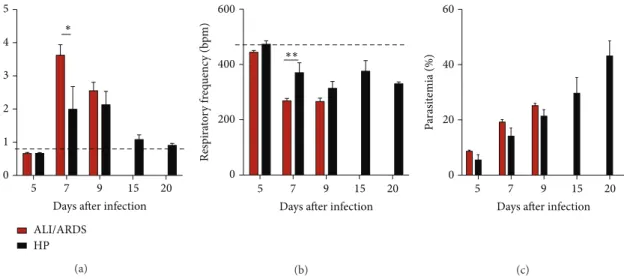
physiopathology during infection, we analyzed the enhanced pause (Penh), respiratory frequency (RF), and parasitemia (EP) levels at ive diferent time points. On the 5th DAI the ALI/ARDS group had breathing patterns and parasitemia similar to the HP group and NI mice. However, by the 7th DAI, the ALI/ARDS group had increased Penh, decreased RF, and a tendency to increase the parasitemia even if not signiicantly, compared with the HP mice (Figures 3(a)– 3(c)). Ater the 9th DAI, the statistical comparison between the ALI/ARDS and HP mice could not be performed in the individual experiments due to the minimal numbers of surviving ALI/ARDS mice. Interestingly, animals that survived for longer periods, that is, animals that did not develop ALI/ARDS and subsequently died by HP, breathing patterns returned nearly to baseline levels. NI mice had no changes in breathing patterns over time.
Parasitemia increased over the course of infection (Figure3(c)). On the day of death, parasitemia in the ALI/ ARDS group was 20.8% (SD±4.6), while parasitemia in mice that died of HP was 40.9% (SD±9.21;� ≤ 0.0001). However, on the 7th DAI, the mice that went on to die of ALI/ARDS had 17.0% (SD±5.0) iRBCs, while the mice that would go on to die with HP were 12.2% (SD±4.5) parasitemic (Supplementary Figure S4a).
Aiming to perform a correlation study examining the res-piratory parameters (Supplementary Figure S4b and S4c) and EP (Supplementary Figure S4a), we conducted experiments on the 7th DAI, when the onset of important pulmonary pathology occurred, rather than using the 5th DAI when we did not observe consistent diferences in respiratory patterns between the study groups.
he high correlation between Penh, RF, and EP and the development of ALI/ARDS or HP (Pearson correlation Penh
versusRF:−0.827,� = 0.001; PenhversusEP:−0.152,� =
100
80
60
40
20
0
0 3 6 9 12 15 18 21
Su
rv
iv
al
(
%
)
Days ater infection
(a)
Days ater infection 50
40
30
20
10
0
0 2 4 6 8 10 12 14 16 18 20
ALI/ARDS HP
P
arasi
temia (%)
(b)
0.0 0.1 0.2 0.3 0.4
ALI/ARDS HP
L
un
g w
eig
h
t (g)
∗∗
(c)
(d) (e) (f)
(g) (h) (i)
(j) (k)
NI-score0 ALI/ARDS-score2 HP-score1 HP-score0
(a)
L
u
n
g o
p
acifica
tio
n in x-ra
y (s
co
re
) 3
2
1
0
ALI/ARDS HP
∗
(b)
ALI/ARDS HP
500
400
300
200
100
0
Pa
O2
/
FiO
2
∗
(c)
ALI/ARDS HP
∗∗∗ 40
38
36
34
32
30
0 5 7 9
Days ater infection
T
em
p
era
tur
e (
∘C)
(d)
Figure 2: Radiography of the lungs, hypoxemia, and body temperature over time. (a) From let to right, X-rays from NI and infected DBA/2 mice that died with ALI/ARDS and HP showing diferent lung opaciication scores on the 7th DAI. (b) Lung opaciication scores on the 7th DAI. Mice that will later die with ALI/ARDS have a higher lung opaciication score compared with mice that will die with HP (� = 12;
∗� ≤ 0.05, Mann-Whitney test of scores taken from two separate experiments). NI mice do not have any lung opaciication and are assigned
score zero. (c) PaO2/FiO2values inP. bergheiANKA-infected mice on the 7th DAI. Results from three grouped experiments (� = 13mice,
∗� ≤ 0.05; Mann-Whitney test). (d) Body temperatures in DBA/2 mice infected withP. bergheiANKA slightly increased on the 5th DAI from
37.1∘C in the NI mice to 37.3 in the ALI/ARDS mice and 37.4∘C in the HP mice). However, the temperatures dropped and the mice became hypothermic (especially the ALI/ARDS mice), with mean temperatures of 33.0∘C on the 7th DAI and 32.8∘C on the 9th DAI. Results are from three grouped experiments (� = 31mice;∗∗∗� ≤ 0.001, two-way ANOVA with Bonferroni post test). Data (d) represents means and SEM. he dashed line represents the mean value of NI mice. NI: noninfected mice; ALI/ARDS: acute lung injury/acute respiratory distress syndrome; HP: hyperparasitemia.
while groups 3 and 4 were dominated by HP, with a diference of 25%. Additionally between the groups with major higher frequencies of one of the two pathologies (groups 1 and 2 or groups 3 and 4), the diference between the analyzed char-acteristics was 10%.
3.5. A Murine Model to Predict Malaria-Associated ALI/ARDS
at an Early Time Point. Mice that die from ALI/ARDS
present altered Penh, RF, and EP values from the mice that die from HP; thus, we hypothesized that those parameters could be used as predictive criteria for thecausa mortis. As described in the materials and methods, for each individual experiment, we used an infected control group (survival group) and established cut-ofs using ROC curves for Penh, RF, and EP (Supplementary Table S1) measured on the 7th DAI and applied these cut-ofs to classify the sacriice group
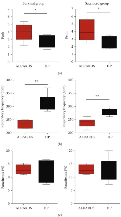
(Supplementary Figure S1). Following this procedure, the mice sacriiced on the 7th DAI could be classiied as likely to die with ALI/ARDS or HP. We compared their respiratory patterns and parasitemia with a survival group that was not sacriiced and for which the cause of death was known. he sensitivity (≤100% and≥67%; average 88.31%; SD±11.95) and speciicity (100%≤and ≥71%; average 90.85%; SD±10.81) were calculated from the survival group for each individual experiment. In addition, we observed that the respiratory patterns and parasitemia were similar between the survival group and the sacriiced group (Figures5(a)–5(c)).
ALI/ARDS HP
∗ 4
2
5 5
3
1
0
7 9 15 20
Days ater infection
Pe
n
h
(a)
Resp
ira
to
ry
f
req
uenc
y (b
p
m
)
600
400
200
0
∗∗
5 7 9 15 20
Days ater infection
(b)
P
arasi
temia (%)
60
40
20
0
5 7 9 15 20
Days ater infection
(c)
Figure 3: Breathing patterns and parasitemia from ALI/ARDS and HP mice over time. (a) and (b) Breathing patterns and (c) parasitemia from DBA/2 mice infected withP. bergheiANKA that developed ALI/ARDS and HP over time. (a) here was no evidence on the 5th and 9th DAI that the ALI/ARDS and HP mice had diferent breathing patterns. However, on the 7th DAI, there was evidence that the ALI/ARDS mice had a higher enhanced pause (Penh) (a) and a lower respiratory frequency (b) than the HP mice. Parasitemia increased over time in both groups (c). Results are representative from three independent experiments (� = 11mice/experiment;∗� ≤ 0.05, two way ANOVA with Bonferroni post test). he dashed line represents the mean value of NI mice; NI: noninfected mice; ALI/ARDS: acute lung injury/acute respiratory distress syndrome; HP: hyperparasitemia.
Group 1 Group 2 Group 3 Group 4
5 10 15 20 25
0
F
req
uenc
y (%)
Figure 4: Breathing patterns and parasitemia could be used to group mice into two main clusters. Ward’s linkage cluster analysis illustrates the distance between the physiological cluster patterns in DBA/2 mice infected with P. berghei ANKA that developed ALI/ARDS and HP. Values were measured on the 7th DAI. he data are from 13 independent experiments;� = 142mice. Group 1 = 88.46% of individuals with ALI/ARDS and 11.53% with HP; group 2 = 57.77% of individuals with ALI/ARDS and 42.22% with HP; group 3 = 19.35% individuals with ALI/ARDS and 80.64% with HP; group 4 = 21.42% of individuals with ALI/ARDS and 78.57% with HP. ALI/ARDS: acute lung injury/acute respiratory distress syndrome; HP: hyperparasitemia.
(Supplementary Figure S2). On the 7th DAI, we were able to group the mice in the ALI/ARDS or HP groups with 91% sensitivity and 76% speciicity in the three grouped experiments (Table1). Among the experiments, the best result was 100% sensitivity and 100% speciicity, and the worst result was 66.6% sensitivity and 60% speciicity. On the 9th DAI, we were not able to group the mice, as no ALI/ARDS mice from the survival group were alive.
3.6. PbANKA-Induced ALI/ARDS Causes Breakdown of the
Alveolar-Capillary Barrier. In a previous study, we
demon-strated that VEGF promotes malaria-associated ALI in mice
Table 1: Conirming the accuracy of the groupings. True and false pathologies checked by the ROC curves from the predictive criteria∗ on the 7th DAI and thecausa mortis.
Pathology
Test ALI/ARDS HP Total
Hits 11 13 24
Errors 1 4 5
Total 12 17 29
∗Penh, respiratory frequency, and parasitemia.
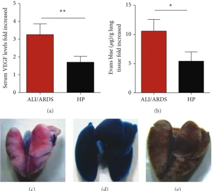
and that expression of this growth factor is increased in mice that died of ALI [14]. Here, using blood samples from mice sacriiced on the 7th DAI, we conirmed that mice classiied as likely to die with ALI/ARDS and HP had VEGF serum levels 3.3-fold and 1.7-fold higher than those in the NI group, respectively (Figure6(a)). Furthermore, the pulmonary vascular permeability measured by Evans blue uptake in the lungs on the 7th DAI was higher in mice predicted to die of ALI/ARDS (10.6-fold higher than NI mice) than in those classiied as sufering from HP (5.5-fold higher than NI mice) (Figures6(b)–6(e)).
4. Discussion
ALI/ARDS HP ∗
Pe
n
h
7
6
5
4
3
2
1
0
ALI/ARDS HP
∗
Pe
n
h
7
6
5
4
3
2
1
0
Survival group Sacrificed group
(a)
ALI/ARDS HP ALI/ARDS HP
∗∗ ∗∗
Resp
ira
to
ry
f
req
uenc
y (b
p
m
)
400
350
300
250
200
Resp
ira
to
ry
f
req
uenc
y (b
p
m
)
400
350
300
250
200
(b)
ALI/ARDS HP ALI/ARDS HP
P
arasi
temia (%)
20
15
10
0
5 P
arasi
temia (%)
20
15
10
0 5
(c)
Figure 5: A murine model to predict malaria-associated ALI/ARDS. (a) Penh (enhanced pause), (b) respiratory frequency, and (c) parasitemia measured on the 7th DAI. he sacriiced mice were classiied according predictive model, using the parameter cut-ofs measured from the survival mice and applied to the sacriiced mice. DBA/2 mice infected withP. bergheiANKA and their breathing parameters were measured in plethysmograph chambers (BUXCO Electronics, USA). Note that these three parameters are similar between the survival group and sacriiced group; (� = 11mice/group;∗� < 0.05,∗∗� < 0.005, Mann-Whitney test). ALI/ARDS: acute lung injury/acute respiratory distress syndrome; HP: hyperparasitemia; bpm: beats per minute. Results are representative of more than 5 independent experiments.
2010). Recently, in accord with he Berlin Deinition, the ALI term was abolished and the ARDS was deined based on oxy-genation: mild ARDS (200 mmHg PaO2/FiO2≤300 mmHg), moderate ARDS (100 mmHg PaO2/FiO2 ≤200 mmHg), and severe ARDS (bellow 100 mmHg PaO2/FiO2, among other criteria [27]. However, he Berlin Deinition was not vali-dated by a later study [28] and ALI is still used for lung injury in mice. Despite the discussion in the ield, some consensual
parameters are used to study ALI in mice such as kinetics of injury, radiographic evaluation, physiological assessment, histological evidence of lung injury, and assessment of increased permeability of the alveolar-capillary membrane [26].
S
er
um VEGF le
ve
ls
fo
ld incr
ea
se
d 5
4
3
2
1
0
ALI/ARDS HP
∗∗
(a)
5 15
0 10
ALI/ARDS HP
E
va
n
s b
lue (
𝜇
g)/g l
un
g
∗
tiss
ue f
o
ld incr
ea
se
d
(b)
(c) (d) (e)
Figure 6: Increased vascular permeability and serum VEGF protein conirmed the predictive criteria for malaria-associated ALI/ARDS. (a) Serum VEGF protein in DBA/2 mice infected withP. bergheiANKA on the 7th DAI was measured by ELISA. he VEGF levels are higher in the mice classiied as likely to die with ALI/ARDS compared with HP mice (according to the proposed predictive criteria). he data represent fold increases in relation to NI mice taken from three experiments; (� = 28mice;∗∗� < 0.005, Mann-Whitney test). (b) Lung vascular permeability in DBA/2 mice infected withP. bergheiANKA 7th DAI, assessed using Evans Blue. he vascular permeability is higher in the mice classiied as likely to die with ALI/ARDS compared with the HP mice (according to the proposed predictive criteria). he data represent fold increased in relation to NI mice taken from three experiments; (� = 51mice;∗� < 0.05, Mann-Whitney test). Bars represent means and SEM. he images represent (c) NI: noninfected mice, (d) ALI/ARDS: acute lung injury acute respiratory distress syndrome, and (e) HP: hyperparasitemia.
such a complex disease [30]. We showed that DBA/2 mice infected withPbANKA constitute a rodent model of malaria-associated ALI/ARDS [14], and here, we approach respiratory and parasitological parameters to devise a mathematical model able to predict development of ALI/ARDS during
PbANKA infection.
his murine model clearly showed edema in ALI/ARDS mice, indicating that these animals had more severe disease and that they were dying during the exudative phase of ALI/ARDS. In addition, we observed local destruction of the alveolar epithelium, with denuded areas covered by ibrin-containing hyaline membranes, a hallmark of ALI/ARDS in humans [23], along with neutrophils, alveolar macrophages, mononuclear cells, and iRBC. he presence of edema observed in the ALI/ARDS mice is also relected by the increased wet weight of the lungs [31], although the increased weight of the lungs could be partially due to cellular accu-mulation [18]. he opaque appearance of the lungs in X-rays and the opaque pulmonary alveolar pattern were sometimes bilateral and other times unilateral. However, we did not observe type II cell hyperplasia in the ALI/ARDS mice or the interstitial ibrosis characteristic of the proliferative phase of ARDS, denoting the early stage of the acute syndrome.
Pleural efusion was used as a gold standard for mice dying of lung injury, as it is a practical nonarbitrary
pheno-type to assess. Even though this inding is infrequent in human malaria, pleural efusion has been observed in malar-ia-associated ARDS in humans and in a nonmalarial setting and has previously been correlated with reduced gas exchange in the lungs [32–34].
Here, we showed that respiratory patterns and parasite-mia difer between the mice that would go on to die with ALI/ARDS or HP in a model of malaria-associated lung injury. We further show that we could use these diferences to accurately group the mice as likely to die with ALI/ARDS as early as the 7th DAI but not on the 5th DAI. On the 7th DAI, there were signiicant diferences in these parameters between the two groups, and the use of a cut-of from the ROC curves enabled us to identify the mice that would die with ALI/ARDS, with 88.5% (SD ± 11.52) sensitivity and 90.7% (SD±11.45) speciicity at this point using the cut-of for Penh, RF, and EP from the survival group as a template for the sacriiced group.
a number of respiratory models, including Penh data that have been used in diferent murine models [35–37]. he high parasitemia that is associated with adverse outcomes has been shown in a number of murine malaria models [38,39], further supporting our choice to use these parameters to distinguish mice sufering from ALI/ARDS. Despite some controversy regarding the use of Penh [20], our results clearly show that this parameter varies between mice that will or will not die of ALI/ARDS.
ARDS in humans causes tachypnea [23]. Nevertheless, the current data showed the ALI/ARDS mice had a lower RF than the HP mice. In addition, our results showed that animals with ALI/ARDS experienced a sharp decline in body temperature, especially on the 7th DAI. Despite malaria being traditionally known as a febrile illness [40,41], murine malaria, including ARDS, can lead to hypothermia [18,42, 43]. his symptom may be an interesting efort to reduce inlammation-mediated damage to the endothelium, as it has been shown that increased temperatures result in increased sensitivity of endothelial cells to proinlammatory factors such as tumor necrosis factor [44]. We suggest that decreased RF may be a side efect of hypothermia; it may also be associated with the increased efort required by the mice to breathe due to the lung damage, edema and/or hypothermia that may have contributed to the development of ALI/ARDS and the death of these animals.
Even though the parasitemia average was higher in the ALI/ARDS group on the 7th DAI in the 13 experiments observed (Supplementary Figure S4a), it was the most vari-able analyzed parameter. In our predictive model, even small diferences oten helped to deine whether an animal would be classiied with ALI/ARDS or HP because the proposed method combines two or three parameters at the same time. he high correlation identiied between the Penh, RF, and EP and the development of ALI/ARDS or HP exhibited in the development of these pathologies by cluster analysis (Figure4) with any diferentiation factors studied under 5% could allow a positive identiication of ALI/ARDS with an accuracy varying from 57.77% to 88.46% or of HP with an accuracy varying from 78.57% to 80.64%. Furthermore, these experiments revealed a large diference in these variables between the groups ranging from 25% to 10% in terms of physiological conditions studied, enabling us to establish parameters to predict the presence or absence of ALI/ARDS or HP with greater accuracy in our study model either with a reinement of the data or with the inclusion of one or more variables.
Previously, it was shown that lung vessel permeability and VEGF levels were signiicantly higher in infected DBA/2 mice exhibiting ALI symptoms when death is imminent [14]. Here, we conirmed similar these results using this new predictive criteria to classify these mice.
How Plasmodium infection causes ALI/ARDS remains
largely unknown. Animal models have the potential to eluci-date the mechanisms of disease and identify prognostic mark-ers and therapeutic targets. he results presented in this paper describe a murine model of ALI/ARDS and, most importantly, describe how it is possible to accurately identify mice with lung injury before death. he study of mechanisms
involved in the genesis of ALI/ARDS on earlier time points is essential for the elucidation of the pathogenic events underlying the development of this severe disease.
Ethical Approval
All experiments were performed in accordance with the ethical guidelines for experiments with mice, and the proto-cols were approved by the Animal Health Committee of the Biomedical Sciences Institute of the University of S˜ao Paulo (CEUA no. 003 page 98 book2) and of the Federal University of S˜ao Paulo (CEP 1712/09). he guidelines for animal use and care were based on the standards established by the he Brazilian College of Animal Experimentation (COBEA).
Conflict of Interests
he authors declare no commercial or other associations that might pose conlict of interests.
Authors’ Contribution
Luana S. Ortolan, Michelle K. Sercundes, Renato Barboza, Oscar Murillo, Daniela Debone, and Stefano C. F. Hagen designed and performed the experiments, discussed the results, and analyzed the data. Jos´e M. Alvarez, Marcos Amaku, Claudio R. F. Marinho, and Sabrina Epiphanio conceived and designed the study, discussed the results, and wrote the paper. Maria Regina D’ Imp´erio Lima and Momtchilo Russo reviewed the paper. Claudio R. F. Marinho and Sabrina Epiphanio funded this work.
Acknowledgments
he authors thank Maria M. Mota and Silvia Portugal for critically reviewing the paper and Bernardo Paulo Albe and Erika Paula Machado Peixoto for their technical support. Financial support was provided by Grants 2009/53256-7 (Sabrina Epiphanio) and 2009/53889-0 (Claudio R. F. Mar-inho) from the S˜ao Paulo Research Foundation (FAPESP) and the Conselho Nacional de Desenvolvimento Cient´ıico e Tecnol´ogico (CNPq) 306668/2012-2 and 470590/2009-2 (Sabrina Epiphanio).
References
[1] World Health Organization,World Malaria Report, 2012.
[2] S. Antinori, L. Galimberti, L. Milazzo, and M. Corbellino, “Biol-ogy of human malaria plasmodia includingPlasmodium know-lesi,”Mediterranean Journal of Hematology and Infectious Dis-eases, vol. 4, no. 1, 2012.
[3] N. J. White, “Plasmodium knowlesi: the ith human malaria parasite,”Clinical Infectious Diseases, vol. 46, no. 2, pp. 172–173, 2008.
[5] L. H. Miller, D. I. Baruch, K. Marsh, and O. K. Doumbo, “he pathogenic basis of malaria,”Nature, vol. 415, no. 6872, pp. 673– 679, 2002.
[6] W. R. J. Taylor, J. Hanson, G. D. H. Turner, N. J. White, and A. M. Dondorp, “Respiratory manifestations of malaria,”Chest, vol. 142, no. 2, pp. 492–505, 2012.
[7] N. J. White, S. Pukrittayakamee, T. T. Hien et al., “Malaria,”he Lancet, vol. 383, no. 9918, pp. 723–735, 2013.
[8] N. Lacerda-Queiroz, M. A. Rachid, M. M. Teixeira, and A. L. Teixeira, “he role of platelet-activating factor receptor (PAFR) in lung pathology during experimental malaria,”International Journal for Parasitology, vol. 43, no. 1, pp. 11–15, 2013.
[9] P. F. Piguet, C. Da Laperrousaz, C. Vesin, F. Tacchini-Cottier, G. Senaldi, and G. E. Grau, “Delayed mortality and attenuated thrombocytopenia associated with severe malaria in urokinase-and urokinase receptor-deicient mice,”Infection and Immunity, vol. 68, no. 7, pp. 3822–3829, 2000.
[10] J. Li, W. Chang, G. Sun et al., “Intercellular adhesion molecule 1 is important for the development of severe experimental malaria but is not required for leukocyte adhesion in the brain,”
Journal of Investigative Medicine, vol. 51, no. 3, pp. 128–140, 2003. [11] N. Favre, C. Da Laperousaz, B. Ryfel et al., “Role of ICAM-1 (CD54) in the development of murine cerebral malaria,”
Microbes and Infection, vol. 1, no. 12, pp. 961–968, 1999. [12] P. F. Piguet, C. D. Kan, C. Vesin, A. Rochat, Y. Donati, and C.
Barazzone, “Role of CD40-CD40L in mouse severe malaria,”
he American Journal of Pathology, vol. 159, no. 2, pp. 733–742, 2001.
[13] G. Senaldi, C. Vesin, R. Chang, G. E. Grau, and P. F. Piguet, “Role of polymorphonuclear neutrophil leukocytes and their integrin CD11a (LFA-1) in the pathogenesis of severe murine malaria,”
Infection and Immunity, vol. 62, no. 4, pp. 1144–1149, 1994. [14] S. Epiphanio, M. G. Campos, A. Pamplona et al., “VEGF
pro-motes malaria-associated acute lung injury in mice,”PLoS Path-ogens, vol. 6, no. 5, Article ID e1000916, 2010.
[15] L. Hee, A. Dinudom, A. J. Mitchell et al., “Reduced activity of the epithelial sodium channel in malaria-induced pulmonary oedema in mice,”International Journal for Parasitology, vol. 41, no. 1, pp. 81–88, 2011.
[16] F. E. Lovegrove, S. A. Gharib, L. Pe˜na-Castillo et al., “Parasite burden and CD36-mediated sequestration are determinants of acute lung injury in an experimental malaria model,” PLoS Pathogens, vol. 4, no. 5, Article ID e1000068, 2008.
[17] K. Deroost, A. Tyberghein, N. Lays et al., “Hemozoin induces lung inlammation and correlates with malaria-associated acute respiratory distress syndrome,”he American Journal of Respi-ratory Cell and Molecular Biology, vol. 48, no. 5, pp. 589–600, 2013.
[18] P. E. van den Steen, N. Geurts, K. Deroost et al., “Immuno-pathology and dexamethasone therapy in a new model for malaria-associated acute respiratory distress syndrome,” Amer-ican Journal of Respiratory and Critical Care Medicine, vol. 181, no. 9, pp. 957–968, 2010.
[19] M. C. Souza, J. D. Silva, T. A. P´adua, V. L. Capelozzi, P. R. M. Rocco, and M. D. G. Henriques, “Early and late acute lung injury and their association with distal organ damage in murine malaria,”Respiratory Physiology and Neurobiology, vol. 186, no. 1, pp. 65–72, 2013.
[20] M. Cotovio, L. Monreal, L. Armengou, J. Prada, J. M. Almeida, and D. Segura, “Fibrin deposits and organ failure in newborn foals with severe septicemia,” Journal of Veterinary Internal Medicine, vol. 22, no. 6, pp. 1403–1410, 2008.
[21] E. Hamelmann, J. Schwarze, K. Takeda et al., “Noninvasive measurement of airway responsiveness in allergic mice using barometric plethysmography,”he American Journal of Respi-ratory and Critical Care Medicine, vol. 156, no. 3 I, pp. 766–775, 1997.
[22] M. Lomask, “Further exploration of the Penh parameter,” Exper-imental and Toxicologic Pathology, vol. 57, supplement 2, pp. 13– 20, 2006.
[23] B. T. hompson and M. Moss,Acute Respiratory Distress Syn-drome, Informa Healh Care, New York, NY, USA, 2nd edition, 2010.
[24] G. Matute-Bello, C. W. Frevert, and T. R. Martin, “Animal mod-els of acute lung injury,”he American Journal of Physiology— Lung Cellular and Molecular Physiology, vol. 295, no. 3, pp. L379–L399, 2008.
[25] G. R. Bernard, A. Artigas, K. L. Brigham et al., “he American-European Consensus Conference on ARDS: deinitions, mech-anisms, relevant outcomes, and clinical trial coordination,”he American Journal of Respiratory and Critical Care Medicine, vol. 149, part 1, no. 3, pp. 818–824, 1994.
[26] G. Matute-Bello, G. Downey, B. B. Moore et al., “An oicial american thoracic society workshop report: features and mea-surements of experimental acute lung injury in animals,”he American Journal of Respiratory Cell and Molecular Biology, vol. 44, no. 5, pp. 725–738, 2011.
[27] V. M. Ranieri, G. D. Rubenfeld, B. T. hompson et al., “Acute res-piratory distress syndrome: the Berlin deinition,”he Journal of the American Medical Association, vol. 307, no. 23, pp. 2526– 2533, 2012.
[28] R. Hernu, F. Wallet, F. hiolli`ere et al., “An attempt to validate the modiication of the American-European consensus deini-tion of acute lung injury/acute respiratory distress syndrome by the Berlin deinition in a university hospital,”Intensive Care Medicine, vol. 39, no. 12, pp. 2161–2170, 2013.
[29] P. E. van den Steen, K. Deroost, J. Deckers, E. van Herck, S. Struyf, and G. Opdenakker, “Pathogenesis of malaria-associated acute respiratory distress syndrome,”Trends in Parasitology, vol. 29, no. 7, pp. 346–358, 2013.
[30] J. A. Bastarache and T. S. Blackwell, “Development of animal models for the acute respiratory distress syndrome,”Disease Models and Mechanisms, vol. 2, no. 5-6, pp. 218–223, 2009. [31] J. C. Parker and M. I. Townsley, “Evaluation of lung injury in rats
and mice,”he American Journal of Physiology—Lung Cellular and Molecular Physiology, vol. 286, no. 2, pp. L231–L246, 2004. [32] M. S. Al-Ibrahim and R. S. Holzman, “Bilateral pleural efusion withPlasmodium falciparuminfection,”he American Journal of Tropical Medicine and Hygiene, vol. 24, no. 6, part 1, pp. 910– 912, 1975.
[33] C. Sirivichayakul, P. Chanthavanich, W. Chokejindachai, K. Pengsaa, K. Kabkaew, and R. Saelim, “Pleural efusion in child-hood falciparum malaria,”Southeast Asian Journal of Tropical Medicine and Public Health, vol. 31, no. 1, pp. 187–189, 2000. [34] S. Luh and C. Chiang, “Acute lung injury/acute respiratory
distress syndrome (ALI/ARDS): the mechanism, present strate-gies and future perspectives of therapies,”Journal of Zhejiang University Science B, vol. 8, no. 1, pp. 60–69, 2007.
[36] X. Ci, X. Chu, X. Xu, H. Li, and X. Deng, “Short-term roxithro-mycin treatment attenuates airway inlammation via MAPK/ NF-�B activation in a mouse model of allergic asthma,” Inlam-mation Research, vol. 61, no. 7, pp. 749–758, 2012.
[37] J. M. Stark, A. M. Khan, C. L. Chiappetta, H. Xue, J. L. Alcorn, and G. N. Colasurdo, “Immune and functional role of nitric oxide in a mouse model of respiratory syncytial virus infection,”
Journal of Infectious Diseases, vol. 191, no. 3, pp. 387–395, 2005. [38] J. V. Harris, T. M. Bohr, C. Stracener et al., “Sequential
Plasmod-ium chabaudi and PlasmodPlasmod-ium berghei infections provide a novel model of severe malarial anemia,”Infection and Immunity, vol. 80, no. 9, pp. 2997–3007, 2012.
[39] K. E. Schmidt, B. Schumak, S. Specht, B. Dubben, A. Limmer, and A. Hoerauf, “Induction of pro-inlammatory mediators in Plasmodium berghei infected BALB/c mice breaks blood-brain-barrier and leads to cerebral malaria in an IL-12 depen-dent manner,”Microbes and Infection, vol. 13, no. 10, pp. 828– 836, 2011.
[40] I. A. Clark, A. C. Budd, L. M. Alleva, and W. B. Cowden, “Human malarial disease: a consequence of inlammatory cytokine release,”Malaria Journal, vol. 5, article no. 85, 2006. [41] C. L. MacKintosh, J. G. Beeson, and K. Marsh, “Clinical features
and pathogenesis of severe malaria,”Trends in Parasitology, vol. 20, no. 12, pp. 597–603, 2004.
[42] M. Hernandez-Valladares, J. Naessens, S. Nagda et al., “Com-parison of pathology in susceptible A/J and resistant C57BL/6J mice ater infection with diferent sub-strains ofPlasmodium chabaudi,”Experimental Parasitology, vol. 108, no. 3-4, pp. 134– 141, 2004.
[43] C. E. Cross and J. Langhorne, “Plasmodium chabaudi chabaudi (AS): inlammatory cytokines and pathology in an erythrocytic-stage infection in mice,”Experimental Parasitology, vol. 90, no. 3, pp. 220–229, 1998.
Submit your manuscripts at
http://www.hindawi.com
Stem Cells
International
Hindawi Publishing Corporationhttp://www.hindawi.com Volume 2014
Hindawi Publishing Corporation
http://www.hindawi.com Volume 2014
INFLAMMATION
Hindawi Publishing Corporation
http://www.hindawi.com Volume 2014
Behavioural
Neurology
Endocrinology
International Journal ofHindawi Publishing Corporation
http://www.hindawi.com Volume 2014
Hindawi Publishing Corporation
http://www.hindawi.com Volume 2014
Disease Markers
Hindawi Publishing Corporation
http://www.hindawi.com Volume 2014
BioMed
Research International
Oncology
Journal ofHindawi Publishing Corporation
http://www.hindawi.com Volume 2014
Hindawi Publishing Corporation
http://www.hindawi.com Volume 2014
Oxidative Medicine and Cellular Longevity
Hindawi Publishing Corporation
http://www.hindawi.com Volume 2014
PPAR Research
The Scientiic
World Journal
Hindawi Publishing Corporation
http://www.hindawi.com Volume 2014
Immunology Research
Hindawi Publishing Corporation
http://www.hindawi.com Volume 2014 Journal of
Obesity
Journal ofHindawi Publishing Corporation
http://www.hindawi.com Volume 2014
Hindawi Publishing Corporation
http://www.hindawi.com Volume 2014 Computational and Mathematical Methods in Medicine
Ophthalmology
Journal ofHindawi Publishing Corporation
http://www.hindawi.com Volume 2014
Diabetes Research
Journal ofHindawi Publishing Corporation
http://www.hindawi.com Volume 2014
Hindawi Publishing Corporation
http://www.hindawi.com Volume 2014
Research and Treatment
AIDS
Hindawi Publishing Corporationhttp://www.hindawi.com Volume 2014
Gastroenterology Research and Practice
Hindawi Publishing Corporation
http://www.hindawi.com Volume 2014
Parkinson’s
Disease
Evidence-Based Complementary and Alternative Medicine
Volume 2014