LETTER TO THE EDITOR
Prone position ventilation, recruitment maneuver
and intravenous zanamivir in severe refractory
hypoxemia caused by influenza A (H1N1)
Jair F. P. Biatto, Eduardo L. V. Costa, Laerte Pastore, Esper G. Kalla´s, Daniel Deheinzelin, Guilherme Schettino
Intensive Care Unit, Hospital Sı´rio-Libaˆnes, Sa˜o Paulo, Sa˜o Paulo, Brazil.
Email: [email protected] Tel.: 55 11 3155-4286
INTRODUCTION
The influenza A (H1N1) resurgence was identified in April of 2009 in North America, 35 years after its initial description.1Since then, cases of influenza A (H1N1) have been reported on all continents, with the clinical presenta-tion ranging from mild symptoms (runny nose, fever, cough, and myalgia) to acute respiratory distress syndrome (ARDS).2-4Approximately 75% of patients with influenza A (H1N1) admitted to an intensive care unit (ICU) required invasive mechanical ventilation,5 one-third of whom pro-gressed to refractory hypoxemia and needed rescue ventilation techniques, including alveolar recruitment man-euvers, ventilation in the prone position, high-frequency ventilation, extracorporeal membrane oxygenation, or inhaled nitric oxide.6-7 In addition to the ventilatory
support, treatment for respiratory failure due to influenza A (H1N1) includes antiviral agents, which should be initiated at the time of clinical suspicion, preferably within 48 hours of the onset of symptoms. We describe herein the case of a patient with ARDS secondary to influenza A (H1N1) on whom recruitment maneuvers and ventilation in the prone position were used for the treatment of refractory hypoxemia, along with corticosteroids, oseltamivir, and intravenous zanamivir.
CASEREPORT
A 63-year-old woman from Campinas, SP, Brazil was admitted to the ICU on August 21, 2009 (Day 1) because of respiratory failure. She had a five-day history of dry cough, myalgia, wheezing, and fever (38
˚
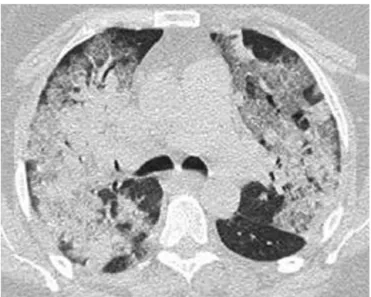
C) and had been using clarithromycin for three days. Her past medical history was significant for hypertension, type 2 diabetes mellitus, obesity (body mass index of 30.1 kg/m2), and total hip replacement 10 months before admission complicated by deep-vein thrombosis (DVT) and pulmonary embolism, both of which were successfully treated. Upon admission, she was placed in respiratory isolation with negative pressure, and nasopharyngeal washings were collected for detection of influenza A (H1N1) by reverse-transcriptasepolymerase chain reaction (RT-PCR). The blood and tracheal aspirate cultures were negative, as was the urinary assessment forPneumococcus andLegionella. Arterial blood gas analysis confirmed severe hypoxemia, and an X-ray computed tomography (CT) scan (Figure 1) of the thorax showed bilateral pulmonary infiltrates.
Orotracheal intubation was performed after a failed attempt at noninvasive ventilation. Oseltamivir (150 mg b.i.d.) administered through the enteral route and intrave-nous ceftriaxone (1 g IV b.i.d.), levofloxacin (500 mg IV q.d.), vancomycin (1 g IV b.i.d.) and methylprednisolone (2 mg/ kg/day) were started. A Doppler ultrasound of the lower limbs was negative for DVT, and an echocardiogram showed a systolic pulmonary artery pressure of 26 mm Hg with no signs of right ventricular dysfunction and a left ventricular ejection fraction of 66%.
Severe hypoxemia (PaO2 55 mm Hg) was present despite ventilation with a positive end-expiratory pressure (PEEP) of 16 cm H2O and pure oxygen, so a recruitment maneuver
was performed for two minutes using a PEEP of 35 cm H2O
and a plateau pressure of 50 cm H2O. After the recruitment,
the PEEP was titrated at 18 cm H2O according to the best
Copyrightß2010CLINICS– This is an Open Access article distributed under the terms of the Creative Commons Attribution Non-Commercial License (http:// creativecommons.org/licenses/by-nc/3.0/) which permits unrestricted non-commercial use, distribution, and reproduction in any medium, provided the original work is properly cited.
Figure 1 –X-ray computed tomography of the thorax showing diffuse, patchy bilateral ground glass opacities and consolidation at ICU admission.
CLINICS 2010;65(11):1211-1213 DOI:10.1590/S1807-59322010001100026
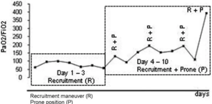
dynamic compliance, but there was no significant improve-ment in the PaO2/FiO2ratio (Figure 2). A decision was then
made to repeat the recruitment maneuver with the patient in the prone position, which resulted in significant improve-ment in gas exchange (Figure 2). Mechanical ventilation in the prone position for an average of 12 hours/day and one daily recruitment maneuver were continued for three consecutive days, with progressive improvement in the gas exchange (Figure 2). At all times, we maintained a protective ventilatory strategy with low tidal volumes (6 mL/kg of ideal body weight) and a plateau pressure of
,30 cm H2O.
On the 12thday of her ICU stay, amantadine was added to
the treatment regimen, and importation of intravenous zanamivir (not approved in Brazil) was requested because the RT-PCR for influenza A (H1N1) remained positive. On the 19thday of her stay in the ICU, intravenous zanamivir
was started, and the RT-PCR for influenza A (H1N1) became negative two days later. Because the patient still had diffuse, patchy ground-glass opacities on chest CT and had signs of incipient interstitial fibrosis, pulse therapy with methylprednisolone (1 g/day) for three consecutive days was given. The patient showed progressive radiological and gas exchange improvement and was released from mechan-ical ventilation 26 days after intubation. She was discharged from the ICU 30 days after admission and discharged home 3 weeks later.
DISCUSSION
We reported the successful use of mechanical ventilation in the prone position combined with recruitment maneuvers as rescue therapies for refractory hypoxemia in a patient with ARDS due to influenza A (H1N1). We also described the use of intravenous zanamivir in a patient with persis-tently positive RT-PCR for influenza A (H1N1).
Because of high suspicion during the southern hemi-sphere epidemic, even nonspecific symptoms of dry cough, fever, and myalgia triggered the application of our protocol for managing influenza A (H1N1) at our institution. The protocol includes respiratory isolation (negative pressure and use of an N95 mask by health professionals) to prevent in-hospital transmission of the virus, collection of respira-tory specimens for RT-PCR, and immediate therapy with oseltamivir. It is noteworthy that the rapid test for antigen in nasopharyngeal secretions was negative for the patient described here. The rapid test results are available in 15 minutes, but they have a low sensitivity (20%–30%), and a negative result should not rule out influenza A (H1N1) or lead to discontinuation of the respiratory
isolation procedures. The RT-PCR (sensitivity of 98%), available within six hours, was positive for the oropharyn-geal washings and tracheal secretions. Alternatively, viral culture could have been performed, which has a sensitivity of 89% and available results in two to three days.4
Treatment with antiviral drugs should be started early in serious cases, even before diagnostic confirmation, prefer-ably within 48 hours of the onset of symptoms, because the delay in instituting such treatment may contribute to increased disease severity.3 The use of neuraminidase inhibitors (oseltamivir and zanamivir) is preferable to the use of adamantanes (rimantadine and amantadine) because of the increased resistance of the virus to the latter class of drugs;2 the combination of an adamantane with a
neur-aminidase inhibitor is recommended by the World Health Organization. In the present case, the RT-PCR remained positive after 12 days of enteral oseltamivir at twice the recommend dose, which led us to add amantadine and later replace oseltamivir with intravenous zanamivir.2
The association of recruitment maneuvers with prone positioning was crucial to the management of the hypox-emia, serving as a bridge to the recovery of the lungs. Alveolar damage associated with necrotizing bronchiolitis with extensive hemorrhage has been described as the major histological pattern for patients with H1N1 pneumonia and respiratory failure,8and we believe that this type of injury can be limited by alveolar recruitment interventions such as RM, prone positioning, and corticosteroid use. Recruitment maneuvers in ARDS, through the application of high inspiratory pressures for short periods of time1, can be used to open collapsed alveoli and allow a more homo-geneous distribution of the ventilation, potentially reducing the lung injury induced by mechanical ventilation. Mechanical ventilation in the prone position can be used as a rescue therapy for patients with refractory hypoxemia.7
Prone positioning leads to better oxygenation by facilitating the recruitment of collapsed alveoli in the dorsal regions of the lungs and through the improvement of the ventilation/ perfusion matching caused by a shift in pulmonary perfusion to the ventral regions. The prone position requires training of the ICU staff, particularly with regard to the proper and safe positioning of patients, tubes, and catheters during position changes. We successfully combined the use of recruitment maneuvers with prone positioning to optimize gas exchange in our patient, a strategy seldom described in the literature.10 Other therapeutic options
would have been inhaled nitric oxide, high-frequency ventilation, and extracorporeal membrane oxygenation.
This case report illustrates the presentation of severe pneumonia caused by influenza A (H1N1) and the ventilatory and pharmacological measures that can be adopted for the treatment of ARDS associated with refractory hypoxemia.
REFERENCES
1. Smith TF, Burgert EO Jr, Dowdle WR, Noble GR, Campbell RJ, Van Scoy RE. Isolation of swine influenza virus from autopsy lung tissue of man. N Engl J Med. 1976;294:708-10, doi: 10.1056/NEJM197603252941308. 2. Kidd IM, Down J, Nastouli E, Shulman R, Grant PR, Howell DC, et al.
H1N1 pneumonitis treated with intravenous zanamivir. Lancet. 2009;374:1036, doi: 10.1016/S0140-6736(09)61528-2.
3. Perez-Padilla R, de la Rosa-Zamboni D, Ponce de Leon S, Hernandez M, Quin˜ones-Falconi F, Bautista R, et al. Pneumonia and respiratory failure from swine-origin influenza A (H1N1) in Mexico. N Engl J Med. 2009;361:680-9, doi: 10.1056/NEJMoa0904252.
Figure 2 –Recruitment maneuver (R); Prone position (P).
Prone position ventilation, recruitment maneuver and intravenous zanamivir
Biatto JFP et al. CLINICS 2010;65(11):1211-1213
4. Gordon SM. Update on 2009 pandemic influenza A (H1N1) virus. Cleve Clin J Med. 2009;76:577-82, doi: 10.3949/ccjm.76a.05009.
5. Rello J, Rodrı´guez A, Iban˜ ez P, So´cias L, Marques A, Guerrero J, et al. Intensive care adult patients with severe respiratory failure caused by Influenza A (H1N1)v in Spain. Crit Care. 2009;13:R148, doi: 10.1186/cc8044. 6. Hodgson C, Keating JL, Holland AE, Davies AR, Smirneos L, Bradley SJ, et al. Recruitment manoeuvres for adults with acute lung injury receiving mechanical ventilation. Cochrane Database Syst Rev. 2009;15:CD006667.
7. Martı´nez O, Nin N, Esteban A. Prone position for the treatment of acute respiratory distress syndrome: a review of current literature. Arch Bronconeumol. 2009;45:291-6, doi: 10.1016/j.arbres.2008.05.010.
8. Mauad T, Hajjar LA, Callegari GD, da Silva LF, Schout D, Galas FR, et al. Lung pathology in fatal novel human influenza A (H1N1) infection. Am J Respir Crit Care Med. 2010;181:72-9, doi: 10.1164/rccm.200909-1420OC.
9. Jain S, Kamimoto L, Bramley AM, Schmtiz AM, Benoti SR, Louie J, et al. Hospitalized Patients with 2009 H1N1 Influenza in the United States, April-June 2009. N Engl J Med. 2009; 361:1935-44, doi: 10.1056/ NEJMoa0906695.
10. Oczenski W, Ho¨rmann C, Keller C, Lorenzi N, Kepka A, Schwarz S, et al. Recruitment maneuvers during prone positioning in patients with acute respiratory distress syndrome. Crit Care Med. 2005;33):54-61, doi: 10. 1097/01.CCM.0000149853.47651.F0.
CLINICS 2010;65(11):1211-1213 Prone position ventilation, recruitment maneuver and intravenous zanamivir Biatto JFP et al.