Maleki M.,BioImpacts, 2015, 5(4), 165-167 doi: 10.15171/bi.2015.24
http://bi.tbzmed.ac.ir/
Stem cell therapy of cataract
Masoud Maleki*Department of Biology, Tabriz Branch, Islamic Azad University, Tabriz, Iran
Introduction
Cataract has been identified as one of the problems affecting clear vision. This disease is caused by multiple reasons including aging, eye injuries, inflammation, and other related diseases. According to a latest assessment, cataract has been the cause of 51% of world blindness cases, affecting about 20 million people.1 The effective
procedure to treat the cataract is the surgical intervention, in which eye’s natural lens is removed and replaced with an artificial lens.2 After the surgery, problem may occur
with the development of posterior capsule opacification (PCO). Because of the growth and proliferation of remainder epithelial cells, slight lines or folds are found on the surface of lens capsule that interfere with the transmission of light.3,4 Therefore, it is important for the
prevention of cataract and PCO.
Research has been conducted to find ways to treat cataract. Cell and tissue- based therapeutic procedures are new strategies for this purpose. O’Connor et al in 2007 showed that the lens epithelial pieces that were paired with their apical surfaces facing each other could grow and differentiate after 43 days induction by the vitreous body. Immunohistochemistry, conventional light, and
electron microscopy showed that these structures show lens- like properties.3 Stem cell therapy is anew candidate
for the treatment of cataract. Accordingly, the selection of appropriate stem cells for therapy is an important issue. In this case, selected stem cells must have potency towards differentiation into the lens fiber cells. To this end, in our previous study, we investigated the differentiation of murine and human umbilical cord stromal stem cells into the lens fiber cells. α and βγ crystallins are the major markers of differentiation of the lens. In the first study, it was found that mouse umbilical cord stromal stem cells could express αA-crystallin and αB-crystallin. In the other study, human Wharton’s jelly stem cells (hWJSCs) were induced to differentiate into the lens fiber cells. Electron microscopy images showed that these cells elongated and aligned with others cells. The expression of αB-, βB1- and βB3-crystallin genes were investigated by RT- PCR analysis.4,5
The lens capsule is a basement membrane that entirely envelops the lens, isolating the lens from other ocular tissues and keeping it from infectious viruses and bacteria.6-8 It shapes the lens and its surface inflection
participates in the accommodation mechanism.9 The
*Corresponding author: Masoud Maleki, Email: [email protected]
© 2015 The Author(s). This work is published by BioImpacts as an open access article distributed under the terms of the Creative Commons Attribution License (http://creativecommons.org/licenses/by-nc/4.0/). Non-commercial uses of the work are permitted, provided the original work is properly cited.
BioImpacts PublishingGroup
TUOMS
ccessPublish Free
Abstract
Introduction: Cataract is recognized as a disease of the lens resulting in many blindness cases, while the only therapeutic procedure is surgery. Thus, to tackle this disease, alternative methods are required. Stem cell therapy is one of the alternative treatment modalities. Paired lens’ epithelial pieces induced by vitreous body were shown to produce lens-like structures. Here, Wharton’s jelly derived stem cells are suggested as the best candidates for this purpose, as these cells have potency for the differentiation into the lens fiber cells.
Hypothesis: It is hypothesized that Wharton’s jelly
derived stem cells could be used as a novel and appropriate source for the treatment of cataract.
Evaluation of Hypothesis: To attain this aim, lens of an animal model of cataract can be removed. Then, the human Wharton’s jelly stem cells (hWJSCs) are injected into a capsule. Finally, the expression of crystalline proteins and vision function are analyzed.
Conclusion: It is hypothesized that the lens capsule could act as a natural scaffold and hWJSCs could be used to restore the lens structure in the empty capsule.
Article Type: Hypothesis
Article History: Received: 11 Apr. 2015 Revised: 22 July 2015 Accepted: 26 July 2015 ePublished: 22 Dec. 2015
Keywords: Cataract
Wharton’s jelly stem cells Crystallin
Lens Cell therapy
Maleki et al
BioImpacts, 2015, 5(4), 165-167
166
molecular adult lens capsules are composed of networks of laminin, type IV collagen, entactin/nidogen, several heparan sulfate proteoglycans including perlecan and collagen XVIII, and possibly collagen XV and agrin.10-17
Hence, the lens capsule can act as a natural scaffold for tissue engineering.18
Hypothesis
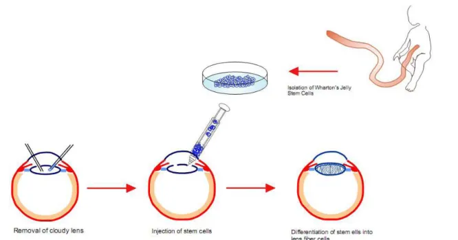
Based on the present information, if the cloudy crystallin lens is removed and hWJSCs injected in the lens capsule, the hWJSCs could be induced by the vitreous body to differentiate into the lens fiber cells, align with other cells, and restore the lens structure (Fig. 1).
Evaluation of hypothesis
In a first step, an animal model such as rabbit or rat could be set up for the induction of cataract by administration of sodium selenite.19 The second step is to prepare the
hWJSCs without any contamination. Preparation of stem cells could be performed according to the method explained by Khatami et al. In the third step, according to one type of cataract surgery, such as phacoemulsification, the cloudy lens could be removed. It should be stated that opening of capsule is considered as one of the major problems. It is thought to be solved by modifying the surgery procedure to create small opening and the set-up of this facility is necessary.In the next step, an approved amount of hWJSCs would be injected into the lens capsule. In the fifth step, examination of vision in experimental and control groups of animals could be performed. In the last step, after the removal of lenses, they can be sectioned and analyzed by electron microscopy for the lens structure and by the immunologic tests for the expression of crystalline proteins.
Empirical data
In our previous study, the expression of βB1- and
βB3-Fig. 1. Schematic diagram of hypothesis.
crystallin genes in hWJSCs was shown and alignment of differentiated cells as well as the lens fiber cells was confirmed by electron microscopy analyses. Therefore, these cells could be directed to form the lens structure with the support of an appropriate scaffold provided by the lens capsule.
Discussion
Cataract is one of the major opthalmologic problems, which affects many people worldwide.1 Therefore, discovery of
an effective therapy for the cataract is an important issue. Stem cell therapy is a new procedure for treating different types of diseases; however, the appropriate source of stem cells selection must be approved. The hWJSCs have been attended in recent years for the cell therapy. These cells represent noteworthy properties such as mesenchymal stem cell markers,20 immunosuppressant,21 anticancer
properties,22 and potency for the differentiation into
numerous cells. The fiber lens cells are responsible for the transmission of light into the retina and the alignment with other cells inside the lens capsule.3 The hWJSCs can
be differentiated into these cells in vitro,5 then used as the
source of stem cells for the designate therapy. Restoring the lens needs a natural scaffold, namely, the lens capsule. After the removal of lens, the hWJSCs could be injected into the empty capsule that is induced by the vitreous body of eye and could start expressing the crystallin.
Conclusion
The lens capsule is composed of laminin, type IV collagen, entactin/nidogen, perlecan and collagen XVIII, and possibly collagen XV and agrin,10-17 hence it can play the
role of the natural scaffold that forms the lens structure. The hWJSCs have the capacity of differentiation into the lens fiber cells and then restoring the lens structure in the empty capsule.
Stem cell therapy of cataract
BioImpacts, 2015, 5(4), 165-167 167
predictions upon the treatment of cataract, even though it should be further studied for its safety and effectiveness through the clinical trials and used as a new method for the treatment of cataract.
Ethical issues
There is none to be disclosed.
Competing interests
There is none to be declared.
References
1. http://www.who.int/blindness/causes/priority/en/index1.html. 2. Snellingen T, Evans J, Ravilla T, Foster A. Surgical interventions
for age-related cataract. Cochrane Database of Systematic Reviews
2002; CD00132.
3. O’Connor MD, Mcavoy JW. In vitro generation of functional lens-like structures with relevance to age-related nuclear cataract. Invest
Ophthalmol Vis Sci2007; 48: 1245-52.
4. Maleki M, Parivar K, Nabiyouni M, Yaghmaei P, Naji M. Induction of Alpha-crystallins expression in umbilical cord mesenchymal stem cell. IJOR2010; 22: 67-71.
5. Khatami S, Zahri S, Maleki M, Hamidi K. Stem Cell Isolation from Human Wharton’s Jelly: A Study of heir Diferentiation Ability into Lens Fiber Cells. Cell Journal (Yakhteh)2014; 15: 364-71. 6. Karkinen-Jääskeläinen M, Saxén L, Vaheri A, Leinikki P. Rubella
cataract in vitro: Sensitive period of the developing human lens. J
Exp Med1975 141: 1238-48
7. Cotlier E, Fox J, Bohigian G, Beaty C, Du Pree A. Pathogenic efects
of rubella virus on embryos and newborn rats. Nature1968; 6: 38-40.
8. Beyer T, Vogler G, Sharma D, O’Donnell FJ. Protective barrier efect of the posterior lens capsule in exogenous bacterial endophthalmitis--an experimental primate study. Invest
Ophthalmol Vis Sci1984; 25: 108-12.
9. Schachar R, Koivula A. he stress on the anterior lens surface during human in vivo accommodation. Br J Ophthalmol2008; 92: 348-50.
10. Ylikärppä R, Eklund L, Sormunen R, Muona A, Fukai N, Olsen
B, et al. Double knockout mice reveal a lack of major functional
compensation between collagens XV and XVIII. Matrix Biol2003; 22: 443-8.
11. Rossi M, Morita H, Sormunen R, Airenne S, Kreiv iM, Wang L, et al. Heparan sulfate chains of perlecan are indispensable in the lens capsule but not in the kidney. EMBO J2003 22: 236-45.
12. Parmigiani C, McAvoy J. Localisation of laminin and ibronectin during rat lens morphogenesis. Diferentiation1984; 28: 53-61. 13. Kelley P, Sado Y, Duncan M. Collagen IV in the developing lens
capsule. Matrix Biol2002; 21: 415-23
14. Fukai N, Eklund L, Marneros A, Oh S, Keene D, Tamarkin L, et al. Lack of collagen XVIII/endostatin results in eye abnormalities.
EMBO J2002 21: 1535-44.
15. Fuerst P, Rauch S, Burgess R. Defects in eye development in transgenic mice overexpressing the heparan sulfate proteoglycan agrin. Dev Biol2007 303: 165-80.
16. Dong L, Chen Y, Lewis M, Hsieh J, Reing J, Chaillet J, et al. Neurologic defects and selective disruption of basement membranes in mice lacking entactin-1/nidogen-1. Lab Invest2002; 82: 1617-30.
17. Cammarata P, Cantu-Crouch D, Oakford L, Morrill A. Macromolecular organization of bovine lens capsule. Tissue Cell
1986 18: 83-97.
18. Gwon AE. Controlled ocular lens regeneration. Google Patents; 2007.
19. Oštádalová I, Babický A, Obenberger J. Cataract induced by administration of a single dose of sodium selenite to suckling rats.
Experienta1978; 34: 222-3
20. Maleki M, Ghanbarvand F, Reza Behvarz M, Ejtemaei M, Ghadirkhomi E. Comparison of mesenchymal stem cell markers in multiple human adult stem cells. Int J Stem Cells2014; 7: 118-26. 21. Zhou C, Yang B, Tian Y, Jiao H, Zheng W, Wang J, et al. Immunomodulatory efect of human umbilical cord Wharton’s jelly-derived mesenchymal stem cells on lymphocytes. Cell
Immunol2011; 272: 33-8.
22. Wu S, Ju G, Du T, Zhu Y, Liu G. Microvesicles derived from human umbilical cord Wharton’s jelly mesenchymal stem cells attenuate bladder tumor cell growth in vitro and in vivo. PLoS One2013 8: e61366.
What is current knowledge?
√ Cataract has been the cause of 51% of blindness cases worldwide.
√ The effective procedure to treat cataract is the surgical intervention such as phacoemulsification.
What is new here?
√ The lens capsule can act as a natural scaffold.
√ HWJSCs could be induced by the vitreous body to differentiate into the lens fiber cells.