ARTICLE DOI: 10.1590/0004-282X20130104
Improvement of motor function
and decreased need for postnatal
shunting in children who had undergone
intrauterine myelomeningocele repair
Melhora em função motora e necessidade reduzida de
shunts
pós-natais em crianças submetidas a cirurgia fetal uterina da mielomeningocele
Tereza Cristina Carbonari de Faria1, Sergio Cavalheiro2, Wagner Jou Hisaba3, Antonio Fernandes
Moron3, Maria Regina Torloni3, Ana Lucia Batista de Oliveira4 , Carolina Peixoto Borges3
ABSTRACT
Objective: To compare neuromotor development between patients who did and those who did not undergo intrauterine myelomenin-gocele repair.Methods:Children with myelomeningocele aged between 3.5 and 6 years who did undergo intrauterine repair (Group A, n=6) or not (Group B; n=7) were assessed for neuromotor development at both anatomical and functional levels, need for orthoses, and cognitive function.Results: Intrauterine myelomeningocele repair signiicantly improved motor function. The functional level was higher than the anatomical level by 2 or more spinal segments in all children in Group A and 2 children in Group B, with a signiicant statistical difference between groups (p<0.05). Five children in Group A and one in Group B were community ambulators. Conclusion: Despite the small sample, it was observed that an improvement of motor function and decreased need for postnatal shunting in the 6 children who had undergone intrauterine myelomeningocele repair.
Key words: myelomeningocele, spinal dysraphism, gait, fetal diseases, surgery.
RESUMO
Objetivo: Comparar o desenvolvimento neuromotor de pacientes submetidos à cirurgia fetal intrauterina da mielomeningocele ao de pacientes não submetidos ao procedimento. Métodos: Foram avaliados: o desenvolvimento neuromotor (descrevendo o nível anatômico e motor funcional), o tipo de marcha, a necessidade de órteses e o nível cognitivo de crianças com mielomeningocele entre 3,5 e 6 anos de idade, submetidas (Grupo A; n=6) ou não submetidas (Grupo B; n=7) à cirurgia fetal intra-uterina. Resultados: A função motora apre-sentou melhora signiicante, com nível funcional mais elevado em dois ou mais segmentos em relação ao nível anatômico em todas as crianças do Grupo A e em duas crianças do Grupo B, com diferença estatística entre os grupos (p<0.05). Cinco crianças do Grupo A e uma do grupo B eram deambuladoras comunitárias. Conclusão: Apesar da pequena amostragem, nos 6 casos de cirurgia prenatal observou-se melhora da função motora e menor necessidade de shunts pós-natais.
Palavras-Chave: meningomielocele, disraismo espinal, marcha, doenças fetais, cirurgia.
Myelomeningocele (MMC) or open spina biida is a common and devastating congenital defect of the central nervous system for which there is no cure. MMC is charac-terized by the protrusion of the meninges and spinal cord through open vertebral arches, leading to lifelong paralysis.
Patients with this condition are often limited by various de-grees of mental retardation, bowel and bladder dysfunction, motor and orthopedic disability and hydrocephalia in 85% of cases1,2. Despite folic acid fortiication, the incidence of MMC in the United States has stabilized at 3.4 per 10,000
1Graduate Program in Neurology and Neurosurgery, Universidade Federal de São Paulo (UNIFESP), São Paulo, SP, Brazil; 2Division of Neurosurgery, UNIFESP, São Paulo, SP, Brazil;
3Department of Fetal Medicine, UNIFESP, São Paulo, SP, Brazil; 4Hospital Israelita Albert Einstein, São Paulo, SP, Brazil;
Correspondence: Tereza Cristina Carbonari de Faria; Rua Adolfo Figueiredo Rodrigues 682; 08770-555 Mogi das Cruzes SP - Brasil; Email: [email protected] Conflict of interest:There is no conlict of interest to declare.
live births3.he aim of postnatal surgery is to cover the exposed spinal cord, prevent infection and treat hydroceph-alus with a ventricular shunt4. However, little progress has been made in the postnatal surgical management of chil-dren with spina biida.
Open repair through a hysterotomy has been performed since 1997. he Management of Myelomeningocele Study (MOMS) began in 2003, which is a multicenter randomized trial funded in the United States of America for ive years by he National Institute of Child Health and Human Develop-ment5. he goal is to compare the safety and eicacy of the intrauterine repair of MMC with standard postnatal repair. he three participating clinical centers are the Children’s Hos-pital of Philadelphia (CHOP), the University of California and Vanderbilt University Medical Center6,7. Adzick et al.1 pub-lished the results of the MOMS with one hundred and ifty eight patients whose children were evaluated for 30 months. he authors concluded that prenatal surgery for MMC re-duced the need for shunting and improved motor outcomes in 30 months, but was associated with an increased risk of preterm delivery and uterine dehiscence at delivery.
Motor evaluations of children with MMC reveal a high degree of functional disability in the majority of cases, especially in acquiring independent ambulation8-11. Assess-ing motor skills, fetal surgery in this population (58 CHOP patients) resulted in better than predicted lower extrem-ity function at birth and ambulatory status at follow up revealed that 66% were independent walkers12. he results of MOMS showed that 42% of patient having undergone prenatal surgery were walking independently without or-thoses in 30 months versus 21% of those having undergone postnatal surgery1. A number of factors inluence neuromo-tor development in children with MMC, such as cognitive impairment, health problems (repeated urinary infections), neurological problems (seizures) and skeletal deformities as well as a lack of rehabilitation treatment and adequate braces13-15.
he aim of the present study was to observe the neuro-motor development of patients with MMC either submitted or not submitted to intrauterine surgery, describe anatomi-cal and functional motor levels and determine ambulation type, the need for orthoses and cognitive levels in these children.
METHODS
he study was approved by the Research Ethics Com-mittee of the Universidade Federal de São Paulo, approval number CEP0864/04, and performed in accordance with the ethical standards of the 1964 Declaration of Helsinki and its subsequent amendments. Written informed consent was obtained from the parents or legal guardians.
Children between 3.5 and 6 years of age with a prenatal diagnosis of myelomeningocele and followed up in the pre-natal phase at the Fetal Medicine Sector of the Universidade Federal de São Paulo (Brazil) were evaluated. Six children had been submitted to intrauterine MMC repair in 2003 and 2004 (Group A). he following inclusion criteria were used for fetal surgery: measurement of fetal ventricular atrium less than 16 mm, gestational age less than 27 weeks, opening of the spine below vertebra T10, absence of malformations in other organs, absence of chromosome alterations, and absence of maternal obstetric or clinical disease. he fetuses of seven pregnant women followed up in the same period without these criteria served as controls (Group B). he fetuses in Group B also had no alterations in other organs or systems and all exhibited dilatation of the ventricular system greater than 16 mm or had a gestational age upon admission to the Fetal Medicine Sector greater than 27 weeks. Six other chil-dren having been submitted to the placement of a ventric-ular-amniotic shunt due to progressive hydrocephalus with important dilatation of the ventricular system were excluded from the study.
he fetal surgeries were performed between 25 and 27 weeks of gestation. After hysterotomy, the fetal dorsum was carefully positioned to coincide with the uterine opening and the neurosurgical team performed the closure of the myelo-meningocele. he pregnancies were followed up on a weekly basis. Twelve births occurred in the São Paulo Hospital and one occurred at the Pró-Matre Paulista Maternity due to premature labor and the proximity of this maternity to the patient’s home (this child had undergone intrauterine myelo-meningocele repair).
he children were routinely followed up initially on a monthly basis and subsequently once per semester. After 2007, the children were called in for a new evaluation of cognitive level and functional level of the lesion at a minimum age of 3.5 years. Anatomical level was characterized through either an x-ray of the spine or magnetic resonance imaging, considering the highest vertebra afected by the closure defect.
he Columbia test was administered for the cognitive evaluation (general reasoning) between the ages of three years six months and nine years 11 months16. his test has received approval from the Brazilian Board of Psychology and was administered by a single psychologist who was un-aware of the surgical history of the patients. he test was administered in the same room without any decorations or instruments that might attract the child’s attention.
• Community ambulators: patients capable of walking in internal and external environments during the majority of their activities and may need braces, crutches or both; such individuals only need a wheelchair for long trips outside the community;
• Household ambulators: patients capable of walking only in internal environments and with the use of braces; such patients are able to transfer themselves from the chair to bed with little or no assistance; they may use a wheel-chair for some household and school activities and use a wheelchair for all other activities in the community; • Non-functional or therapeutic ambulators: patients who
only walk during therapy; such patients use a wheelchair for all their transportation needs;
• Non-ambulators: patients conined to a wheelchair that can transfer them from the chair to the bed, when necessary. • he functional level of the lesion was classiied as
follows18:
• horacic level – no sensation below the hips and power in muscles crossing the hip joint or distal to it; • Upper lumbar level – one or more of the follow-ing: some sensation below hip joint, some power in hip adductors or lexors or knee extensor (iliopsoas strength 0–3 degree);
• Lower lumbar level – one or more of the following (quadriceps strength 4–5 degree);
• Sacral level – one or more of the following: power in the ankle or toe plantar lexors or hip extensor (glu-teus and tibialis anterior strength 4–5 degree).
he chi-square test with continuity correction and Fisher’s exact test were used for the statistical analysis, with the level of signiicance set to 5% (p<0.05).
RESULTS
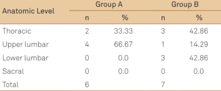
Mean gestational age upon birth among the children submitted to intrauterine MMC repair (Group A) was 32.4 weeks (range: 28 weeks 4 days to 34 weeks 5 days). Mean age in the control group (Group B) was 35.1 weeks (range: 32 weeks 1 day to 36 weeks). he age of the children at the time of the evaluation of cognition and neuromotor func-tion was 3.84 years (range: 3.75 to 4.41 years) in Group A and 4.08 years (range: 3.5 to 6 years) in Group B. Table 1 displays the anatomic level (highest vertebra with closure defect) in both groups. he thoracic level in both groups was at T12 and Table 2 displays the motor level in the groups.
Two children in Group A required cerebrospinal fluid shunts. The first exhibited an increase in the lateral ven-tricles following intrauterine repair and the shunt was placed on the third day of life. The second child exhib-ited signs of intracranial pressure two months following
Table 1. Distribution of anatomical levels in Group A and Group B.
Anatomic Level Group A Group B
n % n %
Thoracic 2 33.33 3 42.86
Upper lumbar 4 66.67 1 14.29
Lower lumbar 0 0.0 3 42.86
Sacral 0 0.0 0 0.0
Total 6 7
Table 2. Distribution of motor levels in Groups A and B.
Motor Level Group A Group B
n % n %
Thoracic 0 0.0 3 42.86
Upper lumbar 1 16.67 0 0.0
Lower lumbar 2 33.33 3 42.86
Sacral 3 50.0 1 14.29
Total 6 7
birth and the shunt was placed with 66 days of life. All children in Group B exhibited dilation of the ventricu-lar system (identified in prenatal ultrasound exams) and were submitted to shunt placement between one and three weeks of life.
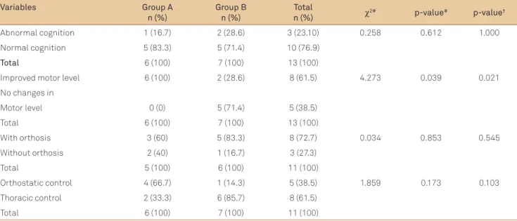
Cognitive abnormality was found in one child in Group A and two children in Group B, with no statistically signii-cant diference between groups (p>0.05; Table 3). he child in Group A did not need a shunt following birth; however, birth in this case occurred at a gestational age of 28 weeks ive days.
DISCUSSION
Intrauterine surgery for the correction of MMC emerged in 1997 as an attempt to minimize the impact of neurologi-cal abnormalities in these patients19.In Brazil, experience with this type of surgery has been limited to six cases, which have been followed up for a period of 40 months.
In an American study, Adzick et al.1 presented the results of the MOMS after 30 months, involving 158 cases divided into two groups (patients having undergone intrauterine surgery and those not having undergone this type of sur-gery) and announced the intension to give continuity to the follow up of this population into adulthood1. In the present study, the decision was made to evaluate the children at a minimum age of 3.5 years. According to Bowman et al.2 the maturation of the central nervous system is virtually com-plete at this age and the gait pattern is similar to that found in the adult population. Moreover, it is possible to perform a cognitive evaluation of children at this age with more reli-able results2.
Preliminary studies have demonstrated the beneits of intrauterine surgery regarding the prevention and even the reversal of dilatation of the ventricular system. Adelberg et al.20 analyzed the impact of the intrauterine repair of MMC regarding enlarged ventricles and found a reduction in the need for a ventriculoperitoneal shuntfollowing birth20. Johnson et al.21 examined the short-term results of the devel-opment of children with MMC with up to two years of age who had undergone intrauterine repair at the Children’s Hos-pital of Philadelphia (USA) and concluded that closure of the lesion results in the reversal of severe herniation of the brain stem, which is correlated with a reduction in or impedance
of the progression of enlarged ventricles as well as a signii-cant reduction in the need for ventriculoperitoneal shunts21. In the present study, two patients (33%) in Group A required the placement of a shunt, whereas all those in Group B needed a shunt. his result is similar to indings described in the literature, especially data from the MOMS, which report that 40% of cases of prenatal surgery require a shunt against 82% of cases of postnatal surgery1,22. It should be pointed out that the two cases in Group A that required a shunt did not exhibit cognitive impairment. he only case with cognitive impairment in this group did not require a shunt, but suf-fered postnatal complications stemming from premature birth and was also the only child restricted to a wheelchair in Group A. In Group B, two cases exhibited cognitive im-pairment (based on the Columbia Test), which is similar to the indings reported in the MOMS, in which only 6% of the cases of postnatal surgery had a score of 85 or more on the Bayley Scale.
In the MOMS, infants in the prenatal surgery group were more likely to have a level of function that was two or more levels better than expected based on the anatomic level
(32 versus 12%, p=0.005) and less likely to have a level of
func-tion that was two or more levels worse than the expected level (13 versus 28%, p=0.03) than infants in the postnatal-
surgery group. In the present study, all children in Group A demonstrated a signiicant improvement, with a functional level of two or more segments higher in relation to the ana-tomic segment.
Danzer et al.12 reported that 66% of the 58 cases having undergone intrauterine surgery for MMC at the CHOP exhibited independent gait, with the acquisition of gait dependent on the motor level of the lesion (the presence
Table 3. Associations between intrauterine surgery and neuromotor assessment outcomes.
Variables Group A
n (%)
Group B n (%)
Total
n (%) χ
2# p-value# p-value†
Abnormal cognition 1 (16.7) 2 (28.6) 3 (23.10) 0.258 0.612 1.000
Normal cognition 5 (83.3) 5 (71.4) 10 (76.9)
Total 6 (100) 7 (100) 13 (100)
Improved motor level 6 (100) 2 (28.6) 8 (61.5) 4.273 0.039 0.021
No changes in
Motor level 0 (0) 5 (71.4) 5 (38.5)
Total 6 (100) 7 (100) 13 (100)
With orthosis 3 (60) 5 (83.3) 8 (72.7) 0.034 0.853 0.545
Without orthosis 2 (40) 1 (16.7) 3 (27.3)
Total 5 (100) 6 (100) 11 (100)
Orthostatic control 4 (66.7) 1 (14.3) 5 (38.5) 1.859 0.173 0.103
Thoracic control 2 (33.3) 6 (85.7) 8 (61.5)
Total 6 (100) 7 (100) 11 (100)
of muscular innervation that provides suicient strength to maintain a standing position and coordinate the loco-motor apparatus during the act of walking). In the MOMS, children in the prenatal surgery group were more likely than those in the postnatal surgery group to walk without orthoses or devices (42 versus 21%, p=0.01). In the present
study, three patients in Group A did not require ortho-ses for gait, but no statistically signiicant diference was found in comparison to the control group due mainly to the small sample size.
All patients in Group A exhibited an improvement with regard to the anatomic level of the lesion, as ive of the six cases were community ambulators and had the motor ca-pacity to maintain a standing position, indicating suicient strength in the gluteus and quadriceps muscles to overcome the action of gravity. According to Adzick et al.1 the better function of the lower limbs and normalization of the position of the brain stem in cases having undergone prenatal sur-gery may represent a primary indication for intrauterine surgery. Despite the neurological beneits of this surgical
procedure, one must consider problems related to prema-ture birth. Indeed, the rate of premaprema-ture birth was 79% in the American trial and 100% in the present study. However, no cognitive alterations were found in these children.
Despite the small sample size in the present study, the six patients having undergone prenatal surgery demonstrated improvements in motor function and had a lesser need for postnatal shunts. Nonetheless, these patients should be fol-lowed up throughout their lives with regard to motor skills, mental capacity, bladder control and quality of living.
Even with the small number of patients evaluated in the present study, the children who had undergone fetal MMC closure had less of a need for shunting in the postoperative period and exhibited improved motor outcomes at 3.5 years. Five of the six patients in Group A achieved a standing pos-ture and were community ambulators. Despite the beneits of intrauterine surgery, problems related to premature birth must be taken into account. Moreover, life-long follow up of these patients is needed to determine the lasting ben-eits of this surgical procedure.
References
1. Adzick NS, Thom EA, Spong CY, et al.A randomized trial of prenatal versus postnatal repair of myelomeningocele. N Engl J Med 2011;364:993-1004.
2. Bowman RM, McLone DG, Grant JA, Tomita T, Ito JA. Spina biida outcome: a 25-year prospective. Pediatr Neurosurg 2001;34: 114-120.
3. Boulet SL, Yang Q, Mai C, et al. Trends in the postfortiication prevalence of spina biida and anencephaly in the United States. Birth Defects Res A Clin Mol Teratol 2008;82:527-532.
4. Adzick NS. Fetal myelomeningocele: natural history, pathophysiology, and in-utero intervention. Semin Fetal Neonatal Med 2010;15:9-14.
5. The National Institute of Child Health and Human Development (NICHD). MOMS Management of myelomeningocele study. Available at http://www.spinabiidamoms.com. Accessed March 26, 2012.
6. Bruner JP. Intrauterine surgery in myelomeningocele. Semin Fetal Neonat Med 2007;12:471-476.
7. Bruner JP, Tulipan N. Intrauterine repair of spina biida. Clin Obstet Gynecol 2005;48:942-955.
8. Bartonek A, Saraste H. Factors inluencing ambulation in myelomeningocele: a cross-sectional study. Dev Med Child Neurol 2001;43:253-260.
9. Padua L, Rendeli C, Rabini A, Girardi E, Tonali P, Salvaggio E. Health-related quality of life and disability in young patients with spina biida. Arch Phys Med Rehabil 2002;83:1384-1388.
10. Sutton LN, Sun P, Adzick NS. Fetal neurosurgery. Neurosurgery 2001;48:124-142.
11. Tubbs RS, Chambers MR, Smyth MD, et al. Late gestational intrauterine myelomeningocele repair does not improve lower extremity function. Pediatr Neurosurg 2003;38:128-132.
12. Danzer E, Gerdes M, Bebbington MW, et al. Lower extremity neuromotor function and short-term ambulatory potential
following in utero myelomeningocele surgery. Fetal Diagn Ther 2009;25:47-53.
13. Davis BE, Daley CM, Shurtleff DB, et al. Long-term survival of individuals with myelomeningocele. Pediatr Neurosurg 2005;41:186-191.
14. Kirpalani HM, Parkin PC, Willan AR, et al. Quality of life in spina bifida: importance of parental hope. Arch Dis Child 2000;83: 293-297.
15. Selber P, Dias L. Sacral-level myelomeningocele: long-term outcome in adults. J Pediatr Orthop 1998;18:423-427.
16. Burgemeister BB, Blum LH, Lorge I. Escala de Maturidade Mental Columbia. São Paulo: Vetor Editora Psicopedagógica; 1977.
17. Hoffer MM, Feiwell E, Perry R, Perry J, Bonnett C. Functional ambulation in patients with myelomeningocele. J Bone Joint Surg Am 1973;55:137-148.
18. McDonald CM, Jaffe KM, Mosca VS, Shurtleff DB. Ambulatory outcome of children with myelomeningocele: effect of lower- extremity muscle strength. Dev Med Child Neurol 1991;33:482-490.
19. Hisaba WJ. Resultado perinatal e tardio em crianças submetidas à correção intra-uterina da mielomeningocele [Thesis]. São Paulo: Universidade Federal de São Paulo; 2008.
20. Adelberg A, Blotzer A, Koch G, et al. Impact of maternal-fetal surgery for myelomeningocele on the progression of ventriculomegaly in utero. Am J Obstet Gynecol 2005;193:727-731.
21. Johnson MP, Gerdes M, Rintoul N, et al. Maternal-fetal surgery for myelomeningocele: neurodevelopmental outcomes at 2 years of age. Am J Obstet Gynecol 2006;194:1145-1150.