C A S E R E P O R T UDC: 616.716.4-089.87-089.844:616.314-76/-77-089.843 DOI: 10.2298/VSP1301080K
Possibilities of reconstruction and implant-prosthetic rehabilitation
following mandible resection
Mogu
ü
nosti rekonstrukcije i implantološko-proteti
þ
ke rehabilitacije nakon
resekcije mandibule
Vitomir S. Konstantinoviü*, Vladimir S. Todoroviü*, Vojkan M. Laziü†
*Clinic of Maxillofacial Surgery, †Clinic of Dental Prosthetics, Faculty of Dental Medicine, University of Belgrade, Belgrade, Serbia
Abstract
Introduction. Mandible reconstruction is still very challeng-ing for surgeons. Mandible defects could be the consequence of ablative surgery for malignancies, huge jaw cysts, infection and trauma. Segmental resection of the mandible may com-promise orofacial function and often lead to patients psy-chological disorders. Despite very frequent use of microvas-cular flaps, autogenous bone grafts are still very reliable tech-nique for mandible reconstruction. Comprehensive therapy means not only mandible reconstruction, but prosthodontic rehabilitation supported by dental implants, which can signifi-cantly improve patients quality of life. The aim of this paper was to evaluate possible techniques of mandible reconstruc-tion and to present a patient who had been submitted to mandible resection and reconstruction with autogenous iliac bone graft and prosthodontic rehabilitation with fixed den-ture anchoraged by disc-shaped implants in early loading protocol. Case report. Mandible reconstruction was per-formed simultaneously with resection. Autogenous iliac bone graft was taken, reshaped and placed in two parts, to the re-quired optimal contour of the mandible. After graft consoli-dation, decision was made for prosthodontics rehabilitation with fixed dentures supported by implants. In addition to the standard preoperative procedures, planning was done based on a biomodel gained by rapid prototyping after CT scan. It offered a real 3D planning to obtain a proper shape, dimen-sion and the position of implants. Conclusion. If bone di-mensions of a reconstructed mandible are insufficient, like in the presented case, the use of basal osseointegrated implants may be a method of choice. Avoiding bone augmentation procedures, as well as early loading protocol for this type of implants, shorten the total rehabilitation time, which is very convenient for patients. Fixed denture supported by dental implants is the best solution for comprehensive rehabilitation after mandible resection.
Key words:
oral surgical procedures; mandibular injuries; reconstructive surgical procedures; dental prothesis, implant-supported; rehabilitation, treatment outcome
Apstrakt
Uvod. Rekonstrukcija mandibule još uvek predstavlja iza-zov za hirurga. Defekti mandibule mogu biti posledica ra-dikalnog hirurškog tretmana malignih tumora, velikih vili-ÿnih cista, infekcija i povreda. Nedostatak dela viliÿne kosti kompromituje sve orofacijalne funkcije, a ÿesti su i psiho-loški poremeýaji. Pored sve uÿestalije primene mikrovas-kularnih režnjeva, slobodni koštani graftovi su još uvek veoma pouzdan metod rekonstrukcije mandibule. Sveobu-hvatna terapija pored rekonstrukcije mandibule podrazu-meva i implantološko-protetiÿku rehabilitaciju, kojom se znatno poboljšava kvalitet života bolesnika. Cilj ovog rada bio je da se kroz pregled literature ocene metode rekons-trukcije mandibule i da se prikaže bolesnica kod koje je nakon segmentalne resekcije mandibule izvršena rehabili-tacija fiksnom zubnom nadokanadom nošenom implanta-tima oblika diska. Prikaz bolesnika. U istom aktu sa re-sekcijom izvršena je i rekonstrukcija slobodnim koštanim ilijaÿnim graftom, koji je preoblikovan i postavljen iz dva dela, kako bi se uspostavio optimalan kontinuitet i forma mandibule. Po konsolidaciji grafta, postavljena je indikacija za izradu fiksne zubne nadoknade nošene implantatima. Pored standardnih preoperativnih procedura, izvršeno je realno trodimenzionalno planiranje na biomodelu dobije-nim softverskom analizom CT podataka. Na taj naÿin od-reĀen je oblik, veliÿina i najpovoljniji položaj implantata. Zakljuÿak. Ukoliko su koštane dimenzije rekonstruisane mandibule nedovoljne, kao kod prikazane bolesnice, pri-mena bazalnih oseointegrišuýih implantata oblika diska može biti metoda izbora. Njihovom upotrebom izbegava se dodatna nadoknada kosti što uz rano optereýenje pred-stavlja pogodnost za bolesnike, jer se znatno skraýuje vre-me rehabilitacije. Izrada fiksne zubne nadoknade nošene implantatima je najbolji naÿin definitivne rehabilitacije bo-lesnika sa rekonstruisanom mandibulom.
Kljuÿne reÿi:
Introduction
Mandible reconstruction has been a challenge for sur-geons for more than a century.Mandible defects resulting in face deformity of various stages are mostly the consequence for ablative surgery for malignancies, huge jaw cysts, infec-tions (osteomyelitis) and trauma, that may compromise oro-facial functions and cause subsequent psychological disor-ders.
Adequate anatomic reconstructing assumes the outcome which should provide satisfactory mandible dimensions, form and shape. Also, muscle attachments which enable normal functioning should be established again. It is neces-sary to consider a definitive prosthetic rehabilitation and to think about the space for the placement of oral implants. In spite of a significant progress achieved, particularly in the last 40 years, none of the existing reconstruction techniques is completely satisfactory 1.
According to the algorithm developed by Takushima et al. 2, mandible defects are classified as either „lateral“ or „anterior“. Soft-tissue defects are classified into three catego-ries: “none” (no or minimal defect on both sides of facial skin and oral mucosa); “skin or mucosal” (only skin or mu-cosal defect); and “and-through” (defect is through-and-through from the oral mucosa to the facial skin). To se-lect a suitable reconstruction method, bone-defect should be considered first, followed by the soft tissue condition. In ac-cordance with this, autologous bone grafts, alloplastic mate-rials and tissue engineered matrix origin cell grafts, are util-ized for the mandible reconstruction 1. Most frequently used are autologous bone grafts which can be applied in three principally different ways, such as: free bone grafts, pedicled bone grafts and microvascular bone grafts (flaps) 3.
The introduction of microvascular surgery has led to a significant progress in mandible defects treatment 1. Micro-vascular bone grafts can be „osteomuscular“ which, apart from the bone, contain the periosteum and the attached muscle, or „osteomusculocutaneous“, which also contain the skin on their surface. These, so-called composite grafts can be taken from the different donor regions: fibula, iliac and scapula. They are indicated for the reconstruction of large bone defects, defects in recipient sites of poor quality (scarred tissue, irradiated tissue, etc.), and when a simulta-neous bone and soft tissue reconstruction is preferred 3. Ac-cording to the literature, the most frequently applied is fib-ula flap 1, 4. The basic advantage of microvascular compos-ite flaps is the possibility of one-stage treatment of both bone and soft tissue defects by using a single donor site, with over 90% efficacy, even in irradiated patients 1, 5, 6. Foster et al. 5 state that the success of implants osseointe-gration in microvascular bone flaps was recorded in 99% cases. Compared with free bone grafts, there is a less risk to develop postoperative complications such as resorption or infection. However, complications of various degrees can be developed in the donor region, such as pain, diffi-culties in walking (limping), pathological fractures, hernia-tion, etc. 7. On the other hand, duration of surgery may im-pose a serious problem with patients with a compromised
general condition 1. It should be also mentioned that such procedures require qualified staff and well-equipped insti-tutions, yet not always possible to provide.
Initial efforts to use free bone grafts date back to the beginning of the 19th century. Owing to the experience gained during the First and Second World War, this tech-nique became widely accepted as standard in treatment of mandible defects. Until 1970s, fixation of these grafts was done with a wire, taking a longer period of intermaxillary fixation with the level of success ranging from 20% to 90%. It is known that the success of free bone grafts depends on the fixation and revascularization of the recipient site. Re-vascularization is very important for the process of resorp-tion and deposiresorp-tion of a new bone,which is referred to as creeping substitution 1. Also, it is known that even micro-movements, if fixation is not enough strong, could jeopardize the viability of a graft or lead to graft failure. There is sur-prisingly little literature about the success rate of free bone grafts fixed with plates and screws as compared to wire fixa-tion. However, fixation is nowadays routinely done with re-constructive plates and screws. The usual donor sites are: the iliac crest, rib or tibia. With regard to the bone quantity and quality, the best characteristics are provided by the iliac crest. The anterior iliac crest is the donor region of choice in most of cases. Iliac grafts could be taken as „cancellous“, „thin cortical“, „corticocancellous“, and „bicorticocancel-lous“ (full thickness) bone grafts 3. The technique of raising free iliac bone grafts is simple one, their shape matches mandible contours and dimensions and they provide enough amount of bone that is very significant for implant place-ment. Generally, patients experience a postoperative course without difficulties, and donor site complications are rare (12%).
A definitive functional reconstruction implies prosthetic rehabilitation, which may be done with mobile restorations which are retained by means of the existing teeth. However, apart from the limited function and discomfort of patient, mobile prosthesis also causes an additional bone resorption. An ideal reconstruction is achieved with fixed dentures an-choraged by dental implants. If bone dimensions are not suf-ficient, bone augmentation or adequate implantation systems can be used.
Disk-shaped dental implants placed in jaw bones by lat-eral approach were described even in 1972. Significantly im-proved in the sense of their design and surface, they have been recently applied as the so-called basal osseointegrated implants 8. Owing to their design which enables bicortical os-seointegration in the basal, the most resoption-resistant part of the jaw bone, they can be also placed even when vertical and horizontal dimensions of the residual alveolar ridge are insufficient, that is a huge advantage over other implantation systems. Moreover, the possibility of early loading, condi-tioned by the achievement of a balanced occlusion, provides a patient with great comfort.
A female patient with mandible resection followed by immediate reconstruction with an autogenous iliac bone graft was presented. After a complete graft integration, in the sec-ond phase, the basal osseointegrated implants were inserted and early loaded with the fixed denture.
Case report
A 55-years-old female patient was admitted to the Clinic of Maxillofacial Surgery, Faculty of Dental Medicine, Univer-sity of Belgrade, with pathological lesion in mandible which was identified during the routine radiographic examination. The patient was not complaining to any discomfort which might indicate the presence of lesion. A radiological finding was unspecific, showing confined multiloccular bone radiolu-cency of the corpus, angulus and ramus of the mandible at the right side (Figure 1). After biopsy and histopathological find-ing the diagnosis of odontogenic keratocyst was made. After preoperative planning, a segmental mandible resection from
the canine to the subcondylar region including a coronoid pro-cess was performed. A total length of the resected part was about 8 cm. Simultaneously, a primary reconstruction with a free bone graft from the iliac bone was done. Due to the size of defect, the graft was reshaped and placed in two parts in order to achieve the most optimal continuity and mandible form. Titanium reconstructive plate and screws (Synthes GmbH, Switzerland) were used for graft fixation (Figure 2).
A postoperative course was uneventful. A control or-thopantomogram, which was made immediately after the op-eration, showed a good position of the graft. Three months after the operation, a complete graft integration with a certain degree of resorption was radiologically confirmed.
On the basis of the control radiography after 10 months, a definite resorption of graft was estimated to be about 20%.
A final decision about prosthetic rehabilitation with fixed dentures supported by implants in the early loading protocol was made in agreement with the patient after having been informed about all the eventual possibilities.
Fig. 1 – Orthopantomograph with odontogenic keratocyst
Based on the orthopantogram, vertical and horizontal bone dimensions were insufficient for placement of conven-tional screw implants. To have an insight into the exact situation, the procedure continued with the 3D planning on the basis of CT data using the software (Mimics, Materialise, Belgium).This software allowed manufacturing of a mandi-ble biomodel by means of 3D copying (rapid prototyping). In the mean time, the impressions of both jaws were taken and plaster study models were obtained. The biomodel served to carefully analyse the available bone, in order to determine the exact shape, size and the position of implants (Figure 3). In addition, a surgical stent which helped in inserting im-plants in the pre-determined specific positions was made.
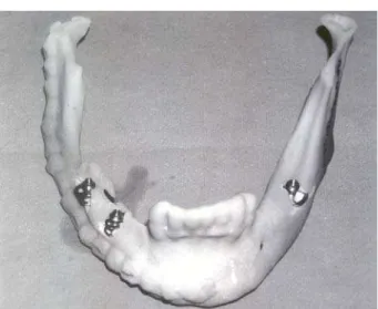
The implantation procedure was performed under gen-eral anesthesia. Intraoperatively, following rising mucoperi-osteal flap, the stability of bone graft was confirmed. After surgical stent adaptation, implantation was done according to the protocol for disk-shaped implants (lateral insertion). In the residual alveolar ridge in the molar region at the left side, an implant was placed, and in the graft at the right side, two disk-shaped implants were inserted (Diskos-ID Brand, Dr. Ihde Dental AG, Switzerland) (Figure 4). Six days after the operation, the impressions were taken, and the implants were loaded with a temporary composite bridge on the day 10.
The control CT and orthopantomographs showed an excel-lent position of implants. Six months later, a complete inte-gration of implants was determined clinically (radiologi-cally), and a definite metalceramic circular bridge was pro-duced. There were no signs of marginal bone loss around the loaded implants after 2 years, which was confirmed by con-trol orthopantomograph (Figure 5).
Fig. 4 – Disk implants inserted in reconstructed mandible
Fig. 5 – Orthopantomograph two years after implantation
Discussion
There are no many papers on mandible reconstruction with free bone grafts and oral implants in the literature. However, there are several hesitations concerning this topic which need further consideration.
First of all, considering the currently available techniques for mandible reconstruction, the question arises about which cases are suitable for the reconstruction with free bone grafts as a method of choice. There are several cru-cial criteria for a definite decision making. As concerns the size of the defect, Goh et al. 1 believe that free bone grafts are still a good option for defects smaller than 5 cm, if the surrounding soft tissues are in good condition. Hotz 9 gives priority to the delayed mandible reconstruction with a free iliac graft for defects smaller than 8 cm. Foster et al. 5 in their comparative study concluded that the use of avascular bone grafts is limited to smaller bone defects (< 5–6 cm), in pa-tients who do not undergo irradiation therapy and/or do not have a compromised general condition. Pogrel et al. 10 share similar opinion indicating the dimension of 6 cm as an upper limit for use of avascular grafts. Contrary to them, Chiapasco et al. 11 in their retrospective study showed that the limiting factor for the use of avascular grafts is not the size of defect but surely the insufficient quality and quantity of surround-ing soft tissues, in the sense of compromised revasculariza-tion. The mentioned study presents successful reconstruc-tions also for defects which spread from the symphyseal part to the condylar region of the mandible.
The majority of authors agree that the iliac crest is the best donor site, because of easy approach and possibility for taking a large amount of bone 3, 9, 10, 12.
One of the dilemmas is whether the reconstruction should be done simultaneously with mandible resection or subsequently. Hotz 9 indicates an important problem of si-multaneous reconstruction in cases with malignancies, be-cause it is not possible to perform a histopathological verifi-cation of the tumour free margins. Also, compared with the postponed reconstruction, the duration of intervention is sig-nificantly prolonged and therefore the risk of postoperative complications is increased.
Since the patient presented in this paper was involved with a benign lesion (the patient was not irradiated), the size of the bone defect was estimated to be about 8 cm, and soft tissues were of a satisfactory quality, the decision was made for a primary reconstruction with a free bone iliac graft. According to the exact dimensions of the resected part of the mandible, the graft was reshaped and fixed in two parts in order to adequately reconstruct the mandible contours.
A contemporary approach to a patient definitive reha-bilitation after mandible resection does not imply an anat-omic reconstruction only, but also a prosthetic rehabilita-tion. In the past, patients were mostly rehabilitated with mobile dentures of limited functional and aesthetic values. The introduction of endosseal implants provided rehabili-tation with fixed dentures showing to be more comfortable, and significantly improving both function and aesthetic.
There are numerous studies describing successful applica-tion of convenapplica-tional screw implants in reconstructed man-dibles 6, 9, 12, 13.
When considering the right timing for implants place-ment, there are two reasons in favour of the delayed implan-tation. The first is in the fact that the successful osseointe-gration depends on osteoblasts capable for osteogenesis, and bone grafts are, so to say “a dead bone” as long as the proc-ess of the so-called “acceptance of the grafts” does not start. Another reason is that the simultaneous implantation is pro-portionally more demanding and rarely meets prosthetic re-quirements. Lekholm et al. 14 have concluded that implanta-tion success is higher with the delayed approach. Lundgren et al. 13 in their research revealed that the delayed procedure not only results in bigger amount of bone on the implant’s surface but also stimulates further remodelling and formation of a new bone. Foster et al. 5 mentioned that it is necessary to wait for 5 to 6 months after reconstruction, so that the im-plantation procedure may be successful. Considering the size of the graft and the time necessary for remodelling and for-mation of osteogenetic potential in the presented case, the decision was made to place the implants subsequently, at least after 6 months.
The basic prerequisite of successful implantation pro-cedure is a sufficient quantity of bone. When free bone grafts are concerned, the expected resorption is 25% in relation to the initial graft volume 15. One study tested the average verti-cal resorption of the graft and the value obtained was 3.53 cm 14. Stošiü16 indicates an average resorption of graft to be 15%–30%. Also, it is important to know whether the patient underwent a postoperative irradiation therapy 17.
A problem might occur with the lack of bone for placement of screw implants. If so, it is possible to apply various augmentation procedures or to use particular implant systems 18, 19. The use of basal osseointegrated (disk) im-plants in immediate or early loading protocols is a useful solution if a bone dimensions are insufficient for placing conventional screw type implants. By means of that, total re-habilitation time could be significantly shortened, which is very convenient to patients.
It is very important to point out that the multidisciplin-ary approach and good planning together with the use of adequate measuring is of crucial importance for an overall favourable outcome 20. A real 3D determination of future im-plant positions on a biomodel obtained on the basis of CT data appears very useful if a limiting anatomic factor is pres-ent. Also, it was possible for the patient to become familiar with the planned procedure.
Conclusion
very comfortable for a patient both in the functional and aesthetic sense.
In case of insufficient bone dimensions of the recon-structed mandible, the use of disk implants in immediate or early loading protocols is an useful optional method of reha-bilitation.
Acknowledgement
A part of this research was financed with the Grant No 175075 of the Ministry of Education, Science and Techno-logical Development of the Republic of Serbia.
R E F E R E N C E S
1. Goh BT, Lee S, Tideman H, Stoelinga PJ. Mandibular reconstruc-tion in adults: a review. Int J Oral Maxillofac Surg 2008; 37(7): 597î605.
2. Takushima A, Harii K, Asato H, Momosawa A, Okazaki M, Na-katsuka T. Choice of osseous and osteocutaneous flaps for mandibular reconstruction. Int J Clin Oncol 2005; 10(4): 234î42.
3. Ehrenfeld M, Hagenmaier C. Autogenous Bone Grafts in Maxil-lofacial Reconstruction. In: Greenberg AM, Prain J, editors. Craniomaxillofacial Reconstructive and Corrective Bone Sur-gery: Principles of Internal Fixation Using the AO/ASIF Technique. New York: Springer; 2002. p. 295î309.
4. Wedler V, Farshad M, Sen M, Koehler C, Hanschin A, Graetz K, et al. Retrospective analysis and clinical evaluation of mandible reconstruction with a free fibula flap. Eur J Plast Surg 2007; 29(6): 285–91.
5. Foster RD, Anthony JP, Sharma A, Pogrel MA. Vascularized bone flaps versus nonvascularized bone grafts for mandibular re-construction: an outcome analysis of primary bony union and endosseous implant success. Head Neck 1999; 21(1): 66î71. 6. Hidalgo DA, Pusic AL. Free-flap mandibular reconstruction: a
10-year follow-up study. Plast Reconstr Surg 2002; 110(2): 438î49; discussion 450î1.
7. Ghassemi A, Ghassemi M, Riediger D, Hilgers RD, Gerressen M. Comparison of donor-site engraftment after harvesting vascu-larized and nonvascuvascu-larized iliac bone grafts. J Oral Maxillofac Surg 2009; 67(8): 1589î94.
8. Ihde S. Principles of BOI, Clinical Scientific, and Practical Guidelines to 4-D Dental Implantology. Berlin, Heidelberg, New York: Springer- Verlag; 2005.
9. Hotz G. Reconstruction of mandibular discontinuity defects with delayed nonvascularized free iliac crest bone grafts and endosseous implants: a clinical report. J Prosthet Dent 1996; 76(4): 350î5.
10. Pogrel MA, Podlesh S, Anthony JP, Alexander J. A comparison of vascularized and nonvascularized bone grafts for reconstruc-tion of mandibular continuity defects. J Oral Maxillofac Surg 1997; 55(11): 1200î6.
11.Chiapasco M, Colletti G, Romeo E, Zaniboni M, Brusati R. Long-term results of mandibular reconstruction with autogenous
bone grafts and oral implants after tumor resection. Clin Oral Implants Res 2008; 19(10): 1074î80.
12.Chiapasco M, Abati S, Ramundo G, Rossi A, Romeo E, Vogel G. Behavior of implants in bone grafts or free flaps after tumor resection. Clin Oral Implants Res 2000; 11(1): 66î75. 13.Lundgren S, Rasmusson L, Sjöström M, Sennerby L. Simultaneous
or delayed placement of titanium implants in free autogenous iliac bone grafts. Histological analysis of the bone graft-titanium interface in 10 consecutive patients. Int J Oral Maxil-lofac Surg 1999; 28(1): 31î7.
14.Lekholm U, Wannfors K, Isaksson S, Adielsson B. Oral implants in combination with bone grafts. A 3-year retrospective mul-ticenter study using the Brånemark implant system. Int J Oral Maxillofac Surg 1999; 28(3): 181î7.
15.Stoll P, Prein J, Bähr W, Wächter R. Considerations in the fixa-tion of bone grafts for the reconstrucfixa-tion of mandibular con-tinuity defects. In: Greenberg AM, Prain J, editors. Craniomaxil-lofacial Reconstructive and Corrective Bone Surgery: Princi-ples of Internal Fixation Using the AO/ASIF Technique. New York: Springer; 2002. p. 317î26.
16.Stošiý S. Mandibular reconstruction. Belgrade: Rubikon; 2005. (Serbian)
17.Ihde S, Kopp S, Gundlach K, Konstantinoviý VS. Effects of radia-tion therapy on craniofacial and dental implants: a review of the literature. Oral Surg Oral Med Oral Pathol Oral Radiol Endod 2009; 107(1): 56î65.
18.Konstantinoviý V. Aspekte der implantologischen Versorgung mit BOI im Bereich des Sinus maxillaris. ZMK 2003; 19: 568î75.
19.Konstantinoviý VS, Laziý VM, Stefan I. Nasal epithesis retained by basal (disk) implants. J Craniofac Surg 2010; 21(1): 33î6. 20.Arsiý S, Periý P, Stojkoviý M, Iliý D, Stojanoviý M, Ajdukoviý Z,
Vu-ÿiý S. Comparative analysis of linear morphometric parameters of the humane mandibula obtained by direct and indirect measurement. Vojnosanit Pregl; 2010; 67(10): 839–46. (Ser-bian)