Research Article
Curcumin Pharmacokinetic and Pharmacodynamic
Evidences in Streptozotocin-Diabetic Rats Support
the Antidiabetic Activity to Be via Metabolite(s)
Vânia Ortega Gutierres,
1Michel Leandro Campos,
2Carlos Alberto Arcaro,
1Renata Pires Assis,
1Helen Mariana Baldan-Cimatti,
2Rosângela Gonçalves Peccinini,
2Silvia Paula-Gomes,
3Isis Carmo Kettelhut,
3Amanda Martins Baviera,
1and Iguatemy Lourenço Brunetti
11Department of Clinical Analysis, School of Pharmaceutical Sciences, S˜ao Paulo State University (UNESP),
Rodovia Araraquara-Ja´u Km 01, 14801-902 Araraquara, SP, Brazil
2Department of Natural Active Principles and Toxicology, School of Pharmaceutical Sciences,
S˜ao Paulo State University (UNESP), Rodovia Araraquara-Ja´u Km 01, 14801-902 Araraquara, SP, Brazil
3Department of Biochemistry and Immunology, School of Medicine, University of S˜ao Paulo (USP),
Avenida Bandeirantes 3900, 14040-900 Ribeir˜ao Preto, SP, Brazil
Correspondence should be addressed to Iguatemy Lourenc¸o Brunetti; brunetti@fcfar.unesp.br
Received 15 December 2014; Revised 16 March 2015; Accepted 17 March 2015
Academic Editor: Yong H. Liao
Copyright © 2015 Vˆania Ortega Gutierres et al. his is an open access article distributed under the Creative Commons Attribution License, which permits unrestricted use, distribution, and reproduction in any medium, provided the original work is properly cited.
his study measures the curcumin concentration in rat plasma by liquid chromatography and investigates the changes in the glucose tolerance and insulin sensitivity of streptozotocin-diabetic rats treated with curcumin-enriched yoghurt. he analytical method for curcumin detection was linear from 10 to 500 ng/mL. he�maxand the time to reach�max(�max) of curcumin in plasma were
3.14±0.9�g/mL and 5 minutes (10 mg/kg, i.v.) and 0.06±0.01�g/mL and 14 minutes (500 mg/kg, p.o.). he elimination half-time was 8.64±2.31 (i.v.) and 32.70±12.92 (p.o.) minutes. he oral bioavailability was about 0.47%. Changes in the glucose tolerance and insulin sensitivity were investigated in four groups: normal and diabetic rats treated with yoghurt (NYOG and DYOG, resp.) and treated with 90 mg/kg/day curcumin incorporated in yoghurt (NC90 and DC90, resp.). Ater 15 days of treatment, the glucose tolerance and the insulin sensitivity were signiicantly improved in DC90 rats in comparison with DYOG, which can be associated with an increase in the AKT phosphorylation levels and GLUT4 translocation in skeletal muscles. hese indings can explain, at least in part, the beneits of curcumin-enriched yoghurt to diabetes and substantiate evidences for the curcumin metabolite(s) as being responsible for the antidiabetic activity.
1. Introduction
Curcumin (diferuloylmethane) is a yellow pigment isolated from the dried rhizomes ofCurcuma longaL. (turmeric). In Asian countries, turmeric is largely used as a dietary spice. Furthermore, traditional Indian and Chinese medicines have used turmeric for the treatment of a diversity of diseases [1]; curcumin has been cited as the main phytochemical respon-sible for the turmeric beneicial efects. Crescent attention has been given to understand the role of curcumin in the
prevention and treatment of various chronic diseases, such as cancer [2, 3], cardiovascular diseases [4], and metabolic disturbances, including obesity [5] and diabetes mellitus [6]. A wide range of biological activities has been attributed to curcumin; however the translation of its experimental biological beneits into clinical trials is diicult due to the low bioavailability of this pigment when administered orally, observed in both rodents [7] and humans [8], which is explained by its poor absorption due to the low solubil-ity in water, limited tissue distribution, and rapid rate of Volume 2015, Article ID 678218, 13 pages
metabolism in liver and intestine followed by the rapid excretion from the body [9]. So the low bioavailability of curcumin appears as a major barrier to reach its adequate cir-culating levels related to desirable pharmacodynamic actions, hindering its clinical approval as a therapeutic agent for several diseases.
Attempting to overcome this situation, several vehicles and associations with curcumin have been tested, such as curcumin-loaded nanoparticles [10], complexing with phos-pholipids [2], microemulsifying [11], and association with drug bioenhancers, for example, piperine [12], among others [13]. Previous study from our laboratory [14] showed that curcumin incorporated in yoghurt improved several physio-logical and biochemical parameters of streptozotocin- (STZ-) diabetic rats treated for 31 days: the food and water intake and the urinary volume were decreased and the body weight gain was increased; the plasma levels of glucose, triglycerides, and transaminases and the urinary levels of glucose, urea, and protein in urine were decreased, when compared with nontreated diabetic rats. hese biomarkers were not changed in nondiabetic rats treated with curcumin-enriched yoghurt. With the objective to increase the antidiabetic activity of curcumin, our laboratory recently investigated its coadmin-istration with piperine, which inhibits the biotransforma-tion processes occurring in liver and intestine, increasing the bioavailability of drugs; notably, the treatment of STZ-diabetic rats with both curcumin and piperine in yoghurt for 45 days did not change or even nulliied, in the dose-dependent way, the antidiabetic and antioxidant activities of curcumin [15], suggesting that the biotransformation is important to these pharmacological actions of curcumin. In light of these data, the investigation of the pharmacokinetic proile of curcumin when administered with yoghurt in diabetic animals became interesting which, as far as we know, has not yet been described.
he aim of this study was to evaluate the pharma-cokinetics of curcumin in diabetic rats when administered intravenously and orally (incorporated in yoghurt) and also to investigate the changes in the glucose tolerance and insulin sensitivity of diabetic rats ater acute (1 day) and subchronic (15 days) treatments with curcumin incorporated in yoghurt.
2. Material and Methods
2.1. Chemical and Reagents. Curcumin (≥77%, 28260,
Fluka-Sigma Aldrich, St. Louis, MO, USA) and analytical standard curcumin (≥98.0%, 08511, Fluka-Sigma Aldrich, St. Louis, MO, USA), streptozotocin (STZ, Sigma Aldrich, St. Louis, MO, USA), dinitrophenol, and dimethyl sulfoxide (DMSO) were obtained from Sigma Aldrich (St. Louis, MO, USA); ketamine and xylazine were obtained from Agener Uni˜ao (Embu-Guac¸u, SP, Brazil); acetonitrile and methanol (HPLC grade) were obtained from J. T. Baker (Mexico City, Mexico); acetic acid was obtained from Qhemis (Indaiatuba, SP, Brazil); and insulin (Biohulin NU-100; 100 units/mL) was obtained from Biobras (Montes Claros, MG, Brazil). Sodium hydroxide was purchased from Cetus (Santo Amaro, SP, Brazil) and plain yoghurt from Nestl´e (Araras, SP, Brazil).
Water was puriied by a Millipore system (EMD Millipore, Darmstadt, Germany).
2.2. HPLC Analysis of Curcumin. he high performance
liquid chromatography (HPLC) system consisted of a Waters 600E with a 50�L sample loop coupled to UV-Vis detector (Waters 2487) set to 420 nm and a computer system for data acquisition (Empower sotware, Waters) was used. he separation was achieved using a reversed phase Symmetry C18 column (4.6 × 250 mm, particle size 5�m). Mobile phase consisting of methanol, acetonitrile, and 5% acetic acid (35 : 50 : 15, v/v) was employed at a low rate of 1.0 mL/min.
2.3. Preparation of the Standard Solutions and Quality Control
of Samples. Standard solutions of curcumin (≥98%, 08511,
Fluka-Sigma Aldrich, St. Louis, MO, USA) were prepared in methanol at six concentration levels (0.10, 0.25, 0.75, 1.25, 2.5, and 5�g/mL). hese solutions were diluted in plasma to reach the inal plasma concentrations of 10, 25, 75, 125, 250, and 500 ng/mL.
2.4. Sample Preparation. An aliquot of 500�L of rat plasma
(blank) or 500�L of rat plasma containing curcumin (con-centrations as described in Section 2.3) were mixed with 10�L of 0.01 M NaOH and 40�L of acetonitrile containing 200�g/mL dinitrophenol; dinitrophenol was used as internal standard (IS). he mixture was vortexed (30 seconds) and 1000�L of ethyl acetate was added and vortexed again, followed by centrifugation at 1000×g for 15 minutes at 10∘C. Ater that, 500�L of supernatant was transferred to a new clean plastic microtube and evaporated under vacuum for 15 minutes at 37∘C in a Mini Vac Sample Concentrator Range-Genevac. Lastly, the dry sample was reconstituted by 125�L of methanol and injected into the HPLC system.
2.5. Method Validation. In order to determine the conidence
limits of the bioanalytical method, the validation was per-formed based on the RDC-27/2012 from Agˆencia Nacional de Vigilˆancia Sanit´aria (ANVISA, Brazil) and theGuidance
for Industry: Bioanalytical Method Validation 2001 from
Food and Drug Administration(FDA, USA). he analytical
methodology was validated regarding selectivity, linearity, accuracy, precision, limit of detection (LOD), lower limit of quantitation (LLOQ), recovery, and stability.
he selectivity evaluation of the method was performed by the analysis of a blank plasma sample in order to deter-mine the presence of interfering peaks in curcumin and IS retention times.
Linearity was achieved by plotting the curcumin/IS ratio versus nominal concentrations. he acceptance criteria for linearity were accuracy as percentage of the nominal concen-tration (%) between 85 and 115%, except at the LLOQ, where it can be between 80 and 120% and linear regression coeicient (�) higher than 0.98.
was carried out in the same day (intraday, � = 5) and in three diferent days (interday,� = 15). he acceptance criterion for precision was a coeicient of variation (%) less than 15%, except at the LLOQ where it can be less than 20%. he accuracy acceptance criterion was a percentage of the nominal concentration (%) between 85 and 115%, except at the LLOQ where it can be between 80 and 120%.
he recovery test, which determines the extraction proce-dure eiciency, was achieved comparing extracted samples to nonextracted samples (100%) of curcumin in HQC and LQC. In order to determine whether the sample can be stored in speciic conditions, the stability studies were performed. Plasma aliquots were prepared at concentrations of 25 and 400 ng/mL in triplicate and analyzed according to the fol-lowing conditions:bench-top stability, where the samples stay at room temperature (30∘C) for 4 h; postprocessing stability, where samples stay at room temperature (30∘C) for 4 h ater extraction;freeze-thaw stability, where samples were frozen at −20∘C and thawed at room temperature over three cycles dur-ing 72 hours;long-term stability, where samples were frozen at−20∘C for seven days. Ater these conditions, samples were analyzed and compared with the results obtained from freshly prepared and analyzed samples.
2.6. Animals. Male Wistar rats (Rattus norvegicus) weighing
140–160 g (6 weeks) were maintained under environmentally controlled conditions of temperature (23±1∘C) and humidity (55 ± 5%) and with a 12 h light/dark cycle, having free access to water and normal lab chow diet (Purina Evialis do Brasil Nutric¸˜ao Animal Ltda., SP, Brazil). Before the beginning of the experiments, the animals were maintained under these conditions for at least 5 days. he experiments were conducted during the light phase and the experimental protocol was approved by the Committee for Ethics in Animal Experimentation of the School of Pharmaceutical Sciences, UNESP, Araraquara (resolution number 37/2012).
2.7. Induction of the Experimental Diabetes Mellitus.
Experi-mental diabetes mellitus was induced by a single intravenous injection of STZ (40 mg/kg b.w.) dissolved in 0.01 M citrate bufer (pH 4.5), in previously 14 h fasted rats. Normal rats received only citrate bufer. Four days ater STZ adminis-tration, diabetic rats with postprandial glycemia values of approximately 400 mg/dL were used in the experiments. Plasma glucose levels were determined by the glucose oxidase method [16] using commercial kit (Labtest Diagnostica SA, Brazil).
2.8. Preparation of Curcumin-Enriched Yoghurt. Curcumin
(≥77%, 28260, Fluka-Sigma Aldrich, St. Louis, MO, USA) was mixed with 0.5 mL of plain yoghurt, in the doses of 45 or 500 mg/kg b.w., with a homogenizer (Metabo, Marconi, Piracicaba, SP, Brazil) operating at 27,000 rpm for 90 seconds at a controlled temperature of 25∘C.
2.9. Pharmacokinetics (PK) of Curcumin in Diabetic Rats.
Twenty-ive diabetic rats (weighing 150–200 g) were divided into two groups: intravenous (i.v.) group (� = 10), of which animals were treated with 10 mg/kg of curcumin (16 mg/mL
in DMSO), and the oral group (� = 15), of which animals were treated with 500 mg/kg of curcumin (single dose) incorporated in 0.5 mL of yoghurt.
Twenty-four hours before the curcumin administration, the animals were submitted to vein and/or artery catheter implantation. hese catheters were used to drug administra-tion or to serial blood collecadministra-tions [17]. Eight hours before the curcumin administration, the animals were deprived of food. he sampling times were 5, 10, 20, 30, 45, and 60 minutes ater the oral administration and 2.5, 5, 7.5, 10, 20, 30, and 45 minutes ater the i.v. administration. Both administrations were performed over a short period of time.
he curcumin PK parameters were calculated based on the concentration in plasma versus time curves. he half-life of elimination (�1/2) was calculated by the graphic method and the half-life of absorption was calculated by the residues method. he constants of absorption (��) and elimination (�el) were calculated by the equation 0.693/�1/2, where �1/2 is the half-life of absorption or elimination, respectively. he area under the curve (AUC) of the plasma concentration versus time proile from time zero to the last sampling time (AUC0-�) was calculated by the trapezoidal rule. he area under the curve from time zero extrapolated to ininity (AUC0-∞) was calculated by the equation AUC0-∞= AUC0-� +�pn/�el(where�pnis the last plasma concentration deter-mined and �el is the elimination constant). he AUC0-∞ was used for the calculation of the total clearance (Cl = dose/AUC0-∞), and the distribution volume (��) was calcu-lated using the equation��= Cl/�el. he mean residence time (MRT) was estimated from AUMC/AUC, where AUMC is area under the irst moment curve. he oral bioavailability (�) of curcumin was calculated by the relation between the AUC obtained in the oral administration and the AUC obtained in the intravenous administration. he maximum plasma concentration (�max) was obtained from the experimental
data as well as the time of the occurrence of�max(�max).
2.10. Pharmacodynamics (PD) of Curcumin Administered with Yoghurt
2.10.1. Treatment of Animals. Forty rats were divided into
four groups: normal rats treated with yoghurt (NYOG) or with 45 mg/kg curcumin-enriched yoghurt twice a day (NC90); diabetic rats treated with yoghurt (DYOG) or with 45 mg/kg curcumin-enriched yoghurt twice a day (DC90). Normal and diabetic rats were treated by gavage at 08:00 h and 17:00 h, for 15 days. Curcumin incorporated in yoghurt (as described inSection 2.8) was administered as a half dose in 0.5 mL, totaling 1.0 mL yoghurt/rat/day of treatment. Con-trol rats received only yoghurt. Glucose and insulin tolerance tests were performed ater 1 and 15 days of treatment. Changes in the insulin signaling pathway (total protein content and phosphorylation levels in serine-473 residue of AKT or protein kinase B) and in the content of glucose transporter type 4 (GLUT4) in plasma membrane were also investigated
ingastrocnemius muscles from NYOG, NC90, DYOG, and
2.10.2. Oral Glucose Tolerance Test (OGTT). OGTT was per-formed in 14 h fasted rats. A glucose solution (2.5 g/kg b.w.) was administered orally, and blood samples were collected from the tip of the tail before (� = 0) and 15, 30, 45, 60, 75, 90, 105, and 120 minutes ater the glucose loading. Results were expressed as mg/dL and the area under the curve (AUC, g/dL/120 min) was calculated.
2.10.3. Insulin Tolerance Test (ITT). For ITT, 6 h fasted
rats received a single intraperitoneal injection of human recombinant insulin (1.5 U/kg b.w.), and blood samples were collected before (� = 0) and 5, 10, 15, 20, 25, and 30 minutes ater the insulin administration for the plasma glucose measurement. Results were expressed as mg/dL. For the estimation of the insulin sensitivity, the constant rate for glucose disappearance (�itt, %/min) was calculated using the equation�itt = 0.693/�1/2, where�1/2represents the half-life of plasma glucose concentration calculated from the slope of least squares analysis in the interval of time in which glycemia declines linearly ater the insulin administration [18].
2.10.4. Western Blotting Analysis. Concerning sample
prepa-ration for AKT protein content and phosphorylation
lev-els, gastrocnemius muscles were homogenized in 50 mM
Tris-HCl bufer (pH 7.4) containing 150 mM NaCl, 1 mM ethylenediaminetetraacetic acid (EDTA), 1% Triton X-100, 1% sodium deoxycholate, 1% SDS, 10 mM sodium pyrophos-phate, 100 mM sodium luoride, 10 mM sodium ortho-vanadate, 5�g/mL aprotinin, 1�g/mL leupeptin, and 1 mM phenylmethylsulfonyl luoride (PMSF). he homogenates were centrifuged at 15,500×g at 4∘C for 40 minutes and the supernatant was used for analysis. Protein levels were determined using bovine serum albumin as standard [19].
Concerning sample preparation for GLUT4 content in plasma membrane, gastrocnemius muscles were homoge-nized in 50 mM Tris-HCl bufer (pH 8.0) containing 0.1% NP-40, 0.5 mM dithiothreitol (DTT), and the same proteases and phosphatases inhibitors used in the above bufer. he homogenates were centrifuged at 1,000×g at 4∘C for 10 minutes. Precipitates were washed twice with the same bufer above described without NP-40 and centrifuged at the same conditions. Precipitates were added in bufer containing 1% NP-40 and then centrifuged at 15,500×g at 4∘C for 20 minutes; the supernatants were considered the plasma membrane fraction [20].
Supernatants (for AKT and GLUT4 studies) were pre-pared with sample bufer (125 mM Tris-HCl, 20% glycerol, 4% SDS, 100 mM DTT, 0.02% bromophenol blue, pH 6.8); equal amounts of protein (20�g) were subjected to SDS-PAGE analysis on 8% acrylamide gels [21] and electroblotted onto nitrocellulose membranes [22].
Ater blocking, proteins were detected by overnight incubation at 4∘C with speciic primary antibodies: AKT (1 : 500, Cell Signaling, Danvers, MA, USA), anti-phospho-[Ser-473]-AKT (1 : 500, Cell Signaling, Danvers, MA, USA), and anti-GLUT4 (1 : 1,000, Santa Cruz Biotech-nology, Santa Cruz, CA, USA). Anti-�-actin (1 : 1,000, Santa Cruz Biotechnology, Santa Cruz, CA, USA) was used as internal control. Primary antibodies were detected by
Curcumin
Dinitrophenol Curcumin
Dinitrophenol
0 100 200 300 400 500 0.0
0.5 1.0 1.5 2.0 2.5 3.0 3.5 4.0 4.5
Ra
ti
o
Nominal concentration (ng/mL)
0.018
0.016 0.014 0.012 0.010 0.008 0.006 0.004 0.002 0.000
1.00 2.00 3.00 4.00 5.00 6.00 7.00 8.00 9.00
(min)
(A
U)
Y = 0.0069X − 0.0085
R2= 0.9999
Figure 1: Chromatogram of plasma analysis containing 25 ng/mL curcumin and IS (5.3�g/mL dinitrophenol) at 420 nm and the linearity of the bioanalytical method.
peroxidase-conjugated secondary antibodies (Cell Signaling, Danvers, MA, USA) and visualized with enhanced chemilu-minescence reagents (SuperSignal West Pico Chemilumines-cent Substrate, hermo Scientiic, Waltham, MA, USA) and detected with C-Digit Blot Scanner (LI-COR, Lincoln, NE, USA). Band intensities were read and analyzed with the LI-COR Image Studio 4.0 Program.
2.11. Statistical Analysis. Data were expressed as mean ±
standard error of mean (SEM). One-way analysis of variance (ANOVA) followed by Student-Newman-Keuls test was used to compare the intergroup diferences and of the AUC. Diferences were considered signiicant at� < 0.05. Statistical analyses were performed using the program Graphpad Instat 3.05 (GraphPad Sotware, USA).
3. Results
3.1. Method Validation. As observed in the chromatogram
Table 1: Intraday (� = 5) and interday (� = 5) accuracy and precision of the bioanalytical method to quantify curcumin in plasma.
Nominal concentration (ng/mL)
Accuracy (%) Precision CV (%)
Intraday
10 113.59 13.52
25 86.05 11.03
125 92.40 2.67
400 112.88 8.68
Interday
10 89.65 7.58
25 91.85 12.46
125 94.52 8.57
400 98.74 9.62
3.2. PK Proile of Curcumin. PK proile was evaluated ater
a single administration of 10 mg/kg b.w. of curcumin in DMSO (i.v.) and 500 mg/kg b.w. of curcumin incorporated in yoghurt (p.o.). Curcumin plasma concentration versus time proiles can be seen inFigure 2. Concentration values were best-itted by a one-compartmental model. Maximum plasma curcumin concentration occurred in about 14 minutes ater the oral administration. he intravenously administered curcumin showed extrapolated�max of3.14 ± 0.90 �g/mL,
whereas oral administration with a dose 50-fold higher showed0.06 ± 0.01 �g/mL (Table 3).
he areas under curve (AUC0-�) were 12.27 ± 2.75 �g/mL/min for intravenous administration and 1.89 ± 0.25 �g/mL/min for oral administration, of which doses were 10 mg/kg and 500 mg/kg, respectively (Table 4).
Although the Cl was not diferent between these adminis-tration routes,��for oral administration was higher than�� for i.v. administration, leading to an increase in the curcumin half-life through oral route (Table 4).
3.3. Glucose Tolerance and Insulin Sensitivity ater Short- and
Long-Term Treatment of Diabetic Rats with Curcumin. In the
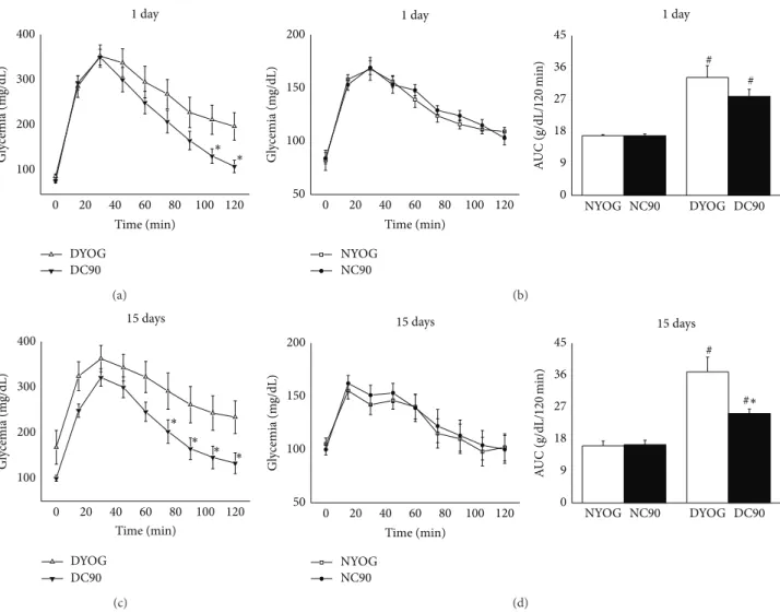
OGTT, the hyperglycemia peek developed by DYOG group 30 minutes ater the glucose overload was approximately 2-fold higher (� < 0.001) than NYOG, in the 1st (Figures3(a) and3(b)) or 15th day (Figures3(c)and3(d)) of experiment. Ater 120 minutes, DYOG did not correct the glycemia with the same eiciency of that observed in NYOG, this last group returning the glycemia to basal values, in the 1st or 15th day of experiment. Consequently, the glucose tolerance was signiicantly impaired in DYOG rats when compared with NYOG rats (Figure 3, AUC, 1 and 15 days). Insulin sensitivity was progressively decreased in diabetic rats, since ater insulin administration the DYOG group was not able to reduce the glycemia levels as eiciently as NYOG, in both 1st
0 10 20 30 40 50 60
10 100 1000 10000
C
o
ncen
tr
at
io
n (n
g/mL)
Time (min)
Curcumin (10mg/kg, i.v.)
Curcumin (500mg/kg, p.o.)
Figure 2: Plasma curcumin concentrations versus time curves in STZ-diabetic rats. Values are expressed as means±SEM.
day (Figures4(a) and 4(b)) and 15th day (Figures4(c) and 4(d)) of experiment.
he treatment of normal, nondiabetic rats with curcumin (NC90) did not change the glucose tolerance in comparison to normal rats treated only with yoghurt (NYOG), ater 1 (Figure 3(b)) or 15 days (Figure 3(d)) of treatment (Figure 3). he ITT was also performed in normal rats ater 1 and 15 days of curcumin treatment, and the constant rate for glucose disappearance (�itt) was calculated to estimate the insulin sensitivity. here were no statistical diferences in the �itt values between NYOG and NC90 rats ater 1 or 15 days of treatment (Figure 4, inserted table, N groups).
Although DYOG and DC90 groups showed similar values of hyperglycemia peek ater the glucose overload, diabetic rats treated with curcumin for 1 day showed a better capacity to reduce the glycemia ater 105 and 120 minutes of the glucose overload (� < 0.05, Figure 3(a)); however the glucose tolerance was not yet diferent when DYOG and DC90 were compared (Figure 3, AUC, 1 day). he insulin sensitivity was not also diferent for the comparison between DYOG and DC90 ater 1 day of treatment, since these groups showed similar rates of glucose disappearance ater insulin administration and similar �itt values (Figure 4, inserted table, D groups).
Table 2: Stability of curcumin in plasma under diferent conditions of storage, temperature, and time intervals (� = 3).
Sample condition
Curcumin nominal concentration
25 ng/mL 400 ng/mL
Accuracy (%) Precision CV (%) Accuracy (%) Precision CV (%)
Bench-top(30∘C, 4 h) 91.85 8.90 105.90 12.50
Postprocessing(30∘C, 4 h) 88.45 7.50 101.56 13.40
Freeze-thaw(−20∘C, 72 h) 89.34 5.40 99.47 11.60
Long time(−20∘C, 7 d) 90.77 6.20 103.24 8.90
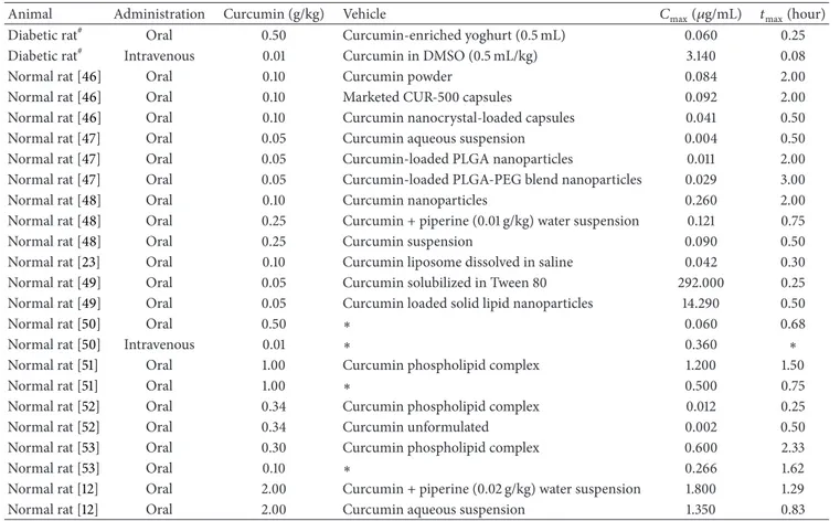
Table 3: Rat plasma/serum levels of curcumin administered in diferent vehicles.
Animal Administration Curcumin (g/kg) Vehicle �max(�g/mL) �max(hour)
Diabetic rat# Oral 0.50 Curcumin-enriched yoghurt (0.5 mL) 0.060 0.25
Diabetic rat# Intravenous 0.01 Curcumin in DMSO (0.5 mL/kg) 3.140 0.08
Normal rat [46] Oral 0.10 Curcumin powder 0.084 2.00
Normal rat [46] Oral 0.10 Marketed CUR-500 capsules 0.092 2.00 Normal rat [46] Oral 0.10 Curcumin nanocrystal-loaded capsules 0.041 0.50 Normal rat [47] Oral 0.05 Curcumin aqueous suspension 0.004 0.50 Normal rat [47] Oral 0.05 Curcumin-loaded PLGA nanoparticles 0.011 2.00 Normal rat [47] Oral 0.05 Curcumin-loaded PLGA-PEG blend nanoparticles 0.029 3.00 Normal rat [48] Oral 0.10 Curcumin nanoparticles 0.260 2.00 Normal rat [48] Oral 0.25 Curcumin + piperine (0.01 g/kg) water suspension 0.121 0.75 Normal rat [48] Oral 0.25 Curcumin suspension 0.090 0.50 Normal rat [23] Oral 0.10 Curcumin liposome dissolved in saline 0.042 0.30 Normal rat [49] Oral 0.05 Curcumin solubilized in Tween 80 292.000 0.25 Normal rat [49] Oral 0.05 Curcumin loaded solid lipid nanoparticles 14.290 0.50
Normal rat [50] Oral 0.50 ∗ 0.060 0.68
Normal rat [50] Intravenous 0.01 ∗ 0.360 ∗
Normal rat [51] Oral 1.00 Curcumin phospholipid complex 1.200 1.50
Normal rat [51] Oral 1.00 ∗ 0.500 0.75
Normal rat [52] Oral 0.34 Curcumin phospholipid complex 0.012 0.25 Normal rat [52] Oral 0.34 Curcumin unformulated 0.002 0.50 Normal rat [53] Oral 0.30 Curcumin phospholipid complex 0.600 2.33
Normal rat [53] Oral 0.10 ∗ 0.266 1.62
Normal rat [12] Oral 2.00 Curcumin + piperine (0.02 g/kg) water suspension 1.800 1.29 Normal rat [12] Oral 2.00 Curcumin aqueous suspension 1.350 0.83
#
his study.
∗Not reported.�
max: peak plasma/serum concentration;�max: time to reach peak plasma/serum concentration.
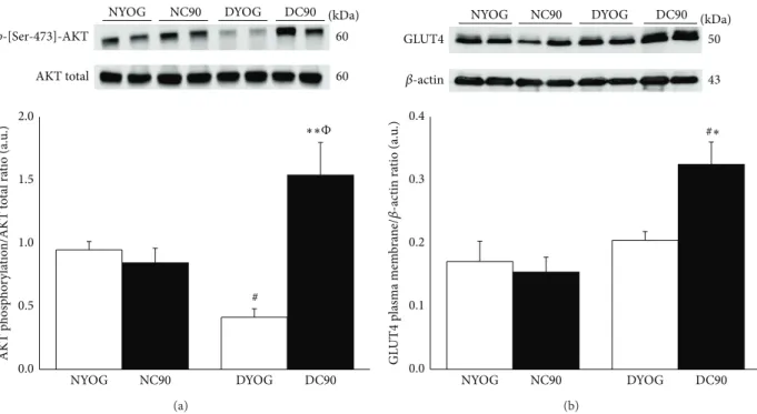
3.4. AKT Phosphorylation Levels and GLUT4 Content in Muscle Plasma Membrane ater Long-Term Treatment of
Diabetic Rats with Curcumin. Phosphorylation levels of AKT
were signiicantly lower (approximately 50%,� < 0.05) in
gastrocnemiusmuscles of diabetic rats treated with yoghurt
(DYOG) when compared with NYOG or NC90 rats. here were no diferences in the AKT phosphorylation levels between NYOG and NC90 rats (Figure 5(a)). On the other hand, ater the glucose overload, the phosphorylation of AKT was signiicantly increased ingastrocnemiusof diabetic rats treated with curcumin when compared with DYOG (3.7-fold, � < 0.01) and with NC90 (82%,� < 0.05) rats (Figure 5(a)).
Ater a glucose overload, the levels of GLUT4 in plasma membrane of NYOG, NC90, and DYOG rats were very sim-ilar (Figure 5(b)). However, GLUT4 content was increased in
plasma membrane ofgastrocnemiusmuscles of diabetic rats treated with curcumin (DC90) when compared with DYOG rats (59%,� < 0.05) and with NYOG and NC90 rats (45 and 38%, resp.,� < 0.05) (Figure 5(b)).
4. Discussion
he values of�maxand�maxfound in the present study ater
0 20 40 60 80 100 120 100
200 300 400
1 day
Time (min)
DYOG DC90
Gl
ycemia (m
g/dL)
∗ ∗
(a)
Time (min)
0 20 40 60 80 100 120
50 100 150 200
1 day
NYOG NC90
Gl
ycemia (m
g/dL)
0 9 18 27 36 45
# # 1 day
NYOG NC90 DYOG DC90
A
UC (g/dL/
120
min)
(b)
0 20 40 60 80 100 120
100 200 300 400
15 days
Time (min)
DYOG DC90
Gl
ycemia (m
g/dL)
∗ ∗
∗ ∗
(c)
0 20 40 60 80 100 120
50 100 150 200
15 days
Time (min)
NYOG NC90
Gl
ycemia (m
g/dL)
0 9 18 27 36 45
# # 15 days
NYOG NC90 DYOG DC90
A
UC (g/dL/
120
min)
∗
(d)
Figure 3: Oral glucose tolerance of normal and STZ-diabetic rats treated with curcumin incorporated in yoghurt. Glycemia levels before
(� = 0) and ater glucose overload in rats treated for 1 day ((a) diabetic; (b) normal) and for 15 days ((c) diabetic; (d) normal) with curcumin.
he insets represent the AUC values (g/dL/120 min). Values are expressed as means±SEM,� = 7-8 per group. Diferences between groups were analyzed with one-way ANOVA followed by Student-Newman-Keuls test (� < 0.05). #: diferences with NYOG and NC90;∗: diferences with DYOG.
can be suggested that the slight diference in the half-life values may be due to diferences in the animal’s body weight, since Yang and colleagues used rats weighing between 280 and 320 g, which could be inluencing the��and increasing the elimination half-life of curcumin.
here are several studies investigating the curcumin pharmacokinetic parameters; however there were some dif-ferences between them, such as the vehicle used for admin-istration, dose, and animal body weight, which make the comparison diicult as can be seen inTable 3; these studies indicate clearly that levels of curcumin are not directly comparable. he�maxand AUC values obtained in the oral
administration of curcumin were signiicantly lower than those found in the i.v. administration, and this fact suggests a low absorption and/or an increased hepatic irst-pass metabolic biotransformation when curcumin is administered orally. he oral bioavailability of curcumin in rats was0.47 ± 0.12% (Table 4).
Several curcumin pharmacokinetic studies in the last decade showed low absorption and fast metabolism, leading to a decrease of its bioavailability. hereby, curcumin plasma levels are extremely low; however it is highly eicient as a therapeutic agent. herefore, it should be reasonable to assume that some of its biological activities are associated with curcumin metabolite(s). In addition, it has been shown that, ater its absorption, curcumin undergoes conjugation to glucuronide or sulfate derivatives [24]. Zhongfa et al. [25] demonstrated that the major curcumin metabolites are curcumin-o-glucuronide, tetrahydrocurcumin (THC), and curcumin-o-sulfate. he curcumin absorption in rats occurs through intestinal cells and most of it is eliminated as curcumin glucuronide or sulfate [26].
1 day
15 days
0 5 10 15 20 25 30
100 200 300 400 500 600
1 day
Time (min)
DYOG DC90
Gl
ycemia (m
g/dL)
Time (min)
0 5 10 15 20 25 30
100 200 300 400 500 600
15 days
DYOG DC90
Gl
ycemia (m
g/dL)
Time (min)
0 5 10 15 20 25 30
40 80 120 160
15 days
NYOG NC90
Gl
ycemia (m
g/dL)
Time (min)
0 5 10 15 20 25 30
40 80 120 160
1 day
NYOG NC90
Gl
ycemia (m
g/dL)
(a) (b)
(c) (d)
DYOG
0.040 ± 0.003
0.012 ± 0.002
DC90
0.045±0.002
NYOG
0.038 ± 0.004
0.040 ± 0.005
NC90
0.040 ± 0.003
0.040 ± 0.003
0.025 ± 0.003∗
kitt(%/min)
NC90
NYOG DYOG DC90
p-[Ser-473]-AKT
AKT total 60
(kDa) 60
0.0 0.5 1.0 1.5 2.0
#
AKT p
h
os
p
h
o
ry
la
tio
n/AKT t
o
ta
l ra
tio (a.u
.)
NYOG NC90 DYOG DC90
Φ ∗ ∗
(a)
NC90
NYOG DYOG DC90
GLUT4
43 50 (kDa)
𝛽-actin
0.0 0.1 0.2 0.3 0.4
NYOG NC90 DYOG DC90
GL
UT
4
pl
asma mem
b
ra
ne/
𝛽
-ac
tin ra
tio (a.u
.) #∗
(b)
Figure 5: AKT activation (a) and plasma membrane GLUT4 content (b) ingastrocnemiusmuscles of normal and STZ-diabetic rats treated with curcumin incorporated in yoghurt for 15 days. AKT activation was evaluated as phosphorylation levels in Ser-473 residues. Results are expressed as means±SEM of the arbitrary units,� = 5-6 per group. Diferences between groups were analyzed with one-way ANOVA followed by Student-Newman-Keuls test. # (� < 0.05): diferences with NYOG and NC90;∗(� < 0.05) and∗∗(� < 0.01): diferences with DYOG;Φ(� < 0.05): diferences between DC90 and NC90.
Table 4: Pharmacokinetic parameters ater oral (500 mg/kg) and i.v. administration (10 mg/kg) of curcumin to STZ-diabetic rats. Values are expressed as means±SEM (� = 5).
Pharmacokinetic parameters Administration route
Oral i.v.
�el(1/min) 0.02±0.01 0.08±0.02 Half-life (min) 32.70±12.92 8.64±2.31 AUC0-�(�g/mL/min) 1.89±0.25 12.27±2.75 AUC0-∞(�g/mL/min) 2.97±0.79 12.45±2.72 Cl (L/kg/min) 0.85±0.24 0.83±0.19
��(L/kg) 37.49±10.46 10.63±4.10
MRT (min) 55.41±20.19 12.46±3.34
��(1/min) 0.29±0.15 —
�max(�g/mL) 0.06±0.01 3.14±0.90
�max(min) 15.00±0.00 5.00±0.00
�(%) 0.47±0.12 100
�el: elimination constant; half-life: time half-life; AUC0-�: area under plasma
concentration/time plot until the last quantiiable value; AUC0-∞: area under
plasma concentration/time plot extrapolated to ininity; Cl: clearance;��:
volume of distribution; MRT: average mean residence time;��: absorption
constant; �max: maximum concentration; �max: time to reach �max; �:
bioavailability.
such as anti-inlammatory [28], hepatoprotective [29], antidi-abetic [27, 30], and antioxidant [31] activities; the most are higher when compared to curcumin efects.
he curcumin half-life was 32.70 ± 12.92 minutes (Table 4); that is, no curcumin should be bioavailable ater
5.45 hours, because of the wash-out in ten half-lives [32]. hus, it is possible to assume that one or more curcumin metabolites are responsible for its antidiabetic activity, since glycemia reduction in STZ-diabetic rats treated with curcumin incorporated in yogurt at intervals of 12 hours was observed [14].
Based on the aforementioned half-life considerations, the evaluation of changes in the glucose tolerance and insulin sensitivity of normal and diabetic rats ater acute (1 day) and subchronic (15 days) treatments with curcumin-enriched yoghurt was performed. For this, we considered that the administration of glucose (OGTT) or insulin (ITT) would be carried out two hours ater the curcumin administration, which means the elapsed time of approximately 4 half-lives; at this time, the curcumin concentration should be in critical levels and so its metabolites would be present and at pharmacological levels [14]. herefore, we could investigate the pharmacodynamic efects on glycemia at intervals of 2–4 hours and 2–2.5 hours for OGTT and ITT, respectively.
Insulin resistance is deined as an attenuated biological response of tissues to physiological or elevated levels of insulin. According to our data, STZ-diabetic rats showed a diminished rate of glucose disappearance ater insulin administration, in the 1st day of the experiment (4 days ater STZ,Figure 4(a)), which was even impaired in the 15th day (19 days ater STZ,Figure 4(c)). Evidences support the insulin resistance observed in STZ-diabetic animals, mainly characterized by a reduction in the tyrosine kinase activity of the insulin receptor (IR), despite the increased IR number [35,36] and decrease in the AKT activation (phosphorylation in serine-473 residue) [37]. According to our data, diabetic rats ater 19 days of the STZ administration and treated with yoghurt showed a proile compatible with insulin resistance, since the muscle AKT phosphorylation was lower than values of normal rats (Figure 5(a)). In addition to the direct disturbances in the insulin signaling of STZ-diabetic rats, the worsening in the insulin responsiveness observed with the progression of the diabetes may be also related to changes in the content of glucose transporters in skeletal muscles. Kahn et al. [38] observed that the levels of glucose trans-porters types 1 and 4 (GLUT1 and GLUT4) are progressively decreased in muscles of STZ-diabetic rats with the extent of the diabetes, although in this present study a reduction in GLUT4 content in muscle plasma membrane of diabetic rats (DYOG) in comparison with normal rats (NYOG) was not found, ater a glucose overload (Figure 5(b)); these diferences may be due to periods of diabetes studied (Kahn study: 7–14 days; present study: 19 days).
he similarity in the rate of glucose disappearance ater a glucose overload (Figures3(b)and3(d)) or insulin admin-istration as well as in the �itt values (Figures 4(b) and 4(d) and inserted table) of normal rats ater 1 and 15 days of treatment represents that the tissue responsiveness to insulin is maintained in these animals over the experimen-tal period. Both pancreas insulin secretion and peripheral insulin responsiveness are well-functioning in the correction of hyperglycemia states, so it is reasonable to understand the absence of efects of curcumin in the glucose tolerance and in the insulin sensitivity of nondiabetic rats. Also, AKT phosphorylation and GLUT4 plasma membrane levels are also similar between NYOG and NC90 rats (Figures 5(a) and5(b)). hese indings corroborate previous data from our laboratory, showing that even a chronic daily treatment (31 days) with curcumin-enriched yoghurt did not change the postprandial glycemia levels of normal rats [14].
Our results showed that one single administration of curcumin was not suicient to improve the ability of STZ-diabetic rats to reverse hyperglycemia. However, in the 1st day of curcumin treatment, it is interesting to note that these rats showed a fast decrease in the glycemia ater 105 and 120 minutes of the glucose overload, when compared with untreated-diabetic rats (Figure 3(a)). his inding reiterates the pharmacokinetic data on the possibility of the beneicial efects of curcumin to be exerting by a metabolite(s), which need an additional time to reach the therapeutic levels and to control the glucose metabolism. In fact, it was observed that the treatment for 15 days with curcumin promoted a most evident beneit on glucose metabolism of diabetic rats,
improving both the glucose tolerance (Figure 3(c)) and the insulin sensitivity (Figure 4(c) and inserted table). Long-term of a daily treatment with curcumin probably culminates in an increased permanence into the circulation of the curcumin metabolite(s), allowing the biological active compound(s) to exert its efects in a most pronounced way. Recent evidence of our laboratory reinforces the possibility of the antidia-betic activity of curcumin to be exerted by metabolite(s): Arcaro et al. [15] found that the treatment of STZ-diabetic rats with yoghurt enriched with 90 mg/kg curcumin and piperine did not increase (20 mg/kg piperine) and even nulliied (40 mg/kg piperine) the antidiabetic and antioxidant activities of curcumin. It is well known that piperine increases the bioavailability of many drugs and compounds, including curcumin, via inhibition of the activity of various metab-olizing enzymes found in liver and intestine, such as aryl hydroxylases, N-demethylases, UDP-glucuronyltransferases, and cytochrome P450 3A4 [39,40]. However, the inhibition of the curcumin biotransformation will not necessarily lead to an increase of its pharmacodynamic actions, and indeed adverse efects can be reached.
Corroborating the hypothesis that curcumin metabo-lite(s) has pivotal importance in determined biological activ-ities when curcumin is administered orally, Neyrinck et al. [41] found that the coadministration of curcuma extract (0.1% of curcumin) and 0.01% of white pepper (which contains piperine) to mice fed a high-fat (HF) diet did not promote any change in the glucose and lipid homeostasis, in compar-ison with nontreated HF mice. Besides, HF mice receiving these phytotherapics showed low levels of proinlammatory cytokines IL-6 and TNF-� in subcutaneous adipose tissue in association with accumulation of THC; the authors sug-gested that this metabolite may be responsible for the anti-inlammatory response.
he indings of the present study showed that diabetic rats treated for 15 days with curcumin incorporated in yoghurt had an increase in the AKT phosphorylation in
gastrocnemiusmuscles (Figure 5(a)) in response to a glucose
STZ-nicotinamide diabetic rats treated for 45 days with curcumin (80 mg/kg) or with THC (80 mg/kg) showed an increased ability for insulin-receptor binding when compared with cells from nontreated diabetic rats, which was associated with the antihyperglycemic efect of these compounds.
5. Conclusion
he present indings in the half-life of curcumin in plasma of diabetic rats (PK) and in the temporal behavior in the glucose tolerance and insulin sensitivity assays ater acute and sub-chronic treatments with curcumin-enriched yoghurt (PD) are inconsistent, which substantiate evidences for curcumin metabolite(s) as being responsible for the antidiabetic activity which may be related, at least in part, to an increase in skeletal muscle glucose uptake due to AKT activation leading to an enhancement in the plasma membrane GLUT4 content.
Conflict of Interests
he authors declare that there is no conlict of interests regarding the publication of this paper.
Authors’ Contribution
Vˆania Ortega Gutierres, Rosˆangela Gonc¸alves Peccinini, Amanda Martins Baviera, and Iguatemy Lourenc¸o Brunetti participated in the research design. Vˆania Ortega Gutier-res, Michel Leandro Campos, Carlos Alberto Arcaro, and Renata Pires Assis performed the experiments. Michel Lean-dro Campos, Helen Mariana Baldan-Cimatti, Rosˆangela Gonc¸alves Peccinini, Silvia Paula-Gomes, Isis Carmo Ket-telhut, and Amanda Martins Baviera contributed to new reagents or analytical tools. Vˆania Ortega Gutierres, Michel Leandro Campos, Helen Mariana Baldan-Cimatti, Rosˆangela Gonc¸alves Peccinini, Silvia Paula-Gomes, Isis Carmo Ket-telhut, Amanda Martins Baviera, and Iguatemy Lourenc¸o Brunetti performed the data analysis. Vˆania Ortega Gutier-res, Michel Leandro Campos, Rosˆangela Gonc¸alves Peccinini, Silvia Paula-Gomes, Isis Carmo Kettelhut, Amanda Martins Baviera, and Iguatemy Lourenc¸o Brunetti wrote or con-tributed to the writing of the paper.
Acknowledgments
he authors wish to thank the Conselho Nacional de Pesquisa e Desenvolvimento (CNPq), Faculdade de Ciˆencias Far-macˆeuticas (FCFAr-UNESP), and Fundac¸˜ao de Amparo a Pesquisa do Estado de S˜ao Paulo (FAPESP) for the inancial support. he authors are also grateful to Vivian Boter Berga-masco for the technical assistance.
References
[1] S. C. Gupta, G. Kismali, and B. B. Aggarwal, “Curcumin, a component of turmeric: from farm to pharmacy,”BioFactors, vol. 39, no. 1, pp. 2–13, 2013.
[2] R. L. hangapazham, A. Puri, S. Tele, R. Blumenthal, and R. K. Maheshwari, “Evaluation of a nanotechnology-based carrier for delivery of curcumin in prostate cancer cells,”International Journal of Oncology, vol. 32, no. 5, pp. 1119–1123, 2008. [3] L. M. Howells, J. Mahale, S. Sale et al., “Translating curcumin
to the clinic for lung cancer prevention: evaluation of the preclinical evidence for its utility in primary, secondary, and tertiary prevention strategies,” Journal of Pharmacology and Experimental herapeutics, vol. 350, no. 3, pp. 483–494, 2014. [4] W. Wongcharoen and A. Phrommintikul, “he protective role
of curcumin in cardiovascular diseases,”International Journal of Cardiology, vol. 133, no. 2, pp. 145–151, 2009.
[5] B. B. Aggarwal, “Targeting Inlammation-induced obesity and metabolic diseases by curcumin and other nutraceuticals,”
Annual Review of Nutrition, vol. 30, pp. 173–199, 2010. [6] D.-W. Zhang, M. Fu, S.-H. Gao, and J.-L. Liu, “Curcumin and
diabetes: a systematic review,”Evidence-Based Complementary and Alternative Medicine, vol. 2013, Article ID 636053, 16 pages, 2013.
[7] M.-H. Pan, T.-M. Huang, and J.-K. Lin, “Biotransformation of curcumin through reduction and glucuronidation in mice,”
Drug Metabolism and Disposition, vol. 27, no. 4, pp. 486–494, 1999.
[8] S. K. Vareed, M. Kakarala, M. T. Ruin et al., “Pharmacokinetics of curcumin conjugate metabolites in healthy human subjects,”
Cancer Epidemiology Biomarkers & Prevention, vol. 17, no. 6, pp. 1411–1417, 2008.
[9] P. Anand, S. G. homas, A. B. Kunnumakkara et al., “Biological activities of curcumin and its analogues (Congeners) made by man and Mother Nature,”Biochemical Pharmacology, vol. 76, no. 11, pp. 1590–1611, 2008.
[10] Y. Gao, Z. Li, M. Sun et al., “Preparation, characterization, phar-macokinetics, and tissue distribution of curcumin nanosuspen-sion with TPGS as stabilizer,”Drug Development and Industrial Pharmacy, vol. 36, no. 10, pp. 1225–1234, 2010.
[11] J. Cui, B. Yu, Y. Zhao et al., “Enhancement of oral absorption of curcumin by self-microemulsifying drug delivery systems,”
International Journal of Pharmaceutics, vol. 371, no. 1-2, pp. 148– 155, 2009.
[12] G. Shoba, D. Joy, T. Joseph, M. Majeed, R. Rajendran, and P. S. S. R. Srinivas, “Inluence of piperine on the pharmacokinetics of curcumin in animals and human volunteers,”Planta Medica, vol. 64, no. 4, pp. 353–356, 1997.
[13] S. Prasad, A. K. Tyagi, and B. B. Aggarwal, “Recent develop-ments in delivery, bioavailability, absorption and metabolism of curcumin: the golden pigment from golden spice,”Cancer Research and Treatment, vol. 46, no. 1, pp. 2–18, 2014.
[14] V. O. Gutierres, C. M. Pinheiro, R. P. Assis, R. C. Vendramini, M. T. Pepato, and I. L. Brunetti, “Curcumin-supplemented yoghurt improves physiological and biochemical markers of experimental diabetes,”British Journal of Nutrition, vol. 108, no. 3, pp. 440–448, 2012.
[15] C. A. Arcaro, V. O. Gutierres, R. P. Assis et al., “Piperine, a natural bioenhancer, nulliies the antidiabetic and antioxidant activities of curcumin in streptozotocin-diabetic rats,” PLoS ONE, vol. 9, no. 12, Article ID e113993, 2014.
[16] P. Trinder, “Determination of blood glucose using 4-amino phenazone as oxygen acceptor,”Journal of Clinical Pathology, vol. 22, no. 2, p. 246, 1969.
gastroulcerogenic efect,”Drug Metabolism Letters, vol. 6, no. 4, pp. 235–241, 2013.
[18] K. Lundbaek, “Intravenous glucose tolerance as a tool in deinition and diagnosis of diabetes mellitus,”British Medical Journal, vol. 1, no. 5291, pp. 1507–1513, 1962.
[19] M. M. Bradford, “A rapid and sensitive method for the quanti-tation of microgram quantities of protein utilizing the principle of protein dye binding,”Analytical Biochemistry, vol. 72, no. 1-2, pp. 248–254, 1976.
[20] S. Nishiumi and H. Ashida, “Rapid preparation of a plasma membrane fraction from adipocytes and muscle cells: appli-cation to detection of translocated glucose transporter 4 on the plasma membrane,”Bioscience, Biotechnology, and Biochem-istry, vol. 71, no. 9, pp. 2343–2346, 2007.
[21] U. K. Laemmli, “Cleavage of structural proteins during the assembly of the head of bacteriophage T4,”Nature, vol. 227, no. 5259, pp. 680–685, 1970.
[22] H. Towbin, T. Staehelin, and J. Gordon, “Electrophoretic trans-fer of proteins from polyacrylamide gels to nitrocellulose sheets: procedure and some applications,”Proceedings of the National Academy of Sciences of the United States of America, vol. 76, no. 9, pp. 4350–4354, 1979.
[23] K. Y. Yang, L. C. Lin, T. Y. Tseng, S. C. Wang, and T. H. Tsai, “Oral bioavailability of curcumin in rat and the herbal analysis from Curcuma longa by LC-MS/MS,”Journal of Chro-matography B: Analytical Technologies in the Biomedical and Life Sciences, vol. 853, no. 1-2, pp. 183–189, 2007.
[24] J.-K. Lin, M.-H. Pan, and S.-Y. Lin-Shiau, “Recent studies on the biofunctions and biotransformations of curcumin,”BioFactors, vol. 13, no. 1–4, pp. 153–158, 2000.
[25] L. Zhongfa, M. Chiu, J. Wang et al., “Enhancement of curcumin oral absorption and pharmacokinetics of curcuminoids and curcumin metabolites in mice,” Cancer Chemotherapy and Pharmacology, vol. 69, no. 3, pp. 679–689, 2012.
[26] A. Asai and T. Miyazawa, “Dietary curcuminoids prevent high-fat diet-induced lipid accumulation in rat liver and epididymal adipose tissue,”Journal of Nutrition, vol. 131, no. 11, pp. 2932– 2935, 2001.
[27] K. Karthikesan, L. Pari, and V. P. Menon, “Combined treatment of tetrahydrocurcumin and chlorogenic acid exerts poten-tial antihyperglycemic efect on streptozotocin-nicotinamide-induced diabetic rats,”General Physiology and Biophysics, vol. 29, no. 1, pp. 23–30, 2010.
[28] Y. Nakamura, Y. Ohto, A. Murakami, T. Osawa, and H. Ohi-gashi, “Inhibitory efects of curcumin and tetrahydrocurcumi-noids on the tumor promoter-induced reactive oxygen species generation in leukocytes in vitro and in vivo,”Japanese Journal of Cancer Research, vol. 89, no. 4, pp. 361–370, 1998.
[29] L. Pari and D. R. Amali, “Protective role of tetrahydrocurcumin (THC) an active principle of turmeric on chloroquine induced hepatotoxicity in rats,”Journal of Pharmacy & Pharmaceutical Sciences, vol. 8, no. 1, pp. 115–123, 2005.
[30] L. Pari and P. Murugan, “Efect of tetrahydrocurcumin on blood glucose, plasma insulin and hepatic key enzymes in streptozotocin induced diabetic rats,” Journal of Basic and Clinical Physiology and Pharmacology, vol. 16, no. 4, pp. 257– 274, 2005.
[31] K. Okada, C. Wangpoengtrakul, T. Tanaka, S. Toyokuni, K. Uchida, and T. Osawa, “Curcumin and especially tetrahy-drocurcumin ameliorate oxidative stress-induced renal injury in mice,”Journal of Nutrition, vol. 131, no. 8, pp. 2090–2095, 2001.
[32] P. L. Toutain and A. Bousquet-M´elou, “Plasma terminal half-life,”Journal of Veterinary Pharmacology and herapeutics, vol. 27, no. 6, pp. 427–439, 2004.
[33] S. Lenzen, “he mechanisms of alloxan- and streptozotocin-induced diabetes,”Diabetologia, vol. 51, no. 2, pp. 216–226, 2008. [34] K. Kagami, H. Morita, K. Onda, T. Hirano, and K. Oka, “Protective efect of cafeine on streptozotocin-induced beta-cell damage in rats,”Journal of Pharmacy and Pharmacology, vol. 60, no. 9, pp. 1161–1165, 2008.
[35] T. Kadowaki, M. Kasuga, Y. Akanuma, O. Ezaki, and F. Takaku, “Decreased autophosphorylation of the insulin receptor-kinase in streptozotocin-diabetic rats,”he Journal of Biological Chem-istry, vol. 259, no. 22, pp. 14208–14216, 1984.
[36] F. Giorgino, J.-H. Chen, and R. J. Smith, “Changes in tyrosine phosphorylation of insulin receptors and a 170,000 molec-ular weight nonreceptor protein in vivo in skeletal muscle of streptozotocin-induced diabetic rats: efects of insulin and glucose,”Endocrinology, vol. 130, no. 3, pp. 1433–1444, 1992. [37] J. J. Hulmi, M. Silvennoinen, M. Lehti, R. Kivel¨a,
and H. Kainulainen, “Altered REDD1, myostatin, and Akt/mTOR/FoxO/MAPK signaling in streptozotocin-induced diabetic muscle atrophy,”he American Journal of Physiology— Endocrinology and Metabolism, vol. 302, no. 3, pp. E307–E315, 2012.
[38] B. B. Kahn, L. Rossetti, H. F. Lodish, and M. J. Charron, “Decreased in vivo glucose uptake but normal expression of GLUT1 and GLUT4 in skeletal muscle of diabetic rats,”he Journal of Clinical Investigation, vol. 87, no. 6, pp. 2197–2206, 1991.
[39] C. K. Atal, R. K. Dubey, and J. Singh, “Biochemical basis of enhanced drug bioavailability by piperine: evidence that piperine is a potent inhibitor of drug metabolism,”Journal of Pharmacology and Experimental herapeutics, vol. 232, no. 1, pp. 258–262, 1985.
[40] K. Srinivasan, “Black pepper and its pungent principle-piperine: a review of diverse physiological efects,”Critical Reviews in Food Science and Nutrition, vol. 47, no. 8, pp. 735–748, 2007. [41] A. M. Neyrinck, M. Alligier, P. B. Memvanga et al., “Curcuma
longaextract associated with white pepper lessens high fat diet-induced inlammation in subcutaneous adipose tissue,”PLoS ONE, vol. 8, no. 11, Article ID e81252, 2013.
[42] Y.-T. Deng, T.-W. Chang, M.-S. Lee, and J.-K. Lin, “Suppression of free fatty acid-induced insulin resistance by phytopolyphe-nols in C2C12 mouse skeletal muscle cells,”Journal of Agricul-tural and Food Chemistry, vol. 60, no. 4, pp. 1059–1066, 2012. [43] T.-C. Cheng, C.-S. Lin, C.-C. Hsu, L.-J. Chen, K.-C. Cheng,
and J.-T. Cheng, “Activation of muscarinic M-1 cholinoceptors by curcumin to increase glucose uptake into skeletal muscle isolated from Wistar rats,”Neuroscience Letters, vol. 465, no. 3, pp. 238–241, 2009.
[44] L.-X. Na, Y.-L. Zhang, Y. Li et al., “Curcumin improves insulin resistance in skeletal muscle of rats,”Nutrition, Metabolism and Cardiovascular Diseases, vol. 21, no. 7, pp. 526–533, 2011. [45] P. Murugan, L. Pari, and C. A. Rao, “Efect of
tetrahydrocur-cumin on insulin receptor status in type 2 diabetic rats: studies on insulin binding to erythrocytes,”Journal of Biosciences, vol. 33, no. 1, pp. 63–72, 2008.
[47] T. H. Marczylo, R. D. Verschoyle, D. N. Cooke, P. Morazzoni, W. P. Steward, and A. J. Gescher, “Comparison of systemic availability of curcumin with that of curcumin formulated with phosphatidylcholine,” Cancer Chemotherapy and Pharmacol-ogy, vol. 60, no. 2, pp. 171–177, 2007.
[48] K. Maiti, K. Mukherjee, A. Gantait, B. P. Saha, and P. K. Mukherjee, “Curcumin-phospholipid complex: preparation, therapeutic evaluation and pharmacokinetic study in rats,”
International Journal of Pharmaceutics, vol. 330, no. 1-2, pp. 155– 163, 2007.
[49] V. Kakkar, S. Singh, D. Singla et al., “Pharmacokinetic appli-cability of a validated liquid chromatography tandem mass spectroscopy method for orally administered curcumin loaded solid lipid nanoparticles to rats,”Journal of Chromatography B: Analytical Technologies in the Biomedical and Life Sciences, vol. 878, no. 32, pp. 3427–3431, 2010.
[50] J. LJi, Y. Jiang, J. Wen, G. Fan, Y. Wu, and C. Zhang, “A rapid and simple HPLC method for the determination of curcumin in rat plasma: assay development, validation and application to a pharmacokinetic study of curcumin liposome,”Biomedical Chromatography, vol. 23, no. 11, pp. 1201–1207, 2009.
[51] J. Shaikh, D. D. Ankola, V. Beniwal, D. Singh, and M. N. V. R. Kumar, “Nanoparticle encapsulation improves oral bioavailabil-ity of curcumin by at least 9-fold when compared to curcumin administered with piperine as absorption enhancer,”European Journal of Pharmaceutical Sciences, vol. 37, no. 3-4, pp. 223–230, 2009.
[52] N. M. Khalil, T. C. F. do Nascimento, D. M. Casa et al., “Pharmacokinetics of curcumin-loaded PLGA and PLGA-PEG blend nanoparticles ater oral administration in rats,”Colloids and Surfaces B: Biointerfaces, vol. 101, pp. 353–360, 2013. [53] R. Ravichandran, “Pharmacokinetic study of nanoparticulate
Submit your manuscripts at
http://www.hindawi.com
Stem Cells
International
Hindawi Publishing Corporationhttp://www.hindawi.com Volume 2014
Hindawi Publishing Corporation
http://www.hindawi.com Volume 2014
INFLAMMATION
Hindawi Publishing Corporation
http://www.hindawi.com Volume 2014
Behavioural
Neurology
Endocrinology
International Journal ofHindawi Publishing Corporation
http://www.hindawi.com Volume 2014
Hindawi Publishing Corporation
http://www.hindawi.com Volume 2014
Disease Markers
Hindawi Publishing Corporation
http://www.hindawi.com Volume 2014 BioMed
Research International
Oncology
Journal ofHindawi Publishing Corporation
http://www.hindawi.com Volume 2014
Hindawi Publishing Corporation
http://www.hindawi.com Volume 2014 Oxidative Medicine and Cellular Longevity Hindawi Publishing Corporation
http://www.hindawi.com Volume 2014
PPAR Research
The Scientiic
World Journal
Hindawi Publishing Corporation
http://www.hindawi.com Volume 2014
Immunology Research Hindawi Publishing Corporation
http://www.hindawi.com Volume 2014
Journal of
Obesity
Journal ofHindawi Publishing Corporation
http://www.hindawi.com Volume 2014
Hindawi Publishing Corporation
http://www.hindawi.com Volume 2014
Computational and Mathematical Methods in Medicine
Ophthalmology
Journal of Hindawi Publishing Corporationhttp://www.hindawi.com Volume 2014
Diabetes Research
Journal ofHindawi Publishing Corporation
http://www.hindawi.com Volume 2014
Hindawi Publishing Corporation
http://www.hindawi.com Volume 2014 Research and Treatment
AIDS
Hindawi Publishing Corporation
http://www.hindawi.com Volume 2014 Gastroenterology Research and Practice
Hindawi Publishing Corporation
http://www.hindawi.com Volume 2014
Parkinson’s
Disease
Evidence-Based Complementary and Alternative Medicine
Volume 2014