ALESSANDRO DE SÁ GUIMARÃES
EPIDEMIOLOGIA DA LINFADENITE CASEOSA OVINA
NO ESTADO DE MINAS GERAIS, BRASIL
Belo Horizonte
Escola de Veterinária – UFMG Dezembro de 2009
Tese apresentada à Universidade Federal de Minas Gerais, Escola de Veterinária, como requisito parcial para obtenção do grau de Doutor em Ciência Animal.
Orientador: Prof. Dr. Marcos Bryan Heinemann
Co-orientadores: Profa. Dra. Aurora Maria Guimarães Gouveia Prof. Dr. Andrey Pereira Lage
G963e Guimarães, Alessandro de Sá, 1971-
Epidemiologia da linfadenite caseosa ovina no estado Minas Gerais, Brasil / Alessandro de Sá Guimarães - 2009.
83 p. : il.
Orientador: Marcos Bryan Heinemann
Co-orientadores: Aurora Maria Guimarães Gouveia, Andrey Pereira Lage, Vasco Ariston de Carvalho Azevedo
Tese (doutorado) – Universidade Federal de Minas Gerais, Escola de Veterinária
Inclui bibliografia
1. Ovino – Doenças – Teses. 2. Ovino – Criação - Teses– 3. Linfadenite caseosa – Teses. 4. Epidemiologia – Teses. I. Heinemann, Marcos Bryan. Gouveia, Aurora Maria Guimarães. III. Lage, Andrey Pereira. IV. Azevedo, Vasco Ariston de Carvalho. V. Universidade Federal de Minas Gerais. Escola de Veterinária. VI. Título.
AGRADECIMENTOS
À Deus pelo dom da vida e pela oportunidade de aprender cada dia mais.
Aos meus pais, Geraldo Nogueira Guimarães e Sônia Maria de Sá Guimarães pelo amor, carinho, ensinamentos e apoio constantes em todos os momentos da vida.
A minha querida filha Amanda Oliveira de Sá Guimarães, fonte de vida e de alegria. A minha irmã Daniela de Sá Guimarães, pela amizade.
Ao amigo Filipe Borges do Carmo, sem o qual esse trabalho seria praticamente impossível. À orientadora Profa. Aurora Maria Guimarães Gouveia pelos ensinamentos durante essa jornada.
Aos orientadores Profs. Andrey Pereira Lage, Marcos Bryan Heinemann e Vasco Ariston de Carvalho Azevedo pelo apoio e ensinamentos durante os quatros anos de trabalho.
Aos amigos e Profs. Alan Maia Borges e Renato Lima Santos pela amizade e valiosos conselhos durante todo o processo.
A todos os alunos de iniciação científica e pós-graduação (mestrado e doutorado) que, direta ou indiretamente, colaboraram na execução desse projeto, em especial aqueles do Laboratório de Bacteriologia Aplicada (LBA), Laboratório de Sanidade de Ovinos e Caprinos (LASOC), ambos da Escola de Veterinária da UFMG, Laboratório de Genética Celular e Molecular (LGCM) do Instituto de Ciências Biológicas (ICB-UFMG) e Laboratório de Imunologia do Instituto de Ciência da Saúde da Universidade Federal da Bahia (ICS-UFBA).
A senhora Maria Anita Guimarães e Fernando Guimarães Gouveia pela amizade e hospitalidade com que me receberam em sua casa.
A Eliane, secretária da professora Aurora pela amizade e apoio.
Ao Dr. Altino Rodrigues Neto, Diretor Presidente do Instituto Mineiro de Agropecuária (IMA), por nos proporcionar o apoio técnico e financeiro, indispensáveis na realização de parte deste trabalho.
A todos os médicos veterinários do Instituto Mineiro de Agropecuária (IMA) pelo apoio na aplicação dos questionários aos ovinocultores de Minas Gerais.
A Superintendência Federal da Agricultura SIPAG/DT-MG, na pessoa do médico veterinário Belfort Rodriguez Dieguez, Fiscal Federal Agropecuário pelo fornecimento dos dados estatísticos relativos ao SIF abate ovinos e por sua paciência em receber-nos com tanta boa vontade.
À Caprileite/ACCOMIG pelo apoio na divulgação das informações por meio de seus veículos de comunicação com os criadores.
A todos os criadores de ovinos que contribuíram para realização deste trabalho.
A todas as pessoas que porventura não tenham sido mencionadas, mas participaram direta ou indiretamente na elaboração deste trabalho.
APOIO FINANCEIRO
Ao Conselho Nacional de Desenvolvimento Científico e Tecnológico (CNPq) pela bolsa concedida ao aluno Alessandro de Sá Guimarães durante o doutorado.
À Fundação de Amparo à Pesquisa do Estado de Minas Gerais (FAPEMIG) e do CNPq pelo apoio financeiro aos laboratórios LASOC – DMVP – EV – UFMG e LGCM – DBG – ICB – UFMG.
Ao Instituto Mineiro de Agropecuária, Superintendência Federal de Agricultura (SFA/MAPA) e Associação dos Criadores de Caprinos e Ovinos de Minas Gerais (Caprileite/ACCOMIG) pelo custeio e organização da etapa de coleta de amostras e aplicação de questionários em criatórios ovinos de Minas Gerais.
À Rio Branco Alimentos (Programa Pif Paf Ovinos) pelo custeio de deslocamentos, hospedagem e alimentação dos dois médicos veterinários pós-graduandos da EV-UFMG, durante seis meses de coleta de amostras na indústria em Patrocínio.
AGRADECIMENTO ESPECIAL
DEDICATÓRIA
Dedico essa tese a minha avó Luci Guimarães (em memória), exemplo de fé e dedicação à família;
Ao meu avô Ilídio de Sá (em memória), exemplo de bom senso e honestidade, homem de palavras certas em momentos certos;
Dedico também a minha avó Maria Nogueira Maia de Sá (em memória);
SUMÁRIO
Pág.
LISTA DE ABREVIATURAS ... 13
RESUMO ... 15
ABSTRACT ... 16
1. INTRODUÇÃO ... 17
1.1 OBJETIVOS ... 18
1.1.1 Objetivo geral ... 18
1.1.2 Objetivos específicos ... 18
2. Capítulo 1 CASEOUS LYMPHADENITIS: EPIDEMIOLOGY, DIAGNOSIS AND CONTROL ... 19
3. Capítulo 2 CASEOUS LYMPHADENITIS IN SHEEP FLOCKS OF THE STATE OF MINAS GERAIS, BRAZIL: PREVALENCE AND MANAGEMENT SURVEYS. ... 39
4. Capítulo 3 LINFADENITE CASEOSA EM OVINOS ABATIDOS EM FRIGORÍFICO DE MINAS GERAIS: CARACTERIZAÇÃO DAS PROPRIEDADES FORNECEDORAS DE OVINOS AO FRIGORÍFICO E SOROEPIDEMIOLÓGICA A PARTIR DE SOROSCOLETADOS ... 51
4.1 INTRODUÇÃO ... 52
4.2 MATERIAL E MÉTODOS... 52
4.2.1 Marco amostral ... 52
4.2.2 Exame e identificação ante mortem dos ovinos e coleta de material no frigorífico ... 54
4.2.3 Teste imunoenzimático (ELISA) para detecção de anticorpos anti-C. pseudotuberculosis ... 55
4.2.4 Coleta de conteúdo de abscessos ... 55
4.2.5 Isolamento bacteriano nas amostras coletadas... 56
4.2.6 Identificação bioquímica dos isolados ... 57
4.2.7 Extração do DNA total a partir de culturas ... 58
4.2.8 Primers e PCR multiplex de material cultivado ... 58
4.3 RESULTADOS ... 59
4.3.1 Resultados obtidos a partir do questionário aplicado em propriedades fornecedoras do frigorífico em Minas Gerais ... 59
4.3.2 Linfadenite caseosa em soros ovinos abatidos em frigorífico de Minas Gerais ... 61
4.3.3 Ocorrência de nódulos caseosos nas carcaças de ovinos em frigorífico de Minas Gerais ... 61
4.3.4 Isolamento e identificação por provas bioquímicas e PCR multiplex (PCRm) de material cultivado ... 61
4.4 DISCUSSÃO ... 62
4.4.1 Caracterização das propriedades fornecedoras para frigorífico em Minas Gerais ... 62
4.4.2 Caracterização soroepidemiológica da linfadenite caseosa em ovinos ... 64
4.4.3 Bacterioteca de isolados de C. pseudotuberculosis de Minas Gerais ... 68
5. Capítulo 4 CONCLUSOES E PERSPECTIVAS ... 69
5.1 Conclusões ... 70
5.2 Perspectivas ... 70
6 REFERENCIAS BIBLIOGRAFICAS... 70
7 ANEXOS ... 79
7.1 Anexos 1 ... 79
7.1.1 Artigos publicados e resumos apresentados em congressos durante o doutorado ... 79
7.1.2 Artigos submetidos em periódicos (no prelo) ... 80
7.2 Anexos 2 ... 81
7.2.1 Soluções para extração de DNA bacteriano... 81
7.3 Anexos 3 ... 82
7.3.1 Questionário aplicado aos fornecedores de ovinos para ... 82
LISTA DE TABELAS Tabela 1 Ocorrência de linfadenite caseosa em ovinos e caprinos no Brasil por ano, unidade da Federação e ferramentas de diagnóstico, 2009. ... 18
Capítulo 1 Tabela 1 Principal phenotypic characteristics of Corynebacterium pseudotuberculosis used for identification. ... 22
Tabela 2 .Principal phenotypic characteristics of Corynebacterium pseudotuberculosis used for identification. ... 29
Capítulo 2 Tabela 1 Frequency distribution of seropositive sheep based on ELISA for Corynebacterium pseudotuberculosis in Minas Gerais, Brazil, 2002. ... 44
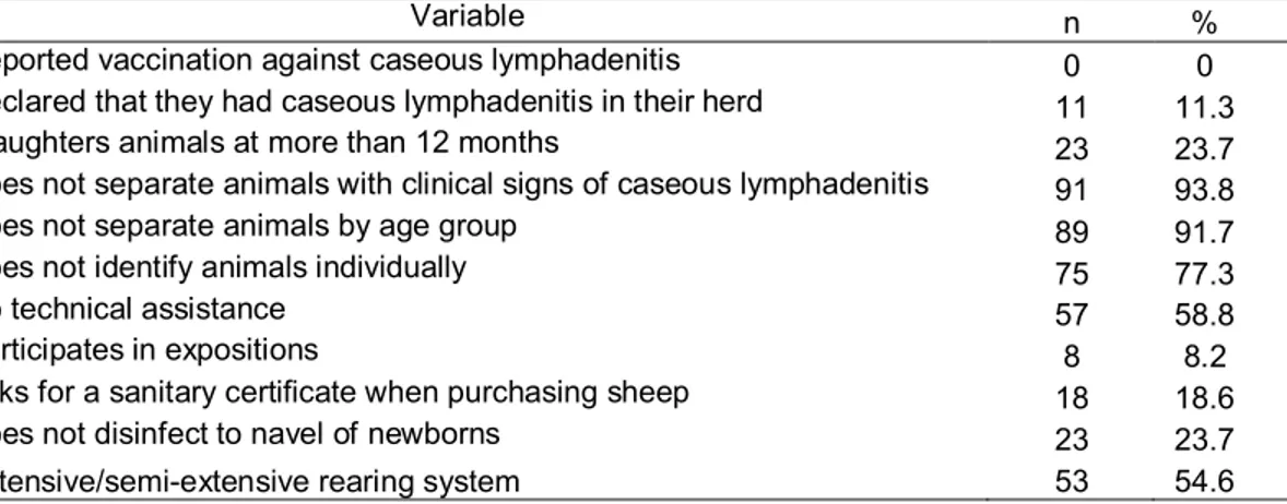
Tabela 2 Principal management practices identified among the 97 sheep herds studied to determine caseous lymphadenitis incidence in the state of Minas Gerais, Brazil, 2002. ... 44
Capítulo 3
Tabela 1 Bactérias utilizadas como controles positivo e negativo para provas bioquímicas, para identificação de C. pseudotuberculosis de Minas Gerais, 2007. ... 57 Tabela 2 Primers de oligonucleotídeos utilizados na PCR multiplex, para
identificação de C. pseudotuberculosis de Minas Gerais, 2007. ... 59 Tabela 3 Distribuição das propriedades fornecedoras de ovinos para frigorífico em
Minas Gerais quanto à anotação dos animais com abscessos, idade em que apresentam abscessos, sua localização e atitude do responsável em caso de recidivas, 2007 ... 59 Tabela 4 Distribuição das propriedades fornecedoras de ovinos para frigorífico em
Minas Gerais quanto ao manejo dos animais com abscessos, destino do material descartado, utilização de dreno no local na incisão e cauterização interna do abscesso com iodo, 2007. ... 60 Tabela 5 Distribuição das propriedades de ovinos em Minas Gerais quanto ao
conhecimento do potencial zoonótico da linfadenite caseosa, destino dos animais e informaçao de perdas durante o abate por linfadenite caseosa no frigorífico, 2007...
60
Tabela 6 Distribuição das propriedades com ovinos em Minas Gerais quanto à presença de assistência veterinária, desinfecção de instalações e tipo de cerca utilizada, 2007. ... 61 Tabela 7 Distribuição de frequência de resultados sorológicos positivos por ELISA
para detecção de anticorpos contra C. pseudotuberculosis em soros de ovinos procedentes de Minas Gerais, coletados em frigorífico, Patrocínio (MG), 2007. ... 61 Tabela 8 Distância, em quilômetros, entre Patrocínio (MG), município sede da
indústria frigorífica, e os municípios de Minas Gerais com propriedades que forneceram ovinos para abate no período de julho a dezembro, 2007 ... 67
LISTA DE FIGURAS
Capítulo 1
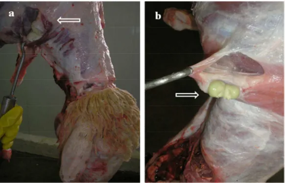
Figura 1 201 countries that reported their sanitary situation to the World Animal Health Organization (OIE), 1996 – 2004. ... 23 Figura 2 Condemnation of sheep carcass at slaughterhouse inspection. A.
Pre-scapular lymph node. B. Superficial lymph node. Arrows indicate pre-scapular lymph nodes with caseous material, characteristic of caseous lymphadenitis in federally inspected slaughterhouse. ... 24 Figure 3 A. Two caeous abscesses, one spontaneous opened and other closed. B.
Ocular and cutaneous infection of sheep with caseous material ... 26 Capítulo 2
Capítulo 3
Figura 1 Municípios em Minas Gerais com propriedades fornecedoras de ovinos para abate inspecionado em Patrocínio (circulado), 2007 ... 53 Figura 2 A. Coleta de soro na mesa de sangria de frigorífico; B. Tubos
devidamente identificados, em repouso para dessoragem, em Patrocínio, Minas Gerais, 2007 ... 54 Figura 3 A. Conteúdo caseoso em linfonodo pré-escapular. B. Corte em cruz no
membro dianteiro indicador de descarte em função de lesão típica de linfadenite caseosa ... 56 Figura 4 Gel de agarose com PCR multiplex do DNA extraído de isolados de C.
LISTA DE ABREVIATURAS
ACCOMIG Associação dos Criadores de Caprinos e Ovinos de Minas Gerais
BA Bahia
BHI Infusão Cérebro Coração
CAMP Christie, Atkins, Munch, Petersen (prova de CAMP)
CE Ceará
DMVP Departamento de Medicina Veterinária Preventiva DNA Ácido desoxirribonucléico
dNTP Desoxinucleotídeo trifosfato DO Densidade ótica
ELISA Ensaio imunoenzimático
Embrapa Empresa Brasileira de Pesquisa Agropecuária
EV-UFMG Escola de Veterinária da Universidade Federal de Minas Gerais FAPEMIG Fundação de Apoio a Pesquisa de Minas Gerais
GEPOC Grupo de Extensão da Pesquisa em Ovinos e Caprinos IBGE Instituto Brasileiro de Geografia e Estatística
ICB Instituto de Ciências Biológicas IDGA Imunodifusão em gel de ágar IHS Inibição da hemólise sinérgica
H hora
HCl Ácido clorídrico
IMA Instituto Mineiro de Agropecuária Kb quilobases (103 pb)
KCl Cloreto de potássio
LASOC Laboratório de Sanidade em Ovinos e Caprinos (EV – UFMG) LGCM Laboratório de Genetica Celular e Molecular (ICB – UFMG)
µL Microlitro
µM Micromolar
M Molar
Mg Miligrama
MG Minas Gerais
MgCl2 Cloreto de magnésio
Min minutos
mL Mililitro
mM milimolar
NaOH Hidróxido de sódio o
C Graus Celsius
OIE Escritório Internacional de Epizootias PAGE Eletroforese em gel de poliacrilamida
pb Pares de bases
PBS Tampão salina fosfato
PBS-T20 Tampão salina fosfato acrescido de Tween 20 PCRm Reação em cadeia da polimerase multiplex RNA Ácido ribonucléico
Rnase Ribonuclease
SIF Serviço de Inspeção Federal SRD Sem raça definida
Taq Thermus aquaticus
U Unidade
RESUMO
A linfadenite caseosa (LC) é uma doença causada pelo Corynebacterium pseudotuberculosis, de distribuição mundial e causa consideráveis prejuízos econômicos que vão desde a condenação de peles e carcaças em razão de abscessos, até expressivas perdas em eficiência na reprodução ou na produção de lã, carne e leite. Diante da necessidade constante de atualização de informações sobre o agente e a doença, decidiu-se fazer artigo de revisão sobre a LC, apresentado nessa tese. A carência de informações relativas ao status sorológico da LC em Minas Gerais motivou a realização de levantamento sorológico com teste de ELISA indireto em 642 soros ovinos coletados em 97 propriedades do Estado no ano de 2002, bem como aplicação de questionário junto aos criadores. A prevalência real encontrada foi de 75,8% nos ovinos testados e 95,9% de propriedades positivas. Não houve relação entre soropositividade e sexo dos animais, mas houve com animais com idade acima de 12 meses e com os puros nacionais, representados pelas raças Santa Inês, Morada Nova e Somalis. Nenhum ovinocultor afirmou utilizar vacina contra LC no rebanho. Apenas 11,3% afirmaram ter LC no rebanho, em contraste com alta soropositividade de animais e de propriedades. A grande maioria, 93,8%, afirmou não separar animais com sinais clínicos de LC; 77,3% não identificam individualmente os animais e 58,8% relatou possuir assistência técnica. Em 2007, foi feita a caracterização das propriedades fornecedoras de ovinos para frigorífico de Patrocínio, Minas Gerais, realizada através da aplicação de questionário aos responsáveis pelas propriedades, em que foi detectado manejo deficiente para controle da LC, pois nenhum criador citou a eliminação do animal com LC clínica, apenas 3,3% queimavam e enterravam o material proveniente da manipulação dos abscessos, 98,3% não utilizavam dreno após tratamento desses, 86,7% não faziam anotação dos animais com sinais clínicos. A visualização dos abscessos foi mais frequente na região da cabeça e pescoço, apenas 1,7% descartavam o animal em casos de recidivas de abscessos, 10% conheciam o potencial zoonótico da LC e 11,7% possuíam assistência veterinária; para a caracterização soroepidemiológica da LC, foram coletados 805 soros de ovinos procedentes de em 23 propriedades e 285 amostras de material caseoso provenientes de 21 propriedades fornecedoras de ovinos para mesmo frigorífico, coletadas em 2007. A positividade para LC foi de 46,8% dos ovinos abatidos e 100% das propriedades fornecedoras; 15,3% (348/2270) das carcaças apresentaram linfonodos superficiais acometidos, 8,9% (203/2270) apresentaram abscessos em órgãos (pulmão, fígado e intestinos) e 0,6% (14/2270) das carcaças foram condenadas. O C. pseudotuberculosis foi isolado e identificado em 72,6% (207/285) das amostras coletadas de material purulento, através de características morfotintoriais, provas bioquímicas e PCR multiplex.
ABSTRACT
Caseous Lymphadenitis (CL) is a diesease of worldwide occurrence caused by Corynebacterium pseudotuberculosis. It inflicts considerable economic losses due to decreased wool, meat, and milk production, impaired reproduction efficiency, and carcass and leather condemnation due to abscesses. A review of the literature concerning CL was made and incorporated into this thesis. The lack of information about the serological status of sheep herds in Minas Gerais, Brazil, motivated the serological survey herein described. Ovine sera (n=642) collected from 97 properties in the State of Minas Gerais, Brazil, in 2002 were analyzed by Indirect ELISA tests. Additionally, questionnaires were filled out for the producers surveyed. The real prevalence of positive animals was 75.8%, with 95.9% of herds positive for CL. There was no relation between positive serology and gender, although age over 12 months and pure Brazilian breeds (Santa Inês, Morada Nova, and Somalis breeds) were related to positive serology. No producer stated to use vaccine against CL. Despite the high positive serology of both animals and herds, only 11.3% of producers stated to have CL in their properties. Most producers (93.8%) affirmed not to segregate animals with clinical signs of CL from the herd, 77.3% don't identify animals individually, and 58.8% reported to have technical assistance available. In 2007, suppliers of sheep to a slaughterhouse in Patrocínio, Minas Gerais, Brazil, were characterized by a questionnaire. Defficient management for control of CL was detected in the answers, since no producer stated to eliminate animals with clinical CL, only 3.3% burned and buried the material from manipulation of abscesses, 98.3% didn't use any drainage after abscess manipulation, and 86.7% of producers kept no record of animals with clinical signs of CL. Moreover, abscesses were visualized most frequently around the head and neck, only 1.7% of producers culled animals with recidiving abscesses, 10% knew the zoonotic potential of CL, and 11.7% of concerns had veterinary assitance available. Still in 2007, a seroepidemiological characterization of CL in this same region was performed. Ovine sera (n=805) from 23 supplier properties and caseous material (n=285) from 21 supplier properties were sampled. All concerns were positive for CL, while 46.8% of slaughtered animals were positive. Superficial lymph nodes were diseased in 15.3% (348/2,270) of carcasses, 8.9% (203/2,270) of viscera (lungs, liver, and intestines) had at least one abscess, and 0.6% (14/2,270) of carcasses were condemned. C. pseudotuberculosis was isolated and identified by morphologic and staining characteristics, biochemical analysis, and multiplex PCR in 72.6% (207/285) of pus samples collected.
1. INTRODUÇÃO
O Corynebacterium pseudotuberculosis, agente etiológico da linfadenite caseosa (LC)é reconhecido como um microrganismo de distribuição mundial e altamente prevalente em países que possuem grandes criações de ovinos. A LC determina expressivas perdas para os ovinocultores e para a indústria de carne ovina. Na Austrália, país que é o maior produtor e consumidor de carne ovina, os prejuízos foram estimados, no ano de 1992, entre 30 a 35 milhões de dólares e para os abatedouros desse país, o custo anual da doença é de aproximadamente hum milhão de dólares, por perdas por condenação de carcaça (Paton, 1997). Estima-se que 75% do tempo dos inspetores de carne ovina sejam gastos conferindo e removendo abscessos nos abatedouros da Austrália Ocidental (Walker, 1996).
Até a década de 80, a LC estava restrita ao Rio Grande do Sul e ao nordeste brasileiro, mas o intenso trânsito de ovinos e de caprinos causou sua disseminação para regiões onde a doença não se mostrava expressiva (norte, sudeste e centro-oeste brasileiros). O conhecimento da epidemiologia da LC no rebanho mineiro, do manejo adotado nas propriedades, bem como a identificação do agente etiológico é o primeiro passo para o controle da LC no rebanho nacional. A região nordeste brasileira, em especial, apresenta alta frequência de LC devido à grande população de ovinos, à existência de vegetação com espinhos que favorece a ocorrência de ferimentos na pele e à falta de informação adequada por parte dos proprietários quanto à sanidade do rebanho. Como essa região é fornecedora de matrizes tipo corte, pode estar ocorrendo a disseminação dessa infecção para outras regiões. Os animais raramente morrem de LC, porém os prejuízos econômicos são significativos e um só abscesso pode
resultar em perda de 40% do valor da pele de ovinos deslanados devido às cicatrizes. Apesar de oferecerem certo grau de proteção, a eficácia das vacinas desenvolvidas até o presente ainda está aquém do desejado (Chaplin et al., 1999). Em face disso, o conhecimento da epidemiologia da doença e das características do agente etiológico é primordial para o desenvolvimento de vacinas e estratégias governamentais eficazes para o controle da LC.
O efetivo ovino no Brasil é de 16.628.571 de cabeças (Censo agropecuário..., 2008). A expansão da demanda por produtos da ovinocultura (carne e derivados) está transformando o cenário produtivo no Brasil. Indústrias frigoríficas nacionais passaram a interessar-se pela matéria prima representada por cordeiros jovens, bem terminados, com adequada cobertura de gordura, para atender as exigências do mercado nacional e internacional. O número de frigoríficos com abates inspecionados tem crescido e a ocorrência da LC pode se apresentar como um dos principais agravos para a condenação de carcaças e depreciação de peles, até então, de importância pouco destacada, em função do predomínio do abate clandestino.
No Brasil, somente um trabalho foi realizado voltado a pesquisa da frequência da LC em frigorífico com abate ovino (Silva et al, 1982), e mesmo não tendo efetuado cultura do agente, com base na frequência e características das lesões encontradas, já naquela época, os autores destacam o papel limitante da LC na produção de carne ovina e sua exportação.
Tabela 1 – Ocorrência de linfadenite caseosa em ovinos e caprinos no Brasil por ano, unidade da Federação e ferramentas de diagnóstico, 2009.
Autores Ano UF Espécie Ferramenta %
(Individual) % (Rebanho)
Silva et al 1974 PE Caprina Questionário - 42,0
Silva et al 1982 RS Ovina Inspeção em
abatedouro - 8,0
Tinoco 1983 BA Caprina Questionário - 82,4
Tinoco 1983 BA Ovina Questionário - 36,5
Magalhães et al 1985 MG Caprina Questionário - 33,4
Souza Neto e Gutierrez 1987 PE Caprina Questionário - 78,0
Baker e Souza 1987 RN Caprina Questionário - 25,0
Cardoso e Schimidt 1987 RS Caprina Relato de caso - Isolamento
Brown et al 1987 CE Caprina IHS - 59,9
Langenegger e Langenegger 1991 RJ Caprina IHS e teste alérgico - 29,4%
Pinheiro et al 2001 CE Caprina Questionário - 66,9
Guimarães e Gouveia 2006 MG Caprina Questionário - 36,2
Guimarães e Gouveia 2006 MG Ovina Questionário - 6,1
Carmo et al 2009 CE Caprina ELISA 26,3 84,5
Carmo et al 2009 SP Ovina ELISA 6,1 -
Carmo et al 2009 DF Ovina ELISA 42,1 50,0
Seyffert et al 2009 MG Caprina ELISA 78,9 98,0
Guimarães et al 2009 MG Ovina ELISA 75,8 95,9
1.1 OBJETIVOS
A ausência de levantamentos sorológicos para LC em MG e no Brasil, de estudos sobre a sua ocorrência em frigorífico, de pesquisas que relacionem sorologia positiva com lesões, de bacterioteca de isolados do C. pseudotuberculosis e o pequeno numero de trabalhos realizados com isolamento e identificação do agente etiológico da LC, justificaram a realização do presente trabalho, que teve os seguintes objetivos:
1.1.1. Objetivo geral
Estudar a epidemiologia da linfadenite caseosa ovina em Minas Gerais.
1.1.2. Objetivos específicos
1. Caracterizar soroepidemiologicamente a linfadenite caseosa ovina em animais e propriedades do Estado de Minas Gerais (2002);
2. Caracterizar as propriedades fornecedoras de ovinos para indústria frigorífica em Minas Gerais (2007);
3. Caracterizar soroepidemiologicamente a linfadenite caseosa ovina em frigorífico no Estado de Minas Gerais;
2 - CAPÍTULO 1
CASEOUS LYMPHADENITIS: EPIDEMIOLOGY, DIAGNOSIS AND CONTROL
Alessandro de Sá Guimarães a,b Filipe Borges do Carmo a,b Rebeca Barbosa Pauletti a, Núbia Seyffert c; Dayana Ribeiro c; Andrey Pereira Lage a,b; Marcos Bryan Heinemann a,b; Anderson Miyoshi c; Vasco Azevedo b,c, Aurora Maria Guimarães Gouveia a,b*
aLaboratório de Sanidade de Ovinos e Caprinos, Departamento de Medicina Veterinária Preventiva, Escola de Veterinária, Universidade Federal de Minas Gerais, UFMG. Belo Horizonte, MG, Brasil
b
Grupo de Extensão da Pesquisa em Ovinos e Caprinos – GEPOC c
Laboratório de Genética Celular e Molecular, Departamento de Biologia Geral, Instituto de Ciências Biológicas, Universidade Federal de Minas Gerais, UFMG. Belo Horizonte, MG, Brasil.
Abstract
Caseous lymphadenitis, caused by Corynebacterium pseudotuberculosis, is one of the most important diseases of sheep and goats, causing considerable losses for herd owners. Due to the chronic and generally subclinical nature of infection, control is difficult and prevalence in animals and herds is high. This review describes the principal characteristics of C. pseudotuberculosis, including pathogenesis, epidemiology and principal manifestations of caseous lymphadenitis, as well as management practices, diagnostic tests and vaccination as disease control tools.
Keywords: caseous lymphadenitis, Corynebacterium pseudotuberculosis, sheep, goat.
Introduction
Caseous lymphadenitis is a chronic and subclinical disease of sheep and goat of worldwide distribuition, presenting high animal and flock prevalences. Corynebacterium pseudotuberculosis, its causal agent, affects sheep and goats, though it can also infect cattle and horses, and rarely, humans; thus, it is considered an occupational zoonosis. The pathogen has been isolated from other species, including pigs, buffaloes, deers, porcupines, llamas, camels and laboratory animals (Willamson, 2001; Dorella et al., 2006). Distributed throughout much of the world, this disease is found in North and South America, Australia, New Zealand, Europe, Asia and Africa; it causes considerable economic losses, from condemnation of skins and carcasses because of abscesses, to expressive losses in reproductive efficiency, and in wool, meat and milk production. It is the main cause of condemnation of sheep carcasses in slaughterhouses in Australia, one of the world’s largest producers of meat and wool (Smith and Sherman, 1994; Stanford et al., 1997; Arsenault et al., 2003; Paton et al., 2003).
This disease is characterized by abscessing of the lymph nodes; both superficial and visceral. In the superficial form, the peripheral lymph nodes swell and abscess, while in the visceral form there are systemic complications that can lead to chronic thinning (Radostits et al., 2002). C. pseudotuberculosis is easily disseminated throughout the herd by normal management practices and by environmental contamination (Brown and Olander, 1987).
Classification of Corynebacterium pseudotuberculosis
biomolecular techniques (Songer et al., 1988; Sutherland et al., 1996; Costa et al., 1998; Connor et al., 2000; Connor et al., 2007).
Corynebacterium pseudotuberculosis is a Gram-positive, nonencapsulated, nonsporing, fímbriated bacterium (Jones and Collins, 1986). The cell wall is composed of mesodiaminopimelic, arabinogalactan and corinomycolic acids (lipids), similar to mycolic acid from Mycobacterium tuberculosis, but it is not acid-alcohol resistant (Jones and Collins, 1986). The attenuation generated by successive passages is due to thinning of this lipid layer (Hard, 1975).
In stained smears, the rods appear isolated and have pleomorphic forms, from coccoids to filamentous rods, grouped in parallel cells or in a format similar to Chinese letters (Quinn et al., 2005). According to Collet et al. (1994), the microorganism, when removed from culture, does not appear pleomorphic; this was also found for 207 strains of C. pseudotuberculosis isolated and identified at the Escola de Veterinária da Universidade Federal de Minas Gerais, obtained from cultures of caseous material collected at a slaughterhouse. The cells are
small (0.5-0.6 µm x 1.0- 3.0 µm), facultative anaerobes and generally contain metachromatic granules (Jones and Collins, 1986; Collet et al., 1994).
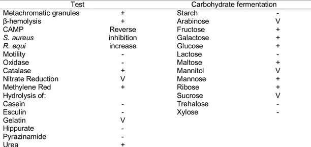
Corynebacterium pseudotuberculosis is identified by its morphology, colony characteristics, and biochemical features, mainly carbohydrate fermentation. It produces catalase, sulfidric acid, phospholipase D (PLD) and hydrolyzes urea. Nitrate reduction varies; it differentiates biovar Ovis, which is nitrate reductase negative, from biovar Equi, nitrate reductase positive (Biberstein et al., 1971). In sheep blood agar, incubated at 37°C, cream-colored colonies, with a -hemolysis zone, are observed after 48 h. It presents a reverse CAMP test, because there is inhibition of β-hemolysis by Staphylococus aureus and synergy with Rhodococcus equi (Jones and Collins, 1986; Quinn et al., 2005). In liquid culture, it forms a surface film, though the culture remains clear; this film is broken by agitation, forming flakes (Jones and Collins, 1986). The principal characteristics of C. pseudotuberculosis that are important for its identification are shown in Table 1 (Jones and Collins, 1986; Quinn et al., 2005).
Table 1. Principal phenotypic characteristics of Corynebacterium pseudotuberculosis used for identification.
Test Carbohydrate fermentation
Metachromatic granules + Starch -
β-hemolysis + Arabinose V
CAMP Reverse Fructose +
S. aureus inhibition Galactose +
R. equi increase Glucose +
Motility - Lactose -
Oxidase - Maltose +
Catalase + Mannitol V
Nitrate Reduction V Mannose +
Methylene Red + Ribose +
Hydrolysis of: Sucrose V
Casein - Trehalose -
Esculin - Xylose -
Gelatin V
Hippurate -
Pyrazinamide -
Urea +
Epidemiology and economic impact
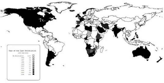
Caseous lymphadenitis is distributed worldwide and generally follows the distribution of sheep and goat herds, though in some regions its prevalence may be under-notified. Dissemination of this disease throughout the world probably occurred through importation of infected animals (Baird and Fontaine, 2007). From 1996 - 2004, among the 201 countries that reported their sanitary situation to the World Animal Health Organization (OIE) (Figure 1), 64
declared that they had animals with caseous lymphadenitis within their borders (OIE, 2009). These countries are distributed in the Americas (19 of 42 countries), Africa (18 of 51), Asia (11 of 43), Europe (14 of 51) and Oceania (2 of 14) (World…, 2009). However, the number of countries that have problems with this disease is probably under-notified, because the declaration to OIE is only done by the official sanitary authorities of each country; some countries that have had this disease reported in scientific papers have not made an official declaration, including Brazil.
Figure 1- 201 countries that reported their sanitary situation to the World Animal Health Organization (OIE), 1996 – 2004.
Prevalences of caseous lymphadenitis as high as 61% were found in Australia (Middleton et al., 1991); however, more recent studies indicate a prevalence of 20 - 30%, after vaccination began (Paton et al., 2003). In the USA, prevalences of up to 43% have been estimated (Stoops et al., 1984), similar to the range of 21 - 36% found among sheep in Quebec province in Canada (Arsenault et al., 2003). In Alberta, also in Canada, vaccination was effective in the
reduction of the prevalence of infection (Stanford et al., 1997). In the United Kingdom, 45% of the producers that were interviewed reported abscesses in their sheep (Binns et al., 2002).
exceeds 30%. In goats, Pinheiro et al. (2001) reported 66.9% of animals to have clinical signs of caseous lymphadenitis in Ceará. In Rio de Janeiro, prevalence in goats was reported to vary from 3.6 - 100% (Langenegger et al., 1991) and in a seroepidemiological ELISA study made by our group, in the State of Minas Gerais, we found prevalence figures of 75.8% for sheep (Guimarães et al., 2009b) and 78.9% for goats (Seyffert et al., 2009). In an ELISA analysis for C. pseudotuberculosis in 805 serum samples from sheep from a federally-inspected slaughterhouse in Minas Gerais; we found 377 positive animals, and a high frequency of alterations in the lymph nodes and internal organs (Figure 2). This confirms the great economical importance of C. pseudotuberculosis infection for the sheep industry due to the high rate of carcass condemnation. Various molecular
techniques have been used to type C. pseudotuberculosis, including RFLP (Restriction Fragment Length Polymorphism) of chromosomal DNA (Songer et al., 1988; Costa et al., 1998), RFLP of ribosomal 16S DNA (Sutherland et al., 1996; Costa et al., 1998), ribotyping (Sutherland et al., 1996; Costa et al., 1998), PFGE (pulsed-field gel electrophoresis) (Connor et al., 2000, Connor et al., 2007) and RAPD (randomly amplified DNA polymorphisms) (Foley et al., 2004; Stefanska et al., 2008). Though these various techniques have been useful for separating the biovars Ovis and Equi, the species C. pseudotuberculosis has been found to be genetically very homogeneous. The two techniques that have given promising results for typing C. pseudotuberculosis strains are PFGE and RAPD.
Pulsed-field electrophoresis was used to characterize 50 strains of C. pseudotuberculosis isolated from goats, sheep and horses in the United Kingdom (Connor et al., 2000); six “pulsetypes” were observed, which allowed the researchers to determine the origin of an outbreak of caseous lymphadenitis. However, in a study of 36 sheep samples and six goat samples from Australia, Canada, Eire, Holland and Northern Ireland, the same research team reported four different “pulsetypes”, with the conclusion that these C. pseudotuberculosis strains, both those from sheep and goats, were quite homogeneous (Connor et al., 2007).
RAPDs were useful in a study of 54 strains of C. pseudotuberculosis isolated from horses in four different states of the USA, identifying 10 different genotypes (Foley et al., 2004). Also, RAPDs made with other initiators made it possible to define eight genotypes among 61 strains of C. pseudotuberculosis isolated from goats in Poland, with a diversity index of 0.539 (Stefanska et al., 2008).
The importance of caseous lymphadenitis in Brazil can be estimated by the increase in the participation of goats and sheep in national animal husbandry and its relationship with the economic impact of this disease. Brazil has 16,239,455 sheep and 9,450,312 goats, totaling 25,689,767 animals (Censo agropecuário…, 2007). The economic losses include decreased milk production, decreased weight gain, reduced value of skins due to scarring, and the cost of the drugs and labor needed to treat superficial abscesses. Losses are increased when the affected lymph nodes are in critical areas (jaw, crural region, udder) negatively affecting chewing, locomotion and milk and meat production; however, economic losses due to this disease have not yet been computed. In industry, losses are due to the lower percent utility of carcasses from affected animals, damage to skins, along with the need for detailed inspection of carcasses. In the Brazilian Northeast, where goat and sheep husbandry are important sources of food and income, the situation is
even more critical because of the type of vegetation (spiny) and the low level of schooling of the farmers (Unanian et al., 1985; Pinheiro et al, 2000). It is also becoming more of a problem in the Southeastern, Northern and Midwestern regions, in which this activity is increasing rapidly, negatively affecting the meat-processing industries (Guimarães et al., 2009a,b).
Sources of infection and form of transmission
The main source of infection is infected animals, with or without clinical symptoms; these animals contaminate the soil, water, feed, pastures and facilities with nasal secretions, feces and pus from abscesses that drain spontaneously (Figure 3). Infected animals that do not present clinical symptoms can eliminate the bacteria through their respiratory tract (O’Reilly et al., 2008). Evaluation of the coefficients of transmission of C. pseudotuberculosis by respiratory tract infection and by pus from spontaneously-draining abscesses, using a mathematical model of transmission, showed that pulmonary abscesses have a small coefficient of transmission, but they are more important for maintaining the infection in the herd (endemic phase) (O’Reilly et al., 2008).
mastitis in Israel (Yeruham et al.; 1996, Braverman et al.; 1999, Spier et al., 2004). In horses, flies have considerable epidemiological importance in the dissemination of C. pseudotuberculosis,
because the higher frequency of infection in this species occurred during periods when there are large populations of flies (Costa et al., 1998).
Figure 3 – A. Two caeous abscesses, one spontaneous opened and other closed. B. Ocular and cutaneous infection of sheep with caseous material
Corynebacterium pseudotuberculosis survives long periods in the soil. Through experimental contaminations of soil and of sheep and goat facilities, it was found that C. pseudotuberculosis can survive up to eight months at various temperatures (Brown and Olander, 1987). In bedding straw, it can remain viable for three weeks, during two months in hay, four months in shearing stalls and for more than eight months in the soil. This bacterium has been isolated after five months in places where there has been contamination with pus (Nairn and Robertson, 1974) and the concentration of viable microorganisms in the purulent material is estimated to be from 106 to 107 bacteria per gram of pus; consequently, environmental contamination due to a leaking abscess is very high and persistent (Brown et al., 1987).
The use of barbed-wire fences or troughs and posts with sharp, cutting edges can cause lesions in the skin of the animals, opening passage for the entry of bacteria (Guimarães et al., 2009a). On farms that rear sheep for wool, the equipment and facilities used for shearing can transmit C. pseudotuberculosis among animals. Immersion baths immediately after shearing can disseminate the infectious agent,
because these solutions can harbor bacteria for up to 24 h (Rizvi et al., 1997). In the Brazilian Northeast, where non-wool sheep predominate almost completely, shearing and tail removal are not common and the sheep are rarely ear tagged (Pinheiro et al., 2000); however, the bacteria can penetrate through the respiratory system, transcutaneously or through skin wounds caused by the caatinga vegetation of this region (Unanian et al., 1985).
sheep flocks are for meat production (Guimarães and Gouveia, 2006).
Corynebacterium pseudotuberculosis is sensitive to common disinfectants, such as hypochlorite, formalin and cresol; however, the surfaces should be cleaned before disinfection, because organic matter interferes with the action of these agents (Ismail and Hamid, 1972). Iiodine is recommended for chemical disinfection of wounds in order to reduce bacterial transmission after surgical draining of the abscesses (Smith and Sherman, 1994).
Pathogenicity and virulence factors
Corynebacterium pseudotuberculosis is a facultative intracellular bacterium, multiplying within macrophages and surviving the action of phagolysosomic enzymes, because of the external lipid layer of the cell wall (Dorella et al., 2006; Baird and Fontaine, 2007). After penetrating into the host, which generally occurs through the oral, nasal and ocular mucosa, or through skin wounds, the agent disseminates freely or within macrophages, mainly through the afferent lymphatic system, to local lymph nodes and internal organs. This process depends on the ability of the agent to infect macrophages, resist phagolysosomes and kill cells, liberating new bacteria and causing necrosis (Batey, 1986). Three minutes after intraperitoneal inoculation in mice, phagocytic vacuoles are observed (Hard, 1969); after an hour, 60-80% of the goat macrophages contain bacteria, and two hours after inoculation, acid phosphatase is present in the vesicles containing the bacteria (Tashjian and Campbell, 1983). A strong local reaction occurs four hours after challenge in sheep (Pépin et al., 1988), and a few hours later macrophages are degenerated and polymorphonuclear cell infiltrates containing bacteria are seen (Hard, 1969; Tashjian and Campbell, 1983; Guilloteau et al., 1990). A day after experimental cutaneous infection, microabscesses develop in draining lymph nodes, and pyogranulomas are formed three to 10 days post-infection (Ellis et al., 1990; Pépin et al., 1997; Radostits et al., 2002).
The lipid cell layer of the bacteria is pyogenic and immunogenic. This same layer makes phagocytosis of the bacteria difficult, increasing its virulence (cytoxicity), and survival inside macrophages; abscesses form through the release of lysosomal enzymes (Williamson, 2001). Besides participating in pathogenicity, mycolic acid appears to be important for the survival of this bacteria in the environment (West et al., 2002).
Phospholipase D (PLD) increases vascular permeability and bacterial survival in the host. It is important for the dissemination of the bacteria from the location of the primary infection (local lymph node) to other organs (lungs, regional lymph nodes, mesenteric limph nodes, etc.), because it lyses mammal cell membranes, rich in phospholipids, causing microhemorrhages and vascular lesions, withincreased vascular permeability (Dorella et al, 2006).
Immune response
Immunity against C. pseudotuberculosis is complex and involves cellular and humoral immune responses (Prescott et al., 2002). Studies point to a greater cellular immune response, chiefly a Th1 response, because of the facultative intracellular nature of the microorganism, with production of gamma-interferon (IFN-γ) and other cytokines that are important for controlling infection (Simmons et al., 1998; Lan et al., 1999; El-Enbaawy et al., 2005). The humoral immune response is observed to present, from 6 to 11 days post-infection, a low production of IFN-γ, which significantly increases thereafter (Paule et al., 2003). Inflammatory cytokines, such as TNF-α and IL-6, are mainly produced at the site of inoculation, while T cell-associated cytokines, such as IFN-γ, are chiefly produced in drainage lymph nodes (Pépin et al., 1997).
Clinical signs
supramammary lymph nodes, while the visceral form is characterized by abscessing of internal organs, such as lungs, liver, kidneys, uterus, spleen and internal lymph nodes, such as the mediastinal and bronchial lymph nodes. These two forms can coexist; however, other less common sites can be involved, such as mammary gland, scrotum, the central nervous system and joints. Internal abscesses are normally associated with weight loss and weakness, known in sheep as thin-ewe syndrome. The mature abscesses easily leak through fistulas, releasing purulent whitish-green discharges into the environment or into the affected organ. Abscesses usually recur, months or years later, in the same animal, due to the failure to eliminate the infection (Williamson, 2001). In some cases, infections produce few characteristic clinical signs, and a post-mortem examination becomes necessary for diagnosis; this makes it difficult to obtain objective data about disease prevalence (Brown et al., 1987).
Differences in the place of the abscesses between sheep and goats have been reported, the visceral form being more frequent among sheep and the superficial form among goats (Brown and Olander, 1987). External abscesses in the lymph nodes of the head and neck are more common in goats, while the subiliac and pre-scapular lymph nodes are more commonly affected in sheep (Brown and Olander, 1987; Smith and Sherman, 1994). Differences in the appearance of abscess content have also been reported between sheep and goats; in sheep the contents have a laminar form when cut, similar to the layers of an onion, caused by the formation of layers of fibrous tissue and thick caseous material, while abscesses in goats have a thin and pasty exudate (Brown and Olander, 1987). However, onion-like abscesses were not always present in sheep. Sheep carcass inspection at a federally inspected slaughterhouse in Minas Gerais, Brazil, showed that most of the abscesses in sheep were located in the head and neck lymph nodes and their content was essentially pasty. Isolation of C. pseudotuberculosis from these materials confirms the infection
status of the animals. It is possible that older abscesses become more consistent, with a tendency towards fibrosis and calcification, progressing to an onion-like appearance, independent of animal species.
In horses, there have been reports of abortions and cases of mastitis associated with visceral abscesses (Quinn et al., 2005). In Israel, this bacterium was isolated from subcutaneous abscesses in milking cows; which could occur in outbreaks and cases of mastitis, affecting the whole mammary gland, resulting in total loss of milk production (Yeruham et al., 2003).
Clinical and laboratory diagnosis
Abscesses in goats and sheep are very suggestive of caseous lymphadenitis, especially if animals of the same lot have similar clinical signs, however bacterial isolation is necessary to identify the causative agent, since other bacteria such as Arcanobacterium pyogenes, Staphylococcus aureus subsp. anaerobius, Actinobacillus licheniformis and Pasteurella multocida, can be found in abscesses (Pekelder, 2003). In animals with respiratory problems, a thoracic X-ray can reveal masses in the pulmonary parenchyma and lymph nodes; which also must be confirmed by culture of tracheal washes (Pugh, 2004). The use of aspirating puncture with a fine needle in the diagnosis of C. pseudotuberculosis was evaluated (Ribeiro et al., 2001). It proved to be easily performed, to have a low cost and to cause little damage to the tissues when compared to histopathology. It allows presumptive cytological diagnosis of the infection, before the affected lymph nodes abscessed, aiding in early adoption of prophylactic measures for the rest of the flock.
inflammatory material from the aspirated lymph nodes, helps in the identification of the infectious agent (Radostits et al., 2002). In order to make a definitive diagnosis of caseous lymphadenitis, the agent should be isolated from purulent material from abscessed lymph nodes samples from live animals. Besides aspirating puncture, the material can be obtained by excision after trichotomy and careful antiseptic cleaning of the skin (Collett et al., 1994; Smith and Sherman, 1994). It can also be collected at necropsy or during slaughter, when internal
abscesses, affecting the liver, lungs, intestine, kidneys, internal lymph nodes and other tissues, become accessible (Riet-Correa et al., 2001).
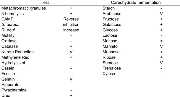
In the laboratory, after isolation, the identification of C. pseudotuberculosisis was done by its morphology, staining characteristics, profile and fermentation of various carbohydrates (Jones and Collins, 1986). The main phenotypic characteristics of Corynebacterium pseudotuberculosis used for identification are shown in Table 1.
Table 2. Principal phenotypic characteristics of Corynebacterium pseudotuberculosis used for identification.
Test Carbohydrate fermentation
Metachromatic granules + Starch -
β-hemolysis + Arabinose V
CAMP Reverse Fructose +
S. aureus inhibition Galactose +
R. equi increase Glucose +
Motility - Lactose -
Oxidase - Maltose +
Catalase + Mannitol V
Nitrate Reduction V Mannose +
Methylene Red + Ribose +
Hydrolysis of: Sucrose V
Casein - Trehalose -
Esculin - Xylose -
Gelatin V
Hippurate -
Pyrazinamide -
Urea +
+: more than 90% positive; v: 21–89% positive; –: more than 90% negative. Adapted from Jones & Collins (1986) and Quinn et al. (2005).
Various diagnostic techniques have been developed for caseous lymphadenitis in goats and sheep, such as serological neutralization for antitoxins, immunodiffusion in agar gel, indirect hemagglutination, complement fixation and hypersensitivity tests (Langenegger et al., 1991; Williamson, 2001; Baird and Fontaine, 2007).
Immunoenzymatic tests (ELISA), using bacterial cells, toxins and secreted proteins
Detection of INF-γ by ELISA, an indicator of cell-mediated immunity, has been used for diagnosis of infection by C. pseudotuberculosis, with a sensitivity of 91% and a specificity of 98%, demonstrating its potential for use in caseous lymphadenitis eradication programs (Prescott et al., 2002; Sunil et al., 2008).
Molecular techniques have also been used for the diagnosis of caseous lymphadenitis. Polymerase chain reaction (PCR), used to identify C. pseudotuberculosis, is an alternative to conventional diagnostic methods, with the advantage of being faster and more specific (Çetinkaya et al, 2002). Multiplex PCR based on amplification of the genes 16S rDNA, rpoB and pld, presented 94.6% diagnostic sensitivity, for C. pseudotuberculosis isolates as well as for clinical material (Pacheco et al., 2007). It facilitates the diagnosis by differentiating C. pseudotuberculosis from other pathogens present in abscesses, chiefly C. ulcerans (Pacheco et al., 2007).
Recently, the genome of two C. pseudotuberculosis strains isolated from goats and sheep has been sequenced by a Minas Gerais Genome Network and Pará Genomic and Proteomic Network. The genomic data will help to identify new specific targets, useful in the diagnosis as well as in the development of drugs and vaccines and in the understanding of C. pseudotuberculosis pathogenicity mechanisms.
Differential diagnosis
Pyogranulomatous lesions, such as found in actinobacillosis, tuberculosis and superficial abscesses caused by Staphylococcus aureus and Actinomyces pyogenes, must be differentiated from caseous lymphadenitis (Collett et al., 1994). The superficial form of the disease should also be differentiated from submandibular edema caused by parasites, Fasciola hepatica and Haemonchus sp., salivary cysts, lymphosarcoma and subcutaneous inoculation of vaccines. The debilitating visceral form can be clinically similar to
chronic parasitism, thinning due to abnormal waste of teeth, alveolar periodontitis, malnutrition and chronic diseases, such as pulmonary adenomatosis, neoplasias and scrapie (Collett et al., 1994).
Pneumonias caused by Mycobacterium bovis, Pasteurella haemolytica, Pasteurella multocida or ovine progressive pneumonia, due to Maedi-Visna virus infection, can make the diagnosis of caseous lymphadenitis even more difficult (Pugh, 2004).
In sheep, orchitis and epidydimitis caused by C. pseudotuberculosis needs to be differentiated from similar lesions caused by Brucella ovis, Actinobacillus seminis, Histophilus ovis and Pasteurella spp. (Collett et al., 1994; Saunders et al, 2007).
Treatment
Treatment of affected animals consists of the drainage of abscesses, followed by cleansing and chemical cauterization, usually with 10% iodine, or even removal of the affected superficial lymph nodes (Nozaki et al, 2000). Although it is an important control measure, this procedure might not be as effective as expected due to the presence of internal abscesses. Drainage of the abscess should be done in a way that avoids environmental contamination, with disinfection of the surgical material before and after the procedure, and all of the disposable materials should be incinerated and buried, including plastics and paper used to cover the area.
Control and prophylaxis
An effective program for the control of caseous lymphadenitis should be based on clinical inspection and periodic serology of all animals in the flock, which includes recently-acquired animals and those that return to the herd, culling the ones that have clinical signs or that are serologically positive. Once infected, an animal hardly eliminates the C. pseudotuberculosis (Campbell et al., 1982). The main source of infection for a flock is introduction of infected or abscessed animals into a herd, which results in a high frequency of abscesses after two or three years. This stresses the importance of employing biosecurity procedures in all flocks, chiefly during the introduction of animals.
Measures designed to reduce the environmental risk of wounding should also be adopted, such as the use of smooth wire fences, troughs and facilities without sharp edges, disinfection of surgical, ear tagging and shearing instruments, systematic use of individual disposable needles, effective control of insects, and disinfection of newborns’ navels and any other wounds with 10% iodine. Although it is not recommended to be applied to swelled lymph nodes because of its irritating and caustic action on tissues (skin, mucosa and lungs), 10% formaldehydeshould be used for disinfection of herd facilities (Brown and Olander,1987; Williamson, 2001; Baird and Fontaine, 2007).
All control programs should be based on sanitary education of herd owners and technical personnel, otherwise success will be compromised. Information about losses throughout the production cycle, as well as concerning the zoonotic potential of C. pseudotuberculosis, should be supplied to the people who work with the herds directly or indirectly (Peel et al, 1997), reinforcing their importance in the success of the control program.
Control measures vary with the prevalence of infection. In countries free of this disease, importation should only be permitted from
herds that have been certified free of caseous lymphadenitis for three years, all animals should be tested by ELISA before importing and they should initially be placed in quarantine. In countries with a low disease prevalence, the clinically affected animals should be separated and submitted to ELISA testing, lambs and kids should be reared away from their mothers, and installations and equipment should be well disinfected. In countries with a high incidence, rigorous sanitary measures should be implemented, associated with vaccination (Brown and Olander, 1987; Collet et al., 1994; Williamson, 2001; Baird and Fontaine, 2007).
Disease eradication can be achieved in endemically-infected herds by initially discarding all animals that have clinical signs and those that are positive in serological tests (Radostits et al., 2002); however, this is difficult to accomplish because of the rapid dissemination of the agent within the herd and the difficulty in identifying animals that have a subclinical form of the disease (Riet-Correa et al., 2001; Çetikaya et al., 2002).
Vaccination
Given that caseous lymphadenitis treatment is ineffective and expensive, the best strategy for control and prevention of the disease is immunization, as it was observed in countries with high prevalence of infection (Patton et al., 2003). The vaccines commercially available have different relevant features that should be considered on their use. Not all of the vaccines licensed for use in sheep have the same efficiency in goats, and normally it is necessary to adjust the vaccination program to the farm conditions and instruct the people who will apply the vaccine. Also, the protection provided by vaccination is only partial, as external and internal abscess development can still occur (Williamson, 2001).
obtained after immunization of goats and sheep with this toxin. Most commercial vaccines against C. pseudotuberculosis use inactivated phospholipase D associated to antigens of various pathogens, such as Clostridium tetani, Clostridium perfringens type D, Clostridium novyi, Clostridium chauvoei and Clostridium septicum, along with some that are associated with the endectocide moxidectin. This formulation is the basis of the Glanvac vaccine (Vetrepharm, Inc London), licensed for use in sheep and goats in Canada, Australia and New Zealand and the Biodectin vaccine (Fort Dodge Austrália PTY LTD), licensed in Brazil for use in sheep.
Glanvac vaccine has been evaluated in various countries (Eggleton et al., 1991). Vaccination of sheep and goats with Glanvac resulted in protection against experimentally-induced infection with C. pseudotuberculosis, evidenced by a decrease in the number of lesions (Fontaine et al., 2006). Another commercial vaccine that has been evaluated, Caseous D-T(PBS Animal Health), has two formulations, one that only contains toxoids (clostridial and from C. pseudotuberculosis) and another that is a combination of clostridial toxoids and the bacterium C. pseudotuberculosis. Preliminary results indicate that this second formulation confers better protection against experimental infection than the first, reducing the number of internal and external lesions (Piontkowski and Shivvers, 1998). The use of PLD toxoid for the immunization of goats can have some negative consequences, including reduced milk production, fever, ventral edema, ataxia and convulsions; consequently, recommendations for use in this species are made with restrictions (Williamson, 2001).
The reasons for the partial protection provided by immunization of goats and sheep with commercial vaccines have to do with the type of immune response. Protection against C. pseudotubeculosis is mainly dependent on a immune response that involves INF-γ production and cytotoxic T-cells. A humoral response alone is insufficient to protect the animal, and a good
cellular response is not achieved with inactivated vaccines (Dorella et al., 2009). Consequently, various attempts have been made to obtain an attenuated vaccine that is effective against caseous lymphadenitis (Hodgson et al., 1994; Simmons et al., 1998). Attenuation can happen naturally or through manipulations using temperature, chemical and genetic (recombinant) agents. With this type of vaccine strategy, the microorganism maintains its capacity to replicate, imitating natural infection and producing humoral and cellular responses. Also, this is the type of vaccine that confers the best and longest-lasting immune response, due to its similarity to natural infection (Dorella et al., 2009). Techniques such as deletion of multiple genes involved in virulence, and insertion of fragments that interrupt these genes in the pathogen, practically eliminate the risk that the pathogen can revert to its virulent form (Jiskoot et al., 2002). Live vaccines that have been attenuated in the laboratory (recombinants) usually have the PLD gene as a target for attenuation, because of its importance as a virulence factor (Carne and Onon, 1978).
In Brazil, the Bahia State Agency for Agricultural Development (EBDA) developed and a laboratory commercializes line 1002, produced from a natural low-virulence strain. It gave significant protection levels of 83% in vaccine trials; however, immunization still presents collateral effects, such as local reactions, and field trials have not been as successful as the initial vaccine tests, with highly variable protection levels (Hage, 2000). Another attenuated live vaccine, LinfoVac (Laboratórios Vencofarma do Brasil Ltda), indicated for use in sheep and goats, was recently made available in Brazil. The results obtained in the field with these attenuated vaccines demonstrate the need to develop a more effective and safe vaccine (Dorella et al., 2009).
Conclusions
production, limiting the net profits of these production systems. The prevalence of this disease is quite high, indicating that specific control measures should be adopted. There are various difficulties that affect the development and application of diagnostic tests that are effective and that can be used within the context of an organized caseous lymphadenitis control program. In order to have useful vaccines, more effective immunogens need to be made available. The intense commercialization and transit of small ruminants without using the necessary security measures, are important obstacles to the control of caseous lymphadenitis. Also, deficient control, with inadequate use of vaccines, lack of hygiene precautions and of serological diagnostic techniques, both on farms and in expositions and fairs, allow the introduction and rapid dissemination of the C. pseudotuberculosis in the herds. Few sheep and goat farmers demand sanitary certificates or use quarantines when they acquire animals or when they return from these events. Breeders and technicians need to make timely diagnoses and implement more effective control of caseous lymphadenitis in order to reduce losses in the properties and in the slaughter houses (Guimarães and Gouveia, 2006; Guimarães et al., 2009b).
Acknowledgements
ASG, APL and VA have scholarships from the Conselho Nacional de Desenvolvimento Científico e Tecnológico – CNPq. This work was supported by the Fundação de Amparo à Pesquisa do Estado de Minas Gerais – FAPEMIG (CVZ APQ 3283-5.04/07) and from CNPq.
References
Arsenault, J.O., Girard, C., Dubreuil, P., Daignault, D. O., Galarneau, J.-R., Boisclair, J., Simard, C., Bélanger, D., 2003. Prevalence of and carcass condemnation from maedi-visna, paratuberculosis and caseous lymphadenitis in culled sheep from Quebec, Canada. Prev. Vet. Med. 59, 67– 81.
Baird, G.J., Fontaine, M.C., 2007. Corynebacterium pseudotuberculosis and its role in ovine caseous lymphadenitis. Journal of Comparative Pathology. 137, 179-210. Batey, R.G., 1986. Pathogenesis of caseous lymphadenitis in sheep and goats. Aust. Vet. J. 63, 269-272.
Biberstein, E.L., Knight, H.D., Jang, S., 1971. Two biotypes of Corynebacterium pseudotuberculosis, Vet. Rec. 89, 691–692. Binns, S.H., Bairley, M., Green, L.E., 2002. Postal survey of ovine caseous lymphadenitis in the United Kingdom between 1990 and 1999. Vet. Rec. 150, 263-268.
Binns, S.H., Green, L. E., Bailey, M., 2007. Development and validation of an ELISA to detect antibodies to Corynebacterium pseudotuberculosis in ovine sera. Vet. Microbiol. 20, 169-179.
Braverman, Y., Chizov-Ginzburg, A., Saran, A., Winkler, M., 1999. The role of houseflies (Musca domestica) in harbouring Corynebacterium pseudotuberculosis in dairy herds in Israel. Revue Scientifique et Technique de l'Office International des Epizooties, 18, 681-690.
Brown C.C., Olander H.J., Alves S.F., 1987. Synergistic hemolysis-inhibition titers associated with caseous lymphadenitis in a slaughterhouse survey of goats and sheep in Northeastern Brazil. Can. J Vet. Res. 51, 46–49.
Brown, C.C., Olander, H.J., 1987. Caseous lymphadenitis of goats and sheep: a review. Vet. Bull. 57, 1-11.
Campbell S.G., Ashfaq M.K., Tashjian J.J., 1982. Caseous lymphadenitis in goats in the USA. In: Proceedings 3rd International Conference on Goat Production and Disease. Tucson. Arizona. 449-454.