EVELYN JARDIM DE OLIVEIRA
EMBRIOGÊNESE SOMÁTICA EM Brachypodium distachyon (L.)
Beauv. (POACEAE): CARACTERIZAÇÃO MORFOANATÔMICA,
HISTOQUÍMICA E EXPRESSÃO DE GENES SERK
Tese apresentada à Universidade Federal de Viçosa, como parte das exigências do Programa de Pós-Graduação em Botânica, para obtenção do título de Doctor Scientiae.
VIÇOSA
Ficha catalográfica preparada pela Biblioteca Central da Universidade Federal de Viçosa - Câmpus Viçosa
T
Oliveira, Evelyn Jardim de, 1958-O48e
2013
Embriogênese somática em Brachypodium distachyon (L.) Beauv. (Poaceae) : caracterização morfoanatômica, histoquímica e expressão de genes SERK / Evelyn Jardim de Oliveira. – Viçosa, MG, 2013.
ix, 90 f. : il. (algumas color.) ; 29 cm.
Orientador: Wagner Campos Otoni.
Tese (doutorado) - Universidade Federal de Viçosa. Inclui bibliografia.
1. Brachypodium distachyon - Morfogênese. 2.
Histoquímica. 3. Ultraestrutura (Biologia). I. Universidade Federal de Viçosa. Departamento de Biologia Vegetal. Programa Pós-Graduação em Botânica. II. Título.
EVELYN JARDIM DE OLIVEIRA
EMBRIOGÊNESE SOMÁTICA EM Brachypodium distachyon (L.)
Beauv. (POACEAE): CARACTERIZAÇÃO MORFOANATÔMICA,
HISTOQUÍMICA E EXPRESSÃO DE GENES SERK
Tese apresentada à Universidade Federal de Viçosa, como parte das exigências do Programa de Pós-Graduação em Botânica, para obtenção do título de Doctor Scientiae.
APROVADA: 14 de outubro de 2013.
_________________________________ ________________________________
Marcelo de Oliveira Santos Aristéa Alves Azevedo
_________________________________ ________________________________ Andréa Dias Koehler Luzimar Campos da Silva
(Coorientadora)
____________________________________________ Wagner Campos Otoni
ii
À minha mãe D. Nice e à memória de meu pai Moacyr.
Às minhas meninas Juliana e Julia.
iii
AGRADECIMENTOS
À Universidade Federal de Viçosa (UFV) e ao Programa de Pós-Graduação em Botânica, pela oportunidade de realização do Doutorado.
À Coordenação de Aperfeiçoamento de Pessoal de Ensino Superior (CAPES), pelo apoio financeiro.
Ao CNPq, pela generosa compreensão e apoio.
Ao Prof. Wagner Campos Otoni, pelo apoio incondicional, pela amizade, por ter me pegado pela mão.
À Dra. Ana Claudia Ferreira da Cruz pela grande ajuda, dedicação e otimismo. À Dra. Andréa Koehler pelo grande auxílio e carinho.
Ao Dr. Elyabe Monteiro de Matos pelo companheirismo desde o começo, pela amizade e disponibilidade.
Ao amigo Diego Rocha pelo apoio, sugestões e críticas dadas ao trabalho.
Aos amigos do LCTII, Cris, Lorena, Marcos, Diego, Marcela e Virgilio por tanta generosidade, companheirismo e boas experiências compartilhadas.
À Dra. Flávia Garcia, pelo apoio, incentivo e amizade sempre presentes. À Profa. Luzimar pela atenção e sugestões valiosas.
Ao Prof. Francisco Tanaka pelo processamento do material por MET. Ao Prof. Fábio Tebaldi Silveira Nogueira (ESALQ/USP) pela gentileza em ceder o material vegetal e pelo incentivo e sugestões valiosas.
Aos professores do Departamento de Biologia Vegetal pelos ensinamentos e pelo estimulante convívio acadêmico.
Ao Instituto de Biotecnologia Aplicada à Agropecuária (BIOAGRO) e ao Departamento de Biologia Vegetal (DBV), pelo suporte a este trabalho.
Ao Núcleo de Microscopia e Microanálise (NMM) da UFV e ao Núcleo de Apoio à Pesquisa em Microscopia Eletrônica Aplicada à Agricultura (NAP/MEPA/ESALQ), pelas análises em microscopia eletrônica realizadas.
Ao Paulo Oliveira, pelo apoio e incentivo.
Aos meus queridos irmãos Braulio e Sandra, por uma vida inteira de amor sincero e lealdade.
À minha filha Juliana, por estar sempre ao meu lado e pelas incontáveis trocas de idéias, e à minha neta Julia, por ter nos presenteado com a sua alegria.
iv BIOGRAFIA
Evelyn Jardim de Oliveira, filha de Moacyr Augusto de Oliveira e Clenilce Jardim de Oliveira, nasceu em 14 de junho de 1958, na cidade de São Paulo, São Paulo.
Graduou-se em Agronomia na Universidade Federal de Viçosa em 1982. Concluiu o Mestrado em Genética e Melhoramento na Universidade Federal de Viçosa em 1989 comăaădissertaçãoăintituladaă―Análise multivariada no estudo da divergência genética entre cultivares de feijão (Phaseolus vulgaris L.)‖.
Em 2009 iniciou o doutorado em botânica na Universidade Federal de Viçosa e submeteuă àă defesaă aă teseă intituladaă ―Embriogênese somática em Brachypodium
distachyon (L.) Beauv. (Poaceae): caracterização morfoanatômica, histoquímica e
v SUMÁRIO
RESUMO ... vii
ABSTRACT ... ix
GENERAL INTRODUCTION ...1
REFERENCES ...10
CHAPTER 1 ...21
SOMATIC EMBRYOGENESIS FROM IMMATURE ZYGOTIC EMBRYOS OF Brachypodium distachyon (POACEAE): MORPHOLOGICAL, HISTOLOGICAL AND HISTOCHEMICAL INVESTIGATIONS ... 21
ABSTRACT ...21
RESUMO ...22
INTRODUCTION ...23
MATERIALS AND METHODS ...25
Plant material ... 25
Zygotic embryo morphology ... 25
Production of compact embryogenic callus (CEC) and plant regeneration ... 26
Microscopy sample preparation ... 27
Light microscopy and histochemical characterization ... 27
Scanning electron microscopy ... 28
RESULTS ...28
General morphology ... 28
Histological examination ... 29
Histochemical analysis of storage compounds ... 30
DISCUSSION...42
REFERENCES ...49
CHAPTER 2 ...59
SOMATIC EMBRYOGENESIS IN Brachypodium distachyon: DYNAMICS OF SERK GENE EXPRESSION. ...59
ABSTRACT ...59
RESUMO ...60
INTRODUCTION ...61
MATERIALS AND METHODS ...63
Extraction of RNA from embryogenic callus ... 63
Synthesis of single-stranded cDNA ... 63
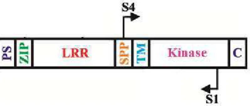
Amplification of the SERK coding sequence ... 64
Purification of DNA after electrophoresis and cloning of fragments ... 65
vi
Collection and preparation of immature zygotic embryos and embryogenic calli for
the analysis of SERK gene expression ... 66
Production of antisense gene probe SERK1 ... 67
Hybridization reaction ... 67
Reaction of post-hybridization and immunological detection ... 67
RESULTS ...68
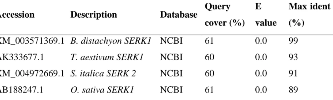
Analysis of the nucleotide sequence of the B. distachyon SERK fragment ... 68
Analysis of the deduced amino acid sequences of BdSERK ... 68
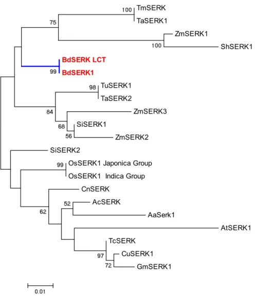
Phylogenetic analysis ... 71
Expression analysis of the B. distachyon SERK fragment ... 73
DISCUSSION...79
REFERENCES ...83
vii RESUMO
OLIVEIRA, Evelyn Jardim de, D.Sc., Universidade Federal de Viçosa, outubro de 2013. Embriogênese somática em Brachypodium distachyon (L.) Beauv. (Poaceae): caracterização morfoanatômica, histoquímica e expressão de genes SERK. Orientador: Wagner Campos Otoni. Coorientadora: Luzimar Campos da Silva.
Brachypodium distachyon (L.) P. Beauv. (Poaceae: Poideae) tem se destacado como
planta modelo para gramíneas de clima temperado e espécies usadas para a produção de biocombustíveis. A linhagem Bd21 de Brachypodium distachyon tem um genoma completamente seqüenciado e montado, além de protocolos de genômica e de transformação bem estabelecidos com base em calos embriogênicos. No entanto, as informações sobre a origem e as alterações celulares que ocorrem durante a diferenciação de embriões somáticos nos estágios iniciais não foi documentada para B.
distachyon. Também não há relatos sobre o uso de abordagens moleculares para
investigar o processo de embriogênese somática nesta espécie. Portanto, os objetivos deste trabalho foram: (1) caracterizar as alterações morfológicas, anatômicas e histoquímicas que ocorrem durante a indução de embriogênese somática a partir de embriões zigóticos imaturos (EZI) da B. distachyon linhagem de referência Bd21 usando microscopia de luz e de varredura em associação com testes histoquímicos e, (2), realizar a clonagem e caracterização de genes SERK (SOMATIC
EMBRYOGENESIS RECEPTOR-LIKE KINASE) e analisar sua expressão na indução de
viii
começaram a acumular nos pró-embrioides após 4 dias em meio de indução e tornaram-se maior e mais abundantes em células do escutelo aos 12 dias em meio de indução. A diferenciação do embrião somático seguiu a mesma sequência de desenvolvimento verificado em outros membros da família Poaceae, ou seja, a passagem pelos estádios globular, escutelar e coleoptilar. O gene SERK tem sido usado como um marcador para as células competentes na embriogênese somática de várias espécies. Neste estudo, utilizando iniciadores degenerados, uma sequência específica homóloga de um fragmento do gene SERK foi amplificada de B. distachyon Bd21. A análise da sequência do fragmento de SERK (766 bp) revelou altos níveis de similaridade com genes SERK relatados em outras espécies. A análise de hibridização in situ mostrou que o gene
SERK estava presente nos tecidos embriogênicos de B. distachyon antes do
desenvolvimento de embriões somáticos e continuou sendo expresso nos estágios globular e escutelar. Estes resultados sugerem que a expressão do gene SERK de B.
distachyon pode estar associada com a indução da embriogênese somática. Este estudo
ix ABSTRACT
OLIVEIRA, Evelyn Jardim de, D. Sc., Universidade Federal de Viçosa, October, 2013. Somatic embryogenesis in Brachypodium distachyon (L.) Beauv. (Poaceae): morphoanatomical and histochemical characterization and analysis of SERK gene expression. Adviser: Wagner Campos Otoni. Co-adviser: Luzimar Campos da Silva.
Brachypodium distachyon (L.) P. Beauv. (Poaceae: Poideae) has been proposed as a
new model for temperate and biofuel grasses. Brachypodium distachyon inbred line Bd21 has a fully sequenced and assembled genome, a series of genomics resources, and well-established somatic embryogenesis-based transformation protocols. However, information about origin and cellular changes occurring during the early differentiation of somatic embryos has not been documented for B. distachyon. There are also no reports on the use of molecular approaches to investigate somatic embryogenesis in B.
distachyon. Therefore, the objectives of this study were (1) to characterize the
x
1
GENERAL INTRODUCTION
Brachypodium distachyon (L.) P. Beauv. is a temperate wild grass native to the
Mediterranean region (Vogel et al., 2009; Catalán et al., 2012) and has successfully invaded disturbed areas of central Europe, Australia, New Zealand, South Africa and North and South America (Garvin et al., 2008; Bakker et al., 2009). It has little agricultural importance and is of no major economic value except for its invasive habit and some varieties that have been used to protect soils of olive orchards from erosion in Spain (Bakker et al., 2009; Hammami et al., 2011).
Brachypodium distachyon is a member of the large grass family Poaceae that
provides most of human and domestic animal nutrition (Kellogg, 2001). Members of the Poaceae subfamilies Ehrhartoideae (rice), Panicoideae (maize, sorghum) and Pooideae (wheat, barley) are the main grain crops throughout the world (Febrer et al., 2010). Furthermore, highly productive grasses of the Panicoideae subfamily (sugarcane, switchgrass, Miscanthus sinensis) are promising sources of sustainable energy (Vogel et al., 2009).
The Brachypodium genus comprises 12 to 15 species with most of them being wide-spread in Mediterranean and Eurosiberian areas (Catalán and Olmstead, 2000). The diploid B. distachyon is the only annual member within the genus, a self-compatible species with chromosome base number x = 5 (Catalán and Olmstead, 2000). Phylogenetic analyses based on plastid and nuclear genes from different Brachypodium lineages indicated a close relationship of B. distachyon to the rhizomatous perennials B.
arbuscula, B. retusum, B. rupestre, B. phoenicoides, B. pinnatum, and B. sylvaticum
(Catalán and Olmstead, 2000; Wolny et al., 2011; Catalán et al., 2012). Most of the perennial species are self-incompatible (except for B. sylvaticum) and have chromosome base numbers ranging from 7 to 9 (Robertson, 1981; Khan and Stace, 1999; Catalán and Olmstead, 2000).
Brachypodium distachyon was first characterized as having three different
2
different taxa, and the 2n = 30 cytotype represents their derived allotetraploid (Hasterok et al., 2006; Idziak et al., 2011; Catalán et al., 2012). The name B. distachyon was kept for the 2n = 10 cytotype and two novel species were described as B. stacei and B.
hybridum for, respectively, the 2n = 20 and 2n = 30 cytotypes (Catalán et al., 2012).
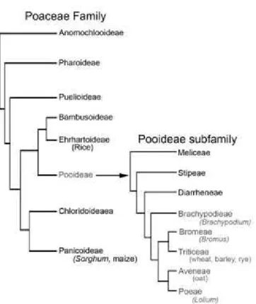
The phylogenetic relationship between the genus Brachypodium and the other grasses has been firmly established (Vogel and Bragg, 2009). The genus Brachypodium belongs to the Brachypodieae tribe, which has consistently been shown to be sister group to the four economically important tribes Triticeae, Aveneae, Poeae, and Bromeae and closely related to rice (Draper et al. 2001; Kellogg 2001; Vogel et al. 2006) (Fig. 1).
Figure 1. Phylogenetic relationship of Brachypodium distachyon to other Poaceae. Figure derived from Hands and Drea (2012).
Brachypodium distachyon has been developed as a model system for the
3
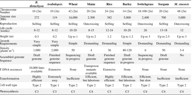
lifecycle) necessary to a model system (Table I). A model grass is needed to answer questions that are specific of the grass biology such as cell wall composition, plant architecture, grain properties, yield, stress tolerance, intercalary meristems, root architecture, development, and plant-pathogen interactions (Vogel and Bragg, 2009; Brkljacic et al., 2011).
Many Pooideae species have large and complex genomes, which makes genome comparisons and biotechnological approaches for crop improvement veryă difficultă ină these crops (Shang et al., 2011). B. distachyon has a genome size smaller than that of rice and is closely related to wheat, barley, and oat, thus it can serve as a structural genomic tool to assist the exploration of the large genomes of these crops (Huo et al., 2009).
Table 1. Comparison of Brachypodium distachyon with other models and crops B.
distachyon Arabidopsis Wheat Maize Rice Barley Switchgrass Sorgum M. sinensis
Chromosome
Number 10 (2n) 10 (2n) 42 (2n) 20 (2n) 24 (2n) 14 (2n) 18-108 (2n) 20 (2n) 48 (2n) Genome size
(Mb) 272 119 16,000 2,300 382 5,000 2,400 700 5,000
Reproduction Selfing Selfing Selfing Outcrossing Selfing Selfing Outcrossing Selfing Outcrossing Life cycle
(weeks) 8-12 8-12 10-20 8-15 12-24 10-20 26 13-18 12
Height (m) 0.3 0.2 Up to 1 Up to 2 1.2 Up to 1.2 Up to 3 Up to 2.5 Up to 3 Growth
Requirements simple Very simple Very Simple Demanding Demanding Simple Demanding Demanding Demanding Density
(plants m-2) 1,000 2,000 50 4 36 80-120 6 50 3-4
Assembled genome Sequence Draft genome Finished genome sequence Sequencing in progress Draft genome Finished genome sequence Draft genome in progress Sequencing in progress Draft genome No
T-DNA resources 10,000 lines
available Extensive None
Transposon mutants available
Extensive None None None None
Transformation efficientHighly Extremely
easy Inefficient
Efficient,
but laborious Highly
efficient but laborious Efficient,
Efficient,
but slow Inefficient Inefficient Cell wall type Type 2 Type 1 Type 2 Type 2 Type 2 Type 2 Type 2 Type 2 Type 2
Photosynthesis C3 C3 C3 C4 C3 C3 C4 C4 C4
Adapted from Opanowicz et al. (2008) and Brkljacic et al. (2011).
Brachypodium distachyon can also be used as a model system for the emerging
energy crops like maize, sugarcane, switchgrass and Miscanthus sinensis with which it shares the typical cell wall of grasses (Opanowicz et al., 2008; Vogel et al., 2009). The large size, large genomes and outcrossing breeding system of these species hinder their use in experimental conditions. In addition, the close relationship within the genus of B.
distachyon with the core of perennial species can be explored to study the perennial life
cycle and self-incompatibility that are also common traits to wild grasses (e.g.
Miscanthus and switchgrass) that are being developed into biofuel crops (Vogel and
4
ofătheăenergyăcropsăuseăaăC4ăphotosyntheticăpathway,ăwhichăisămoreăefficientăunderăhot,ă dry conditions. But even though B. distachyon alone is not suitable to study C4 photosynthesis, it can be used as a model system to study the feasibility of engineering the C4 mechanism into C3 crops such as rice and wheat (Brkljacic et al., 2011).
A remarkable progress has been made in the development of genomic resources for B. distachyon, which include the whole nuclear and chloroplast genome sequences (Bortiri et al., 2008; IBI, 2010); germplasm collections (Garvin et al., 2008; Filiz et al., 2009; Vogel et al., 2009) , genetic markers (Vogel et al., 2009), a genetic linkage map (Garvin et al., 2010), bacterial artificial chromosome (BAC) libraries (Huo et al., 2006; Farrar and Donnison, 2007; Huo et al., 2008), physical maps (Gu et al., 2009; Febrer et al., 2010), mutant collections (Thole et al., 2012), microarrays and databases (comprehensive lists of Internet B. distachyon resources are found in Brkljacic et al., 2011 and Vain, 2011).
Another important aspect considered a key technology for a model plant species is the availability of an easy and efficient transformation system and B. distachyon has proven to be very responsive to in vitro culture and current transformation techniques. Methods for transformation using particle gun (Draper et al., 2001; Christiansen et al., 2005) and Agrobacterium-mediated ( Vogel et al., 2006; P curarăet al., 2008; Vain et al., 2008) have been developed for several genotypes of B. distachyon. Average transformation efficiencies have been reported to range from 30–80% ( P curarăet al., 2008; Vain et al., 2008; Vogel and Hill, 2008; Alves et al., 2009 ).ăTheăhighlyăefficientă transformation procedures established for B. distachyon allow the generation of large collections of T-DNA mutant lines (http://www.brachytag.org/services.htm), the characterization of gene function through over-expression or gene silencing (Olsen et al., 2006; Pacak et al., 2010), and recently, a T-DNA mutation in B. distachyon has been complemented with an Arabidopsis ortholog, bridging dicotyledonous and monocotyledonous models (Vain et al., 2011). In all cases, embryogenic calli were used as the target tissue for transformation.
Bablak et al. (1995) developed, for the first time, a protocol for the induction of embryogenic callus from mature seeds of B. distachyon and the regeneration of fertile plants, making a breakthrough toward developing B. distachyon transformation. In this study, optimum development of embryogenic callus of three diploid accessions of B.
distachyon (B200, B373, and B377) occurred on LS (Linsmaier and Skoog, 1965) and
5
dichlorophenoxyacetic acid (2,4-D). Plant regeneration occurred on several common media and the incidence of albino plantlets was around 10%.
However, the regeneration ability of mature seeds/embryos of most Poaceae species, such as wheat, maize, sorghum and barley, is poor (Schulze, 2007), and the success of the transformation techniques relies on the culture of highly embryogenic callus (Hansen and Wright, 1999). The morphogenetic competence of immature embryos in monocotyledonous species was first described in maize by Green and Philipps (1975). Since then, for most of the important cereals and grasses, plant regeneration systems have been developed based on immature embryos (Eudes et al., 2003; Schulze, 2007).
A considerable improvement in embryogenic callus production and plant regeneration of B. distachyon was achieved by using immature embryos as initial explant. Draper et al. (2001) cultured immature embryos of the diploid ecotype ABR1 to induce callus. They observed that the immature embryos with the greatest potential for plant regeneration via somatic embryogenesis were in the size range of 0.3 to 0.7 mm. When cultured on LS or N6 containing 2.5 mg L−ń 2,4-D the immature embryos formed a mass of embryogenic callus around the edge and on the surface of the scutellum, in general after 10 to 15 d. The level of albinism in plantlets regenerated from the embryonic calli induced on LS medium (7% ) was much lower than from calli formed on N6 medium (45%).
The induction and control of somatic embryogenesis are largely dependent upon plant genotype, tissue-type, physiological conditions of the donor plant, type and level of growth regulators supplemented to culture medium and varied cultural regimes (Toonen and De Vries, 1996). Differences in response of B. distachyon genotypes to in
vitro culture have been reported indicating that the interaction between the genotype
explant source and the culture medium influences the development of B. distachyon in terms of the callus induction and regeneration response (Bablak et al., 1995; Draper et al., 2001; Vogel et al., 2006; Hammami et al., 2011).
6
than 80% when cultured for 8 weeks. They found that longer duration of callus culture resulted in lower regeneration efficiency, with regeneration percentages dropping to 30% and 55% in the accessions BDR017 and BDR030, respectively, when kept for 16 weeks in culture.
The tissue culture capacity of the community standard line Bd21 and its variation line Bd21-3 for the production of embryogenic callus was compared by culturing immature embryos (less than or equal to 0.3 mm in length) on an optimized callus induction medium (Vain et al., 2008). The medium was based on MS salts (Murashige and Skoog, 1962), M5 vitamins, 3% sucrose, 0.2% phytagel and supplemented with 0.6 mgL-1 CuSO4. Embryogenic callus production for the diploid lines Bd21 (68%) and Bd21-3 (94%) was higher than that of tetraploid genotypes (67% for BRD017 and 91% for BRD030) reported by Christiansen et al. (2005). The addition of CuSO4 toătheătissueăcultureămediumăpromotedăefficientăcallusăinductionăandăhigher -frequency plant regeneration from Bd21 calli (Vain et al., 2008). CuSO4 was also reported to decrease the regeneration of albino plants and increase the number of regenerated shoots per embryo in barley (Dahleen, 1995; Bartlett et al., 2008). It has been suggested that some copper enzymes might play important role in regeneration since copper ions are components or activators of many important enzymes involved in electron transport, protein, carbohydrate biosynthesis and polyphenol metabolism (Purnhauser and Gyulai, 1993).
7
Somatic embryogenesis in vitro is an excellent model for studying the theory of plant embryogenesis, determining gene expression and measure substance accumulation at different stages of embryogenesis (Brukhin and Morozova, 2011).
In general, the process of somatic embryogenesis can be divided into two phases: induction and expression. The induction of somatic embryogenesis consists of the end of the current gene expression pattern in the explant tissue, and its replacement with an embryogenic gene expression program, where the somatic cells regain their totipotency (Pasternak et al., 2002; von Arnold et al. 2002). This process involves changes in the morphology, physiology, metabolism and gene expression of the cells, in which the cell must dedifferentiate, acquire embryonic competence, become embryogenically induced and become determined (Namasivayam et al., 2007; Rose et al., 2010). Plant growth regulators (PGRs) and stress factors (e.g. culture conditions and culture medium) are recognized inducers that allow differentiated cells to develop into competent dedifferentiated cells (Fehér, 2005; Potters et al., 2009; Zavattieri et al., 2010). Morphological and anatomical observations indicate that somatic embryos may arise from one cell or a group of embryogenic cells which, in turn, come from cytoplasm-rich, meristematic cells (Kurczynska et al., 2012). However, the events involved in the transition of a somatic cell to an embryogenic-competent cell capable of
forming an embryo are still unclear (Verdeil et al., 2007).
Analysis of histological and ultrastructural changes during SE has contributed to a better understanding of the embryogenic process in dicot and monocot systems (Taylor and Vasil, 1996; Verdeil et al., 2001; Namasivayam et al., 2006; Rocha et al., 2012). During somatic embryogenesis, cells involved in proembryoid formation share distinct features such as dense cytoplasm, increased endoplasmic reticulum, ribosomes and dictyosomes, deep invaginations of the nuclear envelope, increase in cell wall thickness, decreased vacuole size, few amyloplasts and numerous mitochondria, evidencing increased metabolic activity (Fransz and Schel, 1991; Taylor and Vasil, 1996; Verdeil et al., 2001, 2007).
8
et al., 2012). Therefore, a change in the pattern of storage product metabolism can be a good indicator of the acquisition of the embryogenic potential of the tissues (Puigderrajols et al., 2001; Griga et al., 2007).
The morphological, physiological and biochemical changes which occur during the vegetative-to-embryogenic transition in the embryogenic pathway involves differential gene expression and various signal transduction pathways (Santos et al., 2009). Studies have been conducted to identify the genes that have some role in the acquisition of embryogenic competence. Most of the identified genes were structural genes, early or late embryogenesis genes, hormone responsive genes and wound or stress induced genes (Namasivayam and Hanke, 2008). Among the genes expressed in early embryogenesis, Schmidt et al. (1997) identified, in carrot cell suspension cultures, the SOMATIC EMBRYOGENESIS RECEPTOR KINASE (SERK) gene, which was specificallyăexpressedăinăcellsăthatădeveloped into somatic embryos. This gene encodes a leucine-rich transmembrane receptor-like kinase that belongs to the large family of plant receptor kinase genes with roles in signal transduction pathways in plant development, metabolism and defense pathways (Hecht et al., 2001; Nolan et al., 2009).
The SERK gene was also found to be a good molecular marker of cells competent to form somatic embryos in Dactylis glomerata (Somleva et al. 2000) and
Arabidopsis thaliana (Hecht et al. 2001). Since then, in several plant species the
acquisition of embryogenic competence have been correlated with an increase in SERK1 expression, including Medicago truncatula (Nolan et al., 2003), Ocotea catharinensis (Santa-Catarina et al., 2004), Theobroma cacao (de Oliveira Santos et al., 2005),
Triticum aestivum (Singla et al., 2008), Musa acuminata (Huang et al., 2009) and
Brachiaria spp (Koehler, 2010).
Cytological, physiological, biochemical and molecular changes associated with the transition of somatic cells into embryos have been widely studied in the carrot system (Raghavan, 2006). Other model plants such as Arabidopsis thaliana and the legume Medicago truncatula have also been used to investigate the molecular basis of somatic embryogenesis using mutants and functional genomics (Rose and Nolan, 2006).
Brachypodium distachyon with its sequenced genome database and its large number of
9
Theă useă ofă specifică markersă toă distinguishă betweenă competentă andă non -competent cells and/or between embryogenic and non-embryogenic cells or tissues, can help to elucidate the mechanism involved in the determination of cell type. Likewise, an understanding of the origin of somatic embryogenesis, uni- or multicellular, is critical to scientificăandăbiotechnologicalăapplicationsă(Quiroz-Figueroa et al., 2006).
Today, embryogenic callus is the most widely used target tissue for the genetic transformation of B. distachyon (Brkljacic et al., 2011; Vain, 2011). Nevertheless, information about origin and cellular changes occurring during the early differentiation of somatic embryos has not been documented for B. distachyon. Similarly, molecular studies to investigate the SERK gene expression as a potential marker for distinguishing competent cells in early somatic embryogenesis have not been approached in B.
distachyon.
10 REFERENCES
ALVES S. C., WORLAND B., THOLE V., SNAPE J. W., BEVAN M. W., VAIN P. A protocol for Agrobacterium-mediated transformation of Brachypodium distachyon community standard line Bd21. Nature Protocols, v. 4, n. 5, p. 638-649, 2009.
BABLAK P., DRAPER J., DAVEY M. R., LYNCH P. T. Plant regeneration and micropropagation of Brachypodium distachyon. Plant Cell Tissue Organ Culture, v. 42, p. 97–107, 1995.
BAKKER E.G., MONTGOMERY B., NGUYEN T., EIDE K., CHANG J., MOCKLER T.C., LISTON A., SEABLOOM E.W., BORER E.T. Strong population structure characterizes weediness gene evolution in the invasive grass species
Brachypodium distachyon. Molecular Ecology, v. 18, p. 2588–2601, 2009.
BARTLETT J.C., ALVES S.A., SMEDLEY M., SNAPE J.W., HARWOOD W.A. High-throughput Agrobacterium-mediated barley transformation. Plant Methods, v. 4, n. 22, 2008.
BORTIRI E., COLEMAN-DERR D., LAZO G.R., ANDERSON O.D., GU Y.Q. The complete chloroplast genome sequence of Brachypodium distachyon: sequence comparison and phylogenetic analysis of eight grass plastomes. BMC Research Notes, v. 1, p. 61, 2008.
BRKLJACIC J., GROTEWOLD E., SCHOLL R., MOCKLER T., GARVIN D.F., VAIN P., BRUTNELL T., SIBOUT R., BEVAN M., BUDAK H., CAICEDO A. L., GAO C., GU Y., HAZEN S.P., HOLT III B.F., HONG S.-Y., JORDAN M., MANZANEDA A.J., MITCHELL-OLDS T., MOCHIDA K., MUR L.A.J., PARK C.-M., SEDBROOK J., WATT C.-M., ZHENG S.J., VOGEL J.P. Brachypodium as a Model for the Grasses: Today and the Future. Plant Physiology, v. 157, p. 3–13, 2011.
11
CANGAHUALA-INOCENTE G.C., STEINER N., MALDONADO S.B., GUERRA M.P. Patterns of protein and carbohydrate accumulation during somatic embryogenesis of Acca sellowiana. Pesquisa Agropecuária Brasileira, v. 44, p. 217–224, 2009.
CATALÁN P., OLMSTEAD R.G. Phylogenetic reconstruction of the genus
Brachypodium P-Beauv. (Poaceae) from combined sequences of chloroplast ndhF gene
and nuclear ITS. Plant Systematics and Evolution, v. 220, p.1–19, 2000..
CATALÁNăP.,ăMŰLLERăJ.,ăHASTEROKăR.,ăJENKINSăG.,ăMURăL.A.J.,ăLANGDONă T., BETEKHTIN A., SIWINSKA D., PIMENTEL M., LÓPEZ-ALVAREZ D. Evolution and taxonomic split of the model grass Brachypodium distachyon. Annals of Botany, v. 109, p. 385–405, 2012.
CHRISTIANSEN P., DIDION T., ANDERSEN C. H., FOLLING M., NIELSEN K. K. A rapid and efficient transformation protocol for the grass Brachypodium distachyon. Plant Cell Reports, v. 23, p. 751–758, 2005.
CHU C.C., WANG C.C., SUN C.S., HSU C., YIN K.C., CHU C.Y., BI F.Y. Establishment of an efficient medium for anther culture office through comparative experiments on the nitrogen sources. Scientia Sinica, v. 18, p. 659—668, 1975.
CORREIA S., ESTEFANIA CUNHA A. E., SALGUEIRO L., CANHOTO J. M. Somatic embryogenesis in tamarillo (Cyphomandra betacea): approaches to increase efficiencyăofăembryoăformationăandăplantădevelopment.ăPlant Cell Tissue and Organ Culture, v. 109, p. 143–152, 2012.
DAHLEEN, L. S. Improved plant regeneration from barley callus cultures by increased copper levels. Plant Cell Tissue Organ Culture, v. 43, p. 267–269, 1995.
12
EUDES, F., ACHARYA, S., LAROCHE, A., SELINGER, L.B., CHENG, K.J. A novel method to induce direct somatic embryogenesis, secondary embryogenesis and regeneration of fertile green cereal plants. Plant Cell Tissue Organ Culture, v.73, p. 147-157, 2003.
FARRAR, K., DONNISON, I.S. Construction and screening of BAC libraries made from Brachypodium genomic DNA. Nature Protocols, v. 2, p. 1661-1674, 2007.
FEBRER M, GOICOECHEA J.L., WRIGHT J., MCKENZIE N., SONG X., LIN J., COLLURA K., WISSOTSKI M., YU Y., AMMIRAJU J.S.S., et al An integrated physical, genetic and cytogenetic map of Brachypodium distachyon, a model system for grass research. PLoS ONE, v. 5, n. 10, e13461, 2010.
FEHÉR A. Why somatic plant cells start to form embryos? In: MUJID, A.; SAMAJ, J. (Eds). Somatic Embryogenesis. Plant Cell Monographs. Springer: Berlin/Heidelberg, 2:85-101, 2005.
FILIZ E., OZDEMIR B. S., BUDAK F., VOGEL J. P., TUNA M., BUDAK H. Molecular, morphological and cytological analysis of diverse Brachypodium distachyon inbred lines. Genome, v. 52, p. 876–890, 2009.
FRANSZ P. F, SCHEL J. H. N. 1991. Cytodifferentiation during the development of friable embryogenic callus of maize (Zea mays). Canadian Journal of Botany, v. 69, p. 26-33, 1991.
GARVIN D.F., GU Y.Q., HASTEROK R., HAZEN S.P., JENKINS G., et al. Development of Genetic and Genomic Research Resources for Brachypodium
distachyon, a new model system for Grass Crop Research. Plant Genome, v. 48, p. 69–
84, 2008.
13
W., SNAPE J. W. An SSR-based genetic linkage map of the model grass Brachypodium
distachyon. Genome, v. 53, p. 1–13, 2010.
GREEN C. E., PHILLIPS R. L. Plant regeneration from tissue cultures of maize. Crop Science, v. 15, p. 417-421, 1975.
GRIGAăM.,ăHORÁČEKăJ.,ăKLENOTIČOVÁăH.ăProteinăpatternsăassociatedăwithăPisum sativum somatic embryogenesis. Biologia Plantarum, v. 51, n. 2, p. 201-211, 2007.
GU Y. Q., MA Y., HUO N., VOGEL J. P., YOU F. M., LAZO G. R., NELSON W. M., SODERLUND C., DVORAK J., ANDERSON O. D., LUO M-C. A BAC-based physical map of Brachypodium distachyon and its comparative analysis with rice and wheat. BMC Genomics, v. 10, p. 496, 2009.
HAMMAMI R., CUADRADO A., FRIERO E., JOUVE N., SOLER C., GONZÁLEZ J.M. Callus induction and plant regeneration from immature embryos of Brachypodium
distachyon with different chromosome numbers. Biologia Plantarum, v. 55, n. 4, p.
797-800, 2011.
HANDS P., DREA S. A comparative view of grain development in Brachypodium
distachyon. Journal of Cereal Science, v. 56, p. 2-8, 2012.
HANSEN G., WRIGHT M. S. Recent advances in the transformation of plants. Trends in Plant Science, v. 4, p. 226–231, 1999.
HASTEROK R., MARASEK A., DONNISON I.S., et al. Alignment of the genomes of
Brachypodium distachyon andă temperateă cerealsă andă grassesă usingă bacterială artificială
chromosomeă landingă withă fluorescenceăin situ hybridization. Genetics 173: 349–62,
2006.
14
and embryos and enhances embryogenic competence in culture. Plant Physiology, v. 127, p. 803–816, 2001.
HUANG X., LU X. Y., ZHAO J. T., CHEN J. K., DAI X. M., XIAO W., CHEN Y. P., CHEN Y. F., HUANG X. L. MaSERK1 gene expression associated with somatic embryogenic competence and disease resistance response in banana (Musa spp.). Plant Molecular Biology Reports, v. 28, p. 309–316, 2009.
HUO N., GU Y., LAZO G., VOGEL J., COLEMAN-DERR D., LUO M. C., THILMONY R., GARVIN D., ANDERSON O. Construction and characterization of two BAC libraries from Brachypodium distachyon, a new model for grass genomics. Genome, v. 49, p. 1099–1108, 2006.
HUO N., LAZO G. R., VOGEL J. P., YOU F. M., MA Y., HAYDEN D. M., COLEMAN-DERR D., HILL T. A., DVORAK J., ANDERSON O. D., LUO M. C., GU Y. Q. The nuclear genome of Brachypodium distachyon: analysis of BAC end sequences. Functional and Integrative Genomics, v. 8, p.135–147, 2008.
HUO N., VOGEL J.P., LAZO G.R., YOU F.M., MA Y., MCMAHON S., DVORAK J., ANDERSON O.D., LUO M-C., GU Y.Q. Structural characterization of Brachypodium genome and its syntenic relationship with rice and wheat. Plant Molecular Biology, v. 70, p. 47–61, 2009.
IBI – THE INTERNATIONAL BRACHYPODIUM INITIATIVE. Genome sequencing and analysis of the model grass Brachypodium distachyon. Nature, v. 463, p. 763-768, 2010.
IDZIAK D., BETEKHTIN A., WOLNY E., et al.. Painting the chromosomes of
Brachypodium – current status and future prospects. Chromosoma, v. 120, p. 469–479,
2011.
15
KELLOGG E. A. Evolutionary History of the Grasses. Plant Physiology, v. 125, p. 1198–1205, 2001.
KOEHLER A. D., Reprodução em Brachiaria spp.: SERK (Somatic Embryogenesis Receptor-Like Kinase) no desenvolvimento da antera, do ovário e na embriogênese. Tese (Doutorado), CENA, USP. 2010, 108 P.
KURCZYNSKA E. U., POTOCKA I., DOBROWOLSKA I., KULINSKA-LUKASZEK K., SALA K., WROBEL J.. Cellular Markers for Somatic Embryogenesis, Embryogenesis, Dr. Ken-Ichi Sato (Ed.), 2012 InTech, Available from: http://www.intechopen.com/books/embryogenesis/cellularmarkers-for-somatic-embryogenesis
LINSMAIER E.M., SKOOG F. Organic growth factor requirements of tobacco tissue cultures. Physiologia Plantarum, v. 18, p. 100-127, 1965.
MOURA E. F., VENTRELLA M. C., MOTOIKE S. Y. Anatomy, histochemistry and ultrastructure of seed and somatic embryo of Acrocomia aculeata (Arecaceae). Scientia Agricola, v. 67, p. 399–407, 2010.
MURASHIGE T., SKOOG F. A revised medium for rapid growth and bio assays with tobacco tissue cultures. Physiologia Plantarum, v. 15, p. 473–497, 1962.
NAMASIVAYAM, P. Acquisition of embryogenic competence during somatic embryogenesis. Plant Cell, Tissue and Organ Culture, v. 90, n. 1, p. 1-8, 2007.
NAMASIVAYAM P., HANKE D.E. Molecular characterization of Dg3, a cDNA that encodes a novel lipid transfer protein in Brassica napus. Journal of Biological Sciences, v. 8, n. 5, p. 846-856, 2008.
16
NOLAN K. E., IRWANTO R. R., ROSE R. J. Auxin upregulates MtSERK1 expression in both Medicago truncatula root-forming and embryogenic cultures. Plant Physioology, v. 133, p. 218–230, 2003.
NOLAN K. E., KURDYUKOV S., ROSE R. J. Expression of the SOMATIC
EMBRYOGENESIS RECEPTOR-LIKE KINASE1 (SERK1) gene is associated with
developmental change in the life cycle of the model legume Medicago
truncatula. Journal of Experimental Botany, v. 60, p.1759–1771, 2009.
OLSEN P., LENK I., JENSEN C. S., PETERSEN K., ANDERSEN C. H., DIDION T., NIELSEN K. K. Analysis of two heterologous flowering genes in Brachypodium
distachyon demonstrates its potential as a grass model plan. Plant Science, v. 170, p.
1020–1025, 2006.
OPANOWICZ, M., VAIN, P., DRAPER, J., PARKER, D., DOONAN, J.H.
Brachypodium distachyon: making hay with a wild grass. Trends in Plant Science, v.
13, p. 172–177, 2008.
PACAK A., GEISLER K., JORGENSEN B., BARCISZEWSKA-PACAK M., NILSSON L., NIELSEN T.H., JOHANSEN E., GRONLUND M., JAKOBSEN I., ALBRECHTSEN M. Investigations of barley stripe mosaic virus as a gene silencing vector in barley roots and in Brachypodium distachyon and oat. Plant Methods, v.6, p. 26, 2010.
PASTERNAK T.P., PRINSEN E., AYAYDIN F., MISKOLCZI P., POTTERS G., ASARD H., VANONCKELEN H.A., DUDITS D., FEHÉR A. The role of auxin, pH, and stress in the activation of embryogenic cell division in leaf protoplast- derived cells of alfalfa. Plant Physiology, v. 129, n. 4, p. 1807-1819, 2002.
P CURAR,ăD.I.,ăTHORDAL-CHRISTENSEN, H., NIELSEN, K.K., LENK, I. A high-throughput Agrobacterium-mediated transformation system for the grass model species
17
PINTO G., SILVA S., ARAÚJO C., NEVES L., SANTOS C. Histocytological changes and reserves accumulation during somatic embryogenesis in Eucalyptus globulus. Trees, v. 24, p. 763–769, 2010.
POTTERS G., PASTERNAK T.P., GUISEZ Y., JANSEN M.A.K. Different stresses, similar morphogenic responses: integrating a plethora of pathways. Plant, Cell and Environment, v. 32, p. 158–169, 2009.
PUIGDERRAJOLS P., MIR G., MOLINAS M. Ultrastructure of early secondary embryogenesis by multicellular and unicellular pathways in cork oak (Quercus suber L.). Annals of Botany, v. 87, p. 179–189, 2001.
PURNHAUSER L., GYULAI G. Effect of copper on shoot and root regeneration in wheat, triticale, rape and tobacco tissue cultures. Plant Cell Tissue and Organ Culture, v. 35, p.131–139, 1993.
QUIROZ-FIGUEROA F.R., ROJAS-HERRERA R., GALAZ-AVALOS R.M., LOYOLA-VARGAS V.M. Embryo production through somatic embryogenesis can be used to study cell differentiation in plants. Plant Cell Tissue Organ Culture, v. 86, p. 285–301, 2006.
RAGHAVAN V. Can carrot and Arabidopsis serve as model systems to study the molecular biology of somatic embryogenesis? Current Science, v. 90, n. 10 (25), p. 1336-1343, 2006.
ROBERTSON I.H. Chromosome numbers in Brachypodium Beauv. (Gramineae). Genetica, v. 56: p. 55–60, 1981.
18
ROSE R.J., NOLAN K.E. Invited review: genetic regulation of somatic embryogenesis with particular reference to Arabidopsis thaliana and Medicago truncatula. In Vitro Cellular and Developmental Biology—Plant, v. 42, p. 473–481, 2006.
ROSE R.J., MANTIRI F.R., KURDYUKOV S., CHEN S.‐K., WANG X.‐D., NOLAN K.E., SHEAHAN M.B. Developmental biology of somatic embryogenesis. In: PUA E.‐C., DAVEY M.R. (Eds.) Plant developmental biology-biotechnological perspectives, vol 2. Springer:Heidelberg, pp 3‐26, 2010.
SANTA-CATARINA C., HANAI L. R., DORNELAS M.C., VIANA A. M., FLOH E. I. S. SERK gene homolog expression, polyamines and amino acids associated with somatic embryogenic competence of Ocotea catharinensis Mez. (Lauraceae). Plant Cell, Tissue and Organ Culture, v. 79, p. 53–61, 2004.
SANTOS M.O., ARAGÃO F.J.L. Role of SERK genes in plant environmental response. Plant Signaling and Behavior, v. 4, n. 12, p. 1111–1113, 2009.
SANTOS M. O., ROMANO E., YOTOKO K. S. C., TINOCO M. L. P., DIAS B. B. A., ARAGÃO F. J. L. Characterization of the cacao somatic embryogenesis receptor-like kinase (SERK) gene expressed during somatic embryogenesis. Plant Science, v. 168, p. 723–729, 2005.
SHANG Y., MA L., WANG H., FENG W., CHEN P., CAO X., LIU D., WANG X. The evolutionary history of PDR in Brachypodium distachyon polyploids. Molecular Biology Reports, v. 38, p. 2211–2217, 2011.
SCHMIDT E. D., GUZZO F., TOONEN M. A., DE VRIES S. C. A leucine-rich repeat containing receptor-like kinase marks somatic plant cells competent to form embryos. Development, v. 124, p. 2049–2062, 1997.
19
SINGLA B., KHURANA J.P., KHURANA P. Characterization of three somatic embryogenesis receptor kinase genes from wheat, Triticum aestivum. Plant Cell Reports, v. 27, p.833–843, 2008.
SOMLEVA M. N., SCHMIDT E. D. L., DE VRIES S. C. Embryogenic cells in Dactylis
glomerata L. (Poaceae) explants identified by cell tracking and
by SERK expression. Plant Cell Reports, v. 19, p.718–726, 2000.
TAYLOR M.G., VASIL I.K. The ultrastructure of somatic embryo development in pearl millet (Pennisetum glaucum; Poaceae). American Journal of Botany, v. 83, n. 1, p. 28-44, 1996.
THOLE V., PERALDI A., WORLAND B., NICHOLSON P., DOONAN J.H., VAIN P. T-DNA mutagenesis in Brachypodium distachyon. Journal of Experimental Botany, v. 63, n. 2, p. 567–576, 2012.
TOONEN M. A. J., DE VRIES S. C. Initiation of somatic embryos from single cells. In: Wang T. L., Cuming A. (Eds.) Embryogenesis: The Generation of a Plant, Bios Scientific Publishers, Oxford, UK, pp. 173–189, 1996.
VAIN P. Brachypodium as a model system for grass research. Journal of Cereal science, v. 54, p. 1-7, 2011.
VAIN P., THOLE V., WORLAND B., OPANOWICZ M., BUSH M.S., DOONAN J.H. A T-DNA mutation in the RNA helicase, eIF4A, confers a dose-dependentă dwarfingă phenotype in Brachypodium distachyon. The Plant Journal, v. 66, p. 929–940, 2011.
VAIN P., WORLAND B., THOLE V., MCKENZIE N., ALVES S. C., OPANOWICZ M., FISH L. J., BEVAN M. W. SNAPE J. W. Agrobacterium-mediated transformation of the temperate grass Brachypodium distachyon (genotype Bd21) for T-DNA insertional mutagenesis. Plant Biotechnology, v. 6, p. 236–245, 2008.
20
the acquisition of embryogenic competence. Annals of Botany, v. 88, n. 1, p. 9-18, 2001.
VERDEIL, J.L., ALEMANNO, L., NIEMENAK, N., TRAMBARGER, T.J. Pluripotent versus totipotent plant stem cells: dependence versus autonomy? Trends in Plant Science, v. 12, n. 6, p. 245-252, 2007.
VOGEL J., BRAGG J. Brachypodium distachyon, a New Model for the Triticeae. In: FEUILLET C., MUEHLBAUER G. (eds.) Genetics and Genomics of the Triticeae. Springer. vol. 7, p. 427-449, 2009.
VOGEL J. P., M., BUDAK H., HUO N., GU Y. Q., STEINWAND M. A. Development of SSR markers and analysis of diversity in Turkish populations of Brachypodium
distachyon. BMC Plant Biology, v. 9, p. 88, 2009.
VOGEL J. P., GARVIN, D. F., LEONG, O. M., HAYDEN, D. M. Agrobacterium-mediated transformation and inbred line development in the model grass Brachypodium
distachyon. Plant Cell, Tissue and Organ Culture, v. 84, p. 199–211, 2006.
VOGEL J., HILL, T. High-efficiency Agrobacterium-mediated transformation of
Brachypodium distachyon inbred line Bd21–3. Plant Cell Reports, v. 27, p. 471–478,
2008.
VON ARNOLD S., SABALA I., BOZHKOV P., DYACHOK J., FILONOVA L. Developmental pathways of somatic embryogenesis. Plant Cell, Tissue and Organ Culture, v. 69, p. 233–249, 2002.
WOLNY E., LESNIEWSKA K., HASTEROK R., LANGDON T. Compact genomes and complex evolution in the genus Brachypodium. Chromosoma, v. 120, p. 199–212, 2011.
21 CHAPTER 1
SOMATIC EMBRYOGENESIS FROM IMMATURE ZYGOTIC EMBRYOS OF Brachypodium distachyon (POACEAE): MORPHOLOGICAL, HISTOLOGICAL AND HISTOCHEMICAL INVESTIGATIONS
ABSTRACT
This study provides the first description of morphological, histological and histochemical changes underlying the embryogenic process in B. distachyon reference line Bd21 using light and electron microscopic techniques. In vitro regeneration of plantlets derived from somatic embryos occurred by indirect somatic embryogenesis pathway. This process was initiated from zygotic embryo explants cultured 15 days post anthesis. Embryogenic callus and somatic embryos originated from cells of the scutellar epidermis and extended to the periphery of immature zygotic embryos of B. distachyon Somatic embryo had a multicellular origin. The order in accumulation of storage reserves in somatic embryos was similar to that of zygotic embryos. In the embryogenic cultures, storage proteins and lipids were used up in the first 2 days after culture (DAC). The levels of starch increased in the first 2 DAC and then decreased in number of granules that became larger and appeared mainly in the vacuolated cells subtending the proembryonic masses. Small starch granules started accumulating in proembryoids after 4 DAC and became larger and abundant in scutellar cells 12 DAC. Somatic embryo differentiation in B. distachyon proceeded through globular, scutellar and coleoptilar stages, following the morphological pattern of development of that reported in other members of the Poaceae. These results provide important information for the understanding of the developmental processes and the mechanisms that lead to cell differentiation and transition from the somatic to the embryogenic stage that has been lacking in B. distachyon.
22
EMBRIOGÊNESE SOMÁTICA A PARTIR DE EMBRIÕES ZIGÓTICOS DE SEMENTES IMATURAS DE Brachypodium distachyon (POACEAE): INVESTIGAÇÕES MORFOLÓGICAS, HISTOLÓGICAS E HISTOQUÍMICAS.
RESUMO
Este estudo fornece a primeira descrição de alterações morfológicas, histológicas e histoquímicas associadas ao processo de embriogenese somática em B. distachyon linhagem BD21 usando técnicas de microscopia de luz e eletrônica. A regeneração in vitro de plântulas derivadas de embriões somáticos ocorreu por embriogênese somática através da via indireta. Os embriões somáticos tiveram uma origem multicelular e originaram-se de calo embriogênico formado a partir de células da epiderme na região do nó escutelar que se estenderam para a periferia do IZE. A ordem de acúmulo de reservas nos embriões somáticos foi semelhante a dos embriões zigóticos. Nas culturas embriogênicas, proteínas e lipídios de armazenamento foram mobilizados nos primeiros 2 dias em meio de indução (DAC). O teor de amido aumentou nos primeiros 2 DAC e diminuiu em seguida em número de grânulos que se tornaram maiores e apareceram principalmente nas células vacuoladas adjacentes às massas pró-embriogênicas. Pequenos grânulos de amido começaram a acumular nos pró-embrióides após 4 DAC e tornaram-se maior e mais abundantes em células do escutelo aos 12 DAC. A diferenciação do embrião somático seguiu a mesma sequência de desenvolvimento verificado em outros membros da família Poaceae, ou seja, a passagem pelos estádios globular, escutelar e coleoptilar. Estes resultados fornecem informações importantes para a compreensão dos processos de desenvolvimento, e os mecanismos que conduzem à diferenciação celular e da transição das células somáticas para o estágio embriogênico que ainda não foram relatados em B. distachyon.
23 INTRODUCTION
Over the past decade, Brachypodium distachyon has been proposed as a model species for temperate grasses and cereals (Draper et al., 2001; Vogel and Bragg, 2009).
B. distachyon is an ideal system for functional genomic studies, because of its easy
growth requirements, small stature, and rapid life cycle, small genome and self-pollination (Opanowicz et al., 2008). In addition, important genomic resources have been developed for using B. distachyon as a model for grass crops: transformation protocols, large expressed sequence tag (EST) databases, tools for forward and reverse genetic screens, highly refined cytogenetic probes, germplasm collections and, recently, a complete genome sequence has been generated (Vain, 2011; Brkljacic et al., 2011).
Functional genomics studies require from any model plant efficient transformation and regeneration systems. Efficient tissue culture protocols have been established for B. distachyon (Ye and Tao, 2008), but there have been no reports on the literature of basic histological and histochemical studies of the events taking place in the explant cells during the regeneration process. This fundamental knowledge for the understanding of the developmental processes occurring during plant growth and development as well as the mechanisms that lead to cell differentiation and passage from the somatic to the competent stage to form organs or embryos is lacking in B.
distachyon.
Somatic embryogenesis (SE) is the process by which somatic cells differentiate into somatic embryos (von Arnold, 2002). SE plays a very important role in in vitro plant regeneration of various cereal and grass species (Ozias-Akins and Vasil, 1982; Vasil et al., 1985; Brisibe et al., 1993; Taylor and Vasil, 1996; Mariani et al., 1998; Wrobel et al., 2011). When integrated with conventional breeding programs and molecular and genetic engineering techniques, SE provides a valuable tool to enhance genetic improvement of crop species (Quiroz-Figueroa et al., 2006). However, genetic engineering or mutagenesis techniques cannot be successfully achieved if the processes underlying morphogenesis are not well understood (Fortes and Pais, 2000).
24
stimuli to become embryogenic (Namasivayam, 2007). It is not clear how the embryogenic cells originate within the explants and what mechanisms control this process. Cells will change fate and the direction of differentiation by erasing the genetic developmental program and starting a new one. It is unknown how the explant cells do so. Studies indicate that changes in the developmental program occur through the physical isolation of a cell or a group of cells from the surroundings. It has been shown that there are some features of the transition from the somatic to the embryogenic state on the cellular and histological level which allows the recognition of this developmental stage (Kurczynska et al., 2012).
In most embryogenic systems described until now, embryogenic cells show characteristics common to meristematic cells, including a high nucleus:cytoplasm ratio, a dense cytoplasm, and small fragmented vacuoles (Williams and Maheswaran 1986; Fehér et al., 2003). However, meristematic cells have spherically shaped nuclei with several small nucleoli and most of the chromatin exists as heterochromatin, whereas embryogenic cells have irregularly shaped nuclei with invaginations of the nuclear membrane, contain one large nucleolus and thick cell walls (Verdeil et al., 2007).
The morphological, histological and cytological analysis of SE is also an object of studies leading to an understanding of the basis of the totipotency, differentiation, dedifferentiation, transdifferentiation and changes in cell fate and can help in the understanding of the developmental processes taking place during plant growth and development (Quiroz-Figueroa et al., 2006; Sugimoto et al., 2011).
Descriptions of the changes occurring during the transition of somatic cells into embryogenically competent cells and histodifferentiation of somatic embryos were reported for Acrocomia aculeata (Moura et al., 2010), Hordeum vulgari (Wrobel et al., 2011), Musa spp. (Pan et al., 2011) and Passiflora (Rocha et al.,2012). Histochemical tests and ultrastructure analysis have also been used to monitor the synthesis and mobilization of reserves during the embryogenic process (Taylor and Vasil, 1996; Moura et al., 2010; Rocha et al., 2012).
25
formation and monitor, using histochemical methods, reserve mobilization during the induction of somatic embryogenesis from immature zygotic embryos (IZE) of B.
distachyon community standard line Bd21.
MATERIALS AND METHODS
Plant material
Selfed seeds of Brachipodium distachyon (L.) P.Beauv line Bd21 (2n = 10), Poaceae, were provided by Prof. Fabio Tebaldi Silveira Nogueira, Dept of Genetics, Unesp – Botucatu, Brazil, and stored at 4 o
C (dark, low humidity), prior to use.
Seeds were sown in 8 cm plastic pots with a mixture (2:1) of compost and vermiculite and fertilized every week with fertilizer containing micronutrients. To encourage synchronous germination during growth-chamber experiments, seeds were stratified at 4 ºC for 1 week after sowing. Plants were grown for 6 weeks in growth chambers 20 h light:4 h dark photoperiod, 23 ºC with cool-white fluorescent lighting at a level of 150 µE m-2 s-1.
Zygotic embryo morphology
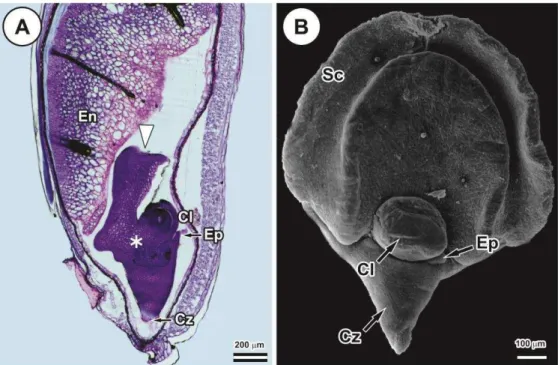
26
Figure 1. Brachypodium distachyon immature embryo 15 d post anthesis. (A) Light micrograph of longitudinal section of immature seed showing the scutellum (white arrow), scutellar node (asterisk), coleoptile enclosing the plumule (Cl), coleorhiza enclosing the primary root (Cz), epiblast (Ep) and endosperm (En). (B) Scanning electron micrograph of immature embryo showing the scutellum (Sc), coleoptile (Cl), coleorhiza (Cz) and epiblast (Ep).
Production of compact embryogenic callus (CEC) and plant regeneration
27
pearly surface were fragmented in 2-4 pieces and transferred (20 callus pieces per plate) onto regeneration medium (MS salts, Fe-EDTA, 30 g L-1 sucrose, 0.2 mg L-1 kinetin, vitamins M5, 2 g L-1 Phytagel - pH 5.8) for 2-3 weeks at 25 ºC under 16-h photoperiod. Shoots (rooted or not) were transferred to jars containing germination medium (40% MS salts, Fe-EDTA, 10 g L-1 sucrose, vitamins B5, 2 g L-1 l Phytagel, 6 g L-1 agar - pH 5.8). Shoots were cultured for 2-3 weeks at 23ºC under 16-h photoperiod. Regenerated plantlets were potted in plastic pots containing a wet compost mixture (3:1 Plantmax®:vermiculite). Plants were grown in a Controlled Environment Room (CER) at 23 °C with a 20-hour photoperiod for 5-6 weeks.
Microscopy sample preparation
For anatomical and histochemical characterization of the process of somatic embryogenesis, immature seeds obtained around 15 days post anthesis (DPA), Day 0, and immature embryos were collected every day from 1 to 6 days after culture in induction medium (DAC), then at days 8, 12 and 21 DAC. The samples were fixed in Karnovsky (1965) solution (glutaraldehyde [2.5%] and paraformaldehyde [4%] in 0.1-M monobasic potassium phosphate buffer [pH 7.2], plus 5 m0.1-M of calcium chloride). Mature seeds (35 DPA) were also prepared for histological and histochemical analyses.
Light microscopy and histochemical characterization
28
Image capture was performed with a light and fluorescence microscope (Olympus AX70TRF, Olympus Optical, Japan) coupled with a digital camera (AxioCam HR, Zeiss).
Scanning electron microscopy
Fixed samples were dehydrated with an increasing acetone series, subjected to critical point drying (CPD 030, Bal-Tec, Balzers, Germany), and coated with gold (SCD 050, Bal-Tec, Balzers, Germany). The analyses were conducted using a scanning electron microscope (LEO 435-VP, Cambridge, England) at the Center for the Support of Research in Electron Microscopy (NAP/MEPA) of the Luiz de Queiroz School of Agriculture (ESALQ/USP) and all images were processed digitally.
RESULTS
General morphology
After 24 h in culture, the coleorhiza and coleoptile had begun to elongate and epidermală hairsă hadă grownă onă theă coleorhiza.ă Duringă theă firstă 2-3 days in culture, a progressive swelling of the scutellum was observed and the surface of the periphery and scutellar node started to bulge (Figs. 2A, B). At day 4, nodular structures started to develop on the surface of the scutellar node (Figs. 2C, D). After 6-8 days, embryos had formed a mass of callus on the surface of the scutellum with areas of creamy-white embryogenic tissue (denser, with a pale, translucent, nodular appearance, fast growing) (Fig. 2E). The nonembryogenic callus was friable, pale yellow, soft, translucent and slower growing (tissue indicated by arrow in Fig. 2E).
The embryogenic potential of the nodular calli produced in induction medium was confirmedă byă theă doubleă stainingă techniqueă withă Evan‘să blueă andă acetocarmine.ă The embryogenic cells of the nodular structures at Day 8 (Fig. 2F) with large nuclei and dense cytoplasms stained intense bright red with acetocarmine. The large and vacuolated cells, with small nuclei, of the nonembryogenic callus stained blue with Evan‘săblue.ăAt day 21 (Fig. 2G), these calli showed a deeply folded appearance with formation of scutellar embryos.
29
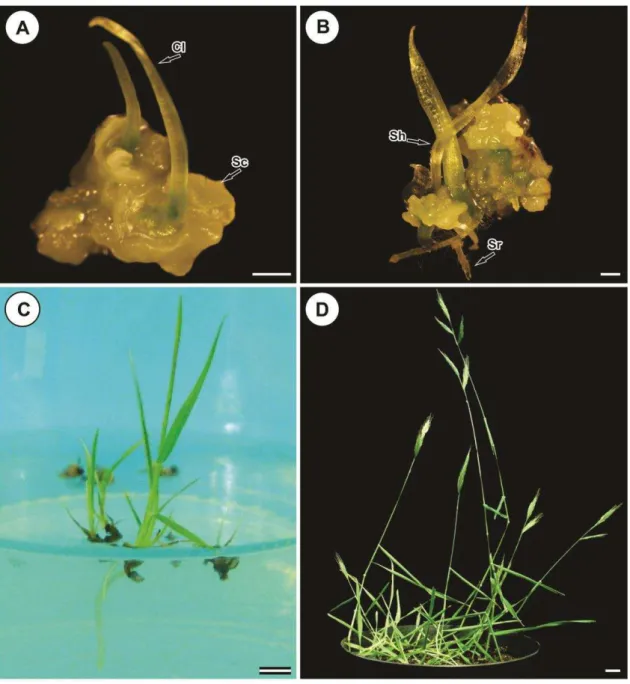
scutella (Figs. 3A, C, D). Localized chlorophyll also occurred in some zones of the callus (Fig. 3D). Coleoptiles emerged from the coleoptilar pore in the scutellum of fully developed somatic embryos (Figs. 3B, C, D). Coleoptiles were also creamy-white but more translucent and had trichome hairs on the surface (Fig. 3D). Development of coleorhiza and coleoptile was observed by SEM examination (Fig. 3E), indicating a bi-polarity of somatic embryo development. Somatic embryos germinated (Fig. 4A) and shoot-root axis growth occurred in regeneration medium and developed green plantlets (Fig. 4B). Occasionally, root development was delayed and the plantlet remained connected to the callus or produced small roots. These plantlets further developed following subculture for 2-3 weeks on the germination medium (Fig. 4C). Rooted plants were established successfully in compost mixture, produced fertile flowers and set viable seeds (Fig. 4D).
Histological examination
Sections through immature embryos, at day 1 DAC, showed that the scutellar tissue is composed of a single epidermal layer, consisting of richly cytoplasmic cells, and the ground tissue consists of relatively large, compact parenchyma cells (Fig. 5A). The abaxial epidermal scutellar cells were densely stained and markedly distinguishable from scutellar ground cells (Fig. 5A). At day 3 DAC, after several mitotic divisions, there was the differentiation of a region with embryogenic cells initiating nodular callus (Fig. 5B). The cells in the scutellar ground layers increased in size (Figs. 5B, C).
From days 4-5 onwards, mitotic divisions were also observed in the ground tissue, in cells associated with the vascular bundle in the mesocotyl of the zygotic embryo (Fig. 5C). Meristematic cells were produced adjacent to the scutellar vascular bundle, which is connected to the embryo provascular tissue in the scutellar node and extend to the scutellum parenchyma (Fig. 5D). No proembryoids were observed arising from these cells. Cells of the shoot and root meristems in the embryo axis remained without modifications.
30
the somatic embryo is established by the formation of the scutellum and zones of cytoplasm-rich cells are clearly visible (Figs. 5F, G). Later (28 days in culture), in regeneration medium, these zones developed into shoot and root meristems and formed the shoot-root axis that established the apical/basal polarity of the somatic embryo (Fig. 3E).
While embryogenic cells were being produced in the surface layer, cells of the scutellar ground tissue, coleorhiza, embryo axis and coleoptile became enlarged, highly vacuolated and with small nucleus (Figs. 5B, C, F). The progressive vacuolation of these cells and the formation of large inter-cellular spaces led to their degradation and fragmentation, and the consequent detachment of the inner layers of the callus and, from 8 to 21 days of culture, the embryogenic tissue was separated from the original explant (Figs. 5 F, G).
Embryogenic callus maintained in culture showed progressive growth and the cells of the inner parts of these calli increased in size and also became vacuolated, restarting the process of proembryoid separation. Staining with Lugol's iodine solution indicated the presence of starch in these cells (Fig. 5H).
Histochemical analysis of storage compounds
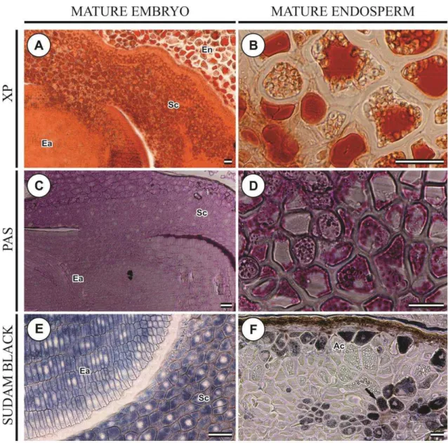
The histochemical tests confirmed the presence of storage reserves in the scutellum of the immature zygotic embryos used to initiate the embryogenic cultures.
The scutellum of the immature embryo (15 DPA) used to initiate the embryogenic callus (Day 0) showed positive reaction to xylidine Ponceau, though rather less intense (Fig. 6A). However, after 2 days in culture on somatic embryogenesis induction medium, cells of the immature scutellum did not react to xylidine Ponceau and the test was unable to detect storage proteins as the embryogenic callus developed and the epidermal cells were actively dividing to form the proembryonic cell masses (Figs. 6D, G, J, M, P).
31
granules were also present in the epidermal cells that were actively dividing. From Day 4, starch granules had increased in size and appeared more localized, but in smaller numbers, in the vacuolated cells near the proembryonic cell masses (Figs. 6H, K, N). This pattern of starch accumulation was observed until Day 21. From Day 4 to 8 in culture, small starch granules became apparent scattered through the cells of the proembryoids (Figs. 6H, K). From Day 12-21, these small granules enlarged and became abundant in the cells of the proembryoids (Fig. 6Q).
The embryonic and scutellar cells of the dissected immature zygotic embryo (15 DPA) at Day 0 showed negative reaction with Sudan black for lipid bodies (Fig. 6C). From 2-21 days of culture on somatic embryogenesis induction medium, there was also no reaction of cells of the immature scutellum and embryo axis with Sudan black B (Figs. 6F, I, L, O, R).
The histochemical tests confirmed the presence of protein, starch and lipids in the mature seeds.
The storage endosperm of mature seeds of B. distachyon consists of vacuolated cells with thick cell walls (Figs. 7B, D, F). At 35 days post-anthesis (DPA), small protein bodies deeply stained with xylidine Ponceau appeared in large amounts in the scutellar parenchyma cells, but none were found in embryo axis cells (Fig. 7A). In endosperm cells, protein appeared coalesced into a single mass surrounded by small starch granules (Fig. 7B).
Starch accumulation in the endosperm of mature seeds of B. distachyon was confirmedă byă lightă microscopyă usingă PASă stainingă (Fig.ă 7D). At 30 DPA, the endospermăcellsăwereămainlyăoccupiedăbyălargeăvacuolesăfilledăwithăproteinsă(Fig.ă7B) and small even-sized starch granules concentrated in the free spaces (Fig. 7D). No starch granules were detected with PAS staining in the embryonic and scutellar cells of mature seeds (Fig. 7C).
32
34
36
37
Figure 5. Induction of embryogenic culture from immature zygotic embryos of
Brachypodium distachyon. Light micrographs of longitudinal sections. (A) Immature
38
39
41