Effects of an Inspiratory Muscle Rehabilitation Program in the
Postoperative Period of Cardiac Surgery
Paulo Eduardo Gomes Ferreira
1, Alfredo José Rodrigues
1,2, Paulo Roberto Barboza Évora
1,2Hospital das Clínicas da Faculdade de Medicina de Ribeirão Preto da Universidade de São Paulo, Serviço de Fisioterapia Cardiorrespiratória1; Faculdade de Medicina da Universidade de São Paulo de Ribeirão Preto, Departamento de Cirurgia e Anatomia, Divisão de Cirurgia Cardiovascular e Torácica2, Ribeirão Preto, SP - Brazil
Summary
Background: Respiratory muscles are affected after cardiac surgeries.
Objective: To verify whether the preoperative conditioning of the inspiratory muscles might help to decrease postoperative respiratory dysfunction.
Methods: Thirty volunteers of both genders and with a minimum age of 50 years, while waiting for myocardial revascularization and/or cardiac valve surgery, were randomly assigned to two groups. Fifteen patients were included in a domiciliary program of at least 2 weeks of preoperative training of the inspiratory muscles, using a device with a load corresponding to 40% of the maximum inspiratory pressure. The other 15 patients received general advice and did not train the inspiratory muscle. Spirometry, before and after the training program, as well as the evolution of the arterial blood gases and of the maximum inspiratory and expiratory pressure, before and after the operation were evaluated in both group.. The clinical outcomes of the two groups were also compared.
Results: We observed that inspiratory muscle training increased the forced vital capacity, the maximum voluntary ventilation and the ratio between the forced expired volume during the first second and the forced vital capacity. The evolution of the arterial blood gases and of the maximum inspiratory and expiratory pressures before and after the operation was similar in both groups, with the outcomes also being similar.
Conclusion: We concluded that our domiciliary program of inspiratory muscle training was safe and improved the forced vital capacity and the maximum voluntary ventilation, although the clinical benefits of this program were not clearly demonstrable in the present study. (Arq Bras Cardiol 2009;92(4):261-268)
Key words: Respiratory muscles; thoracic surgery; postoperative care; physical therapy modalities; rehabilitation.
Mailling address: Paulo Eduardo Gomes Ferreira •
Av. Afonço Valera, 25, casa 11, Recreio das Acácias, 14.098-561 - Ribeirão Preto, SP - Brazil
E-mail: [email protected], [email protected]
Manuscript received January 23, 2008; revised manuscript received March 03, 2008; accepted March 06, 2008.
respiratory complications arising in cardiac postoperative period11-13.
The aim of this investigation was to evaluate if a preoperative training program for the inspiratory muscles, conducted at home and designed to improve respiratory functions, would contribute to reduce morbidity and/or mortality in adult patients submitted to myocardial revascularization surgery and/or valvuloplasty.
Patients and Methods
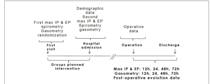
Figure 1 shows the timeline of the interventions and data collection.
Patients
The study included 30 voluntary patients of both sexes, without race discrimination, under 50 years of age, who were submitted to myocardial revascularization surgery or valvuloplasty. All patients signed the Free and Informed Consent Form and the investigation was approved by the
Introduction
Respiratory complications are among the most common causes of morbidity and mortality after cardiac surgery1-3.
Respiratory dysfunctions in cardiac postoperative are usually multifactorial and alterations due to cardiopathies4, to surgery
itself 5-7 and to limitations of the pulmonary functional reserve
due to aging8,9 have a preponderant role. Several studies
suggest that although multifactorial, the morbidity and mortality due to pulmonary causes in the postoperative period could be reduced by training the respiratory musculature altered by cardiopathies and aging10. However, this is a
Figure 1 - Timeline of the interventions and data collection.
Committee on Ethics in Research of the University Hospital of the Faculty of Medicine of Ribeirão Preto, University of São Paulo (HCFMRP-USP).
The patients were randomly divided into two groups, Control Group (CG) and Respiratory Incentive Group (RI). Both groups received general information, but only the RI group was submitted to respiratory incentive.
The exclusion criteria were: patients with unstable angina pectoris at the moment of selection or during the program, congestive decompensated heart failure, lack of physical or intellectual capacity to adequately perform the prescribed exercises, complex ventricular and uncontrolled arrhythmia, uncontrolled high blood pressure (>140/90mm/Hg), myocardial infarction or cerebrovascular accident (CVA) less than three years ago, high grade atrioventricular blockade or exercise-induced bronchial spasm. Patients submitted to surgical procedures before completing a minimum of two weeks of respiratory muscle training were also excluded.
The program was discontinued for patients complaining of dyspnea, before or after exercise; abdominal or precordial discomfort or pain, or any other symptom self-characterized as anginous; dizziness, vision darkening or any other type of physical or mental discomfort; vomiting or nausea; tachycardia or bradycardia; tiredness severe enough to prevent continuous exercise; cyanosis or mucosal-skin paleness; vertigo, dizziness or loss of consciousness.
Physical therapeutic procedures
Patients in both groups were submitted to the following procedures: a) Manovacuometry; b) Spirometry; c) Collection of arterial blood for arterial gasometry.
Manovacuometry
For manovacuometry evaluations, peak inspiratory and expiratory pressures, MaxIP and MaxEP, respectively, were determined from the residual volume (RV) and total pulmonary capacity14 (TPC) measured with a
manovacuometer, model MV-150/300 (Ger-Ar Comércio Equipamentos Ltda. São Paulo, Brazil). Three consecutive and technically acceptable measurements were made with minimal intervals of one minute. Determinations producing values with differences >10% were discarded and new ones repeated in order to have three adequate measurements14.
The first MaxIP and MaxEP determination (D0) was carried out at the start of the program, the second, soon after hospital admission for the programmed surgery (DInt) and the others at 12, 24, 48 and 72 hours, (12h PS, 24h PS, 48h PS, 72h PS respectively) after postoperative extubation. The patients were in the bed-seated position and wearing a nasal clip for all postoperative measurements.
Pulmonary function tests
Both groups of patients were submitted to spirometry tests at the start of the program (D0) and at hospital admission, immediately before surgery (DInt). All spirometric determinations were carried out at the Pulmonary Function Laboratory, at the University Hospital of the School of Medicine of Ribeirao Preto, University of São Paulo, by the same professional, in a constant-temperature environment (220C)
using a 9.0 liter spirometer, model GS Plus (Collins, USA).
Arterial gasometry
Blood samples were collected at the first spirometry determination (D0) before the exercise program and again at hospital admission (DInt). Other samples were collected after surgery, respectively, soon after orotracheal extubation and 12, 24 48 and 72 hours after postoperative extubation.
General advice for pre-surgery
own limits were also suggested to the patients. The same postoperative physical therapeutic procedures were employed in all patients from both groups, according to the necessity and routine programs established by the Physical Therapy Section of the Thoracic and Cardiovascular Surgery Division of the HCFMRP-USP.
Training program for the inspiratory musculature
Patients were instructed to perform five series of 10 calm and deep inspirations with at least one-minute intervals between the series, without feeling tired or sick, with the incentive of a respiratory instrument “Threshold IMT” (Respironics, Cedar Grove, NJ USA), with a load of 40% of MaxIP (D0)15. The series were to be repeated thrice daily,
while waiting for the surgery.
Statistical analysis
All values are expressed as means ± standard deviations or percentages. The Shapiro-Wilks test was employed to determine normal distributions. The paired Student "t" test for paired and non-paired values, as indicated, was used when variables were normally distributed and continuous. If not, the Mann-Whitney or Wilcoxon tests were used. The Fisher exact test was used to compare proportions. The "Two-Way ANOVA" test compared three or more repeated measurements, intra and inter-groups.
Results
None of the patients had to leave the program due to adverse events. There were no deaths during the program; one patient left the training voluntarily and no one was excluded due to surgery anticipation before at least 15 days of participation. There were no missing values for any collected data.
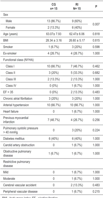
Demographic data
Table 1 shows the demographic data of patients from both groups. Except for a male prevalence in the control group (p=0.007) there were no other significant differences between groups.
Operative data
Duration of aorta clamping was significantly higher in the RI group (92.07±31.03 versus 73.5±26.98,p=0.034). There were no differences in the periods of extra-corporeal circulation (111.6±74.06 versus 112.3±4.5, p=0.696) and in the number of distal anastomoses in revascularizations (2.8±0.77 versus 3.0±0.87, p=0.656). Myocardial revascularization was performed in 73.3% of the control group patients and in 60.0% of the RI ones (p=0.700).
Respiratory function
Regarding the RI group, the time period between patients’ inclusion in the program and admission for surgery, that is, the time available for respiratory musculature training, was 154.0±87.4 days.
Table 1 - Demographic data of patients in the control (CG) and
respiratory incentive (RI) groups. Data expressed as means ± SD or percentages
CG n= 15
RI
N= 15 P
Sex
Male 13 (86.7%) 9 (60%)
0.007 Female 2 (13.3%) 6 (40%)
Age (years) 63.07± 7.93 62.47± 6.06 0.818
BMI 28.34 ± 3.16 26.60 ± 5.17 0.815
Smoker 1 (6.7%) 3 (20%) 0.598
Ex-smoker 4 (26.7%) 4 (26.7%) 1.000
Functional class (NYHA)
Class I 10 (66.7%) 7 (46.7%) 0.462
Class II 3 (20%) 5 (33.3%) 0.682
Class III 2 (13.3%) 2 (13.3%) 1.000
Class IV 0 (0%) 1 (6.7%) 1.000
EF < 35 0 (0%) 2 (13.3%) 0.483
Chronic atrial ibrillation 3 (20%) 3 (20%) 1.000 Arterial hypertension 10 (66.7%) 10 (66.7%) 1.000
Heart failure 0 1 (6.7%) 1.000
Previous myocardial
infarction 7 (46.7%) 4 (26.7%) 0.256
Pulmonary systolic pressure
> 40 mmHg 0 3 (20%) 0.224
Diabetes mellitus 6 (40%) 6 (40%) 1.000
Carotid artery obstruction 0 1 (6.7%) 1.000
Obstructive pulmonary
disease 1 (6.7%) 1 (6.7%) 1.000
Restrictive pulmonary disease
Mild 0 1 (6.7%) 1.000
Moderate 0 1 (6.7%) 1.000
Cerebral vascular accident 0 2 (13.3%) 0.483
Peripheral vascular disease 0 1 (6.7%) 0.215
BMI - body mass index; EF - ejection fraction.
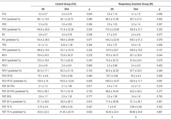
Spirometry
Manovacuometry
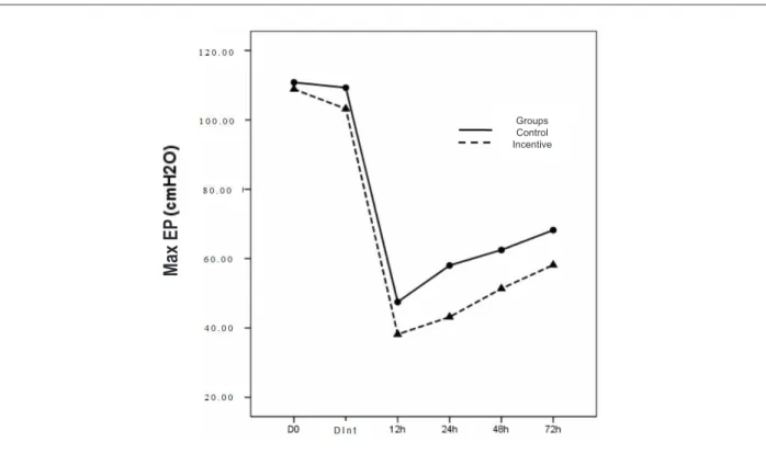
There was no significant difference between groups concerning Max IP and Max EP. However, in both groups, Max IP was significantly increased when comparing values at hospital admission with the ones determined on D0, considered as control values in each group (Figure 2). All determinations of postoperative MaxIP were significantly lower (p<0.001). MaxEP evolution was similar to MaxIP, as demonstrated in Figure 3, except that there was no difference between the first determination (D0) and the one on the day of hospital admission. However, the decrease in the postoperative determinations was significant, when compared to D0 (p<0.001).
Gasometry
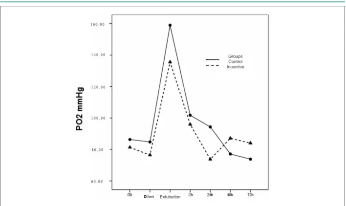
Figure 4 shows that the temporal evolution of arterial PO2 was not different between the two groups. There was a significant PO2 increase (p<0.001) in both groups, immediately after extubation when compared to D0, due to routine oxygen supplementation to all patients at this
Table 2 - Spirometry values for both groups of patients, CG and RI, measured at the start of the program (D0) and at the time of hospital
admission (DInt). Values are expressed as means ±SD. The paired Student “t” test was employed to compare D0 and DInt for each group
Control Group (CG) Respiratory Incentive Group (RI)
D0 DInt p D0 Dint p
FVC 3.2 ± 0.7 3.2 ± 0.74 0.843 2.9 ± 1.14 3.1 ± 1.6 0.006
FVC (predicted %) 88.1 ± 13.8 92.1 ± 23.73 0.480 86.3 ± 21.06 95.7 ± 21.5 0.002
FRC 3.3 ± 0.8 3.5 ± 0.82 0.369 3.5 ± 1.03 3.3 ± 1.4 0.307
FRC (predicted %) 104.8 ± 25.9 111.6 ± 22.36 0.235 115.3 ± 23.69 105.9 ± 31.7 0.230
RV 2.8 ± 0.7 3.0 ± 0.78 0.548 2.7 ± 0.51 2.4 ± 0.8 0.077
RV (predicted %) 143.4 ± 36.0 148.4 ± 29.99 0.571 144.2 ± 22.04 109.3 ± 51.3 0.079
TPC 6.1 ± 1.4 6.23 ± 1.36 0.364 5.8 ± 1.57 5.6 ± 1.8 0.208
TPC (predicted %) 106.8 ± 19.4 112.1 ± 19.15 0.224 107.6 ± 20.7 100.8 ± 19.5 0.157
MVV 79.4 ± 24.5 72.9 ± 30.37 0.278 76.0 ± 34.37 83.1 ± 35.9 0.044
MVV (predicted %) 75.5 ± 18.8 70.1 ± 22.42 0.343 73.9 ± 22.72 81.8 ± 23.4 0.075
FEV1 2.4 ± 0.6 2.4 ± 0.61 0.800 2.3 ± 0.89 2.4 ± 0.9 0.453
FEV1 (predicted %) 92.3 ± 17.1 93.3 ± 21.19 0.743 92.9 ± 22.29 97.7 ± 23.0 0.246
FEV1/FVC 75.1 ± 9.6 73.8 ± 9.56 0.460 79.7 ± 5.68 76.2 ± 6.2 0.008
FEV1/FVC (predicted %) 103.4 ± 14. 103.0 ± 12.04 0.925 109.0 ± 10.07 102.9 ± 11.7 0.097
FEF 25-75% 2.1 ± 1.0 2.1 ± 1.06 0.817 2.4 ± 1.14 2.2 ± 1.3 0.216
FEF 25-75% (predicted %) 78.6 ± 38.3 75.7 ± 33.16 0.720 88.6 ± 34.45 83.0 ± 40.2 0.377
FEF 50% 2.6 ± 1.7 2.5 ± 1.35 0.777 3.5 ± 1.56 3.4 ± 1.8 0.750
FEF 50 % (predicted %) 57.1 ± 36.6 55.0 ± 28.11 0.815 71.5 ± 28.96 72 .0 ± 36.7 0.951
FEF 75 % 0.74 ± 0.4 0.68 ± 0.34 0.427 1 ± 0.47 0.94 ± 0.48 0.532
FEF 75 % (predicted %) 43.6 ± 22.3 41.45 ± 20.74 0.623 50.44 ± 22.4 49.66 ± 24.8 0.861
3 9
FVC - forced vital capacity; FRC - functional residual capacity; RV - residual volume; TPC - total pulmonary capacity; MVV - maximal voluntary ventilation; FEV1 - forced expiratory volume after one second; FEF - forced expiratory low.
time. However, values returned to levels similar to D0 in further determinations during the postoperative period. Temporal evolution of PCO2 shown in Figure 5 does not indicate differences between groups. In addition, the PCO2 arterial values were kept in the normal range and developed in similar manner. In both groups a significant decrease of arterial PCO2 was evident after 48 and 72 hours, in comparison to D0, but the values were still within the normality range.
Postoperative clinical evolution
Figure 2 - Variations in the peak inspiratory pressure (Max IP) at the start of the program (D0), at the day of hospital admission and 12, 24, 48 and 72 hours postoperatively
in both CG and RI groups. * signiicant difference (p<0.001) in relation to D0 in the same group.
Groups Control Incentive
M
ax
IP
Figure 3 - Variations in the peak expiratory pressure (Max EP) at the start of the program (D0), at the day of hospital admission and 12, 24, 48 and 72 hours postoperatively
in both CG and RI groups. * signiicant difference (p< 0.001) in relation to D0 in same group.
Groups Control Incentive
M
ax
E
Figure 4 - PO2 temporal evolution at the start of the program (D0), on the hospital admission day (DInt), soon after orotracheal extubation and 12, 24, 48 and 72 hours
postoperatively in both control (CG) and respiratory incentive (RI) groups . * signiicant difference (p<0.001) in relation to D0 in the same group.
Groups Control Incentive
Extubation
Figure 5 - PCO2 temporal evolution at the start of the program (D0), on the hospital admission day (DInt), soon after orotracheal extubation and 12, 24, 48, and 72 hours
postoperatively in both control (CG) and respiratory incentive (RI) groups. * signiicant difference (p<0.001) in relation to D0 in the same group.
Extubation
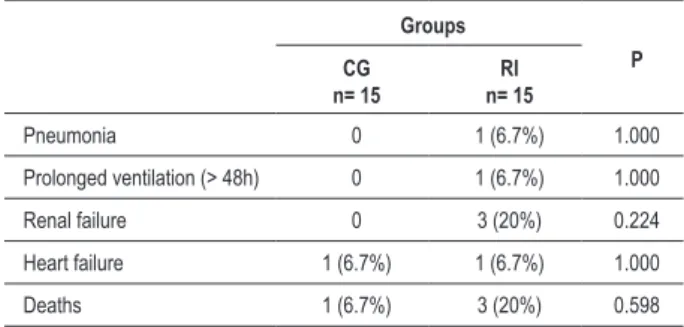
Table 3 - Postoperative clinical evolution Groups P CG n= 15 RI n= 15
Pneumonia 0 1 (6.7%) 1.000
Prolonged ventilation (> 48h) 0 1 (6.7%) 1.000
Renal failure 0 3 (20%) 0.224
Heart failure 1 (6.7%) 1 (6.7%) 1.000
Deaths 1 (6.7%) 3 (20%) 0.598
period (ejection fraction ≤0.35 and/or Functional Class
III/IV of NYHA).
Discussion
Although the results of the preoperative muscular inspiratory training did not show apparent clinical benefits, it significantly improved the ventilation function, as judged by the increased forced vital capacity and the maximum voluntary ventilation.
As expected, the inspiratory musculature training did not influence MaxEP. However, the increase in MaxIP was significant at the hospital admission day in comparison to the first value (D0) determined in both groups. Although the only group that showed ventilatory function benefits was the one receiving training, as shown by the spirometry values at hospital admission, the improvement in the MaxIP regardless of training suggests that a previous methodological knowledge by the patients could have contributed to better testing conditions at hospital admission. Similar results were reported by Newall et al16, in their control patients,
although their argumentation was that high-intensity exercises without load performed by the control patients could have improved the inspiratory function expressed by high Max IP also in this group.
In the present study, the control group did not have a specific training of the inspiratory musculature, but they were equally advised to stop smoking, practice daily walking exercises, in addition to receiving instructions for deep respiratory exercises, consisting of 3 series of 10 repetitions, daily, without apparatus.
It is possible that these measurements and exercises, even without loads, may have contributed to the improvement in the MaxIP in the control group. The lack of alterations in the spirometric values in this group reinforces the hypothesis that previous knowledge of the manovacuometry technique influenced the obtaining of Max IP values.
The positive effects of the inspiratory musculature training on pulmonary ventilation were demonstrated by the elevated forced vital capacity (FVC) and maximal voluntary ventilation (MVV), although clinical benefits were not apparent. However, considering that several of the voluntary patients did not present pulmonary dysfunction, it is difficult to show clinical benefits. Nevertheless, other studies11,12,17 have
suggested the preoperative training as a preventive factor of respiratory complications, as well as in clinical situations such as heart failure, improving dyspnea sensations, with the possibility of increasing walking distances and resulting in a better quality of life18.
One aspect of the program, which cannot be controlled by the organizers, is the time period from the start of the program to hospital admission for surgery. As in many other Brazilian institutions, the repressed demand for cardiac surgeries funded by the Federal Health Services (SUS) is very high, resulting in long waiting periods and decreased motivation among the patients. Some authors have considered16,18 that the effects of respiratory training
may last for several weeks after finishing the training and this may explain the positive spirometry results in a population whose adherence to the program diminished with time.
The significant postoperative reduction in the respiratory musculature performance was expected, as reflected by lower values in MaxEP and IP in both groups. Pain, the effects of cardiac surgery on muscular function and the effect of pain relievers are probable contributors to these findings. As mentioned before, the high PO2 at extubation was probably due to oxygen supplementation within the first 12 hours after surgery. The arterial PCO2 reduction observed 48 and 72 hours postoperatively has no clinical value, as it is within the normality range.
Perioperative respiratory training is considered beneficial by several surgeons and physical therapists, especially in the postoperative period, although there is no consensus on its prophylactic efficacy. Contrarily, Pasquina et al19, concluded
in a recent review that the prophylactic respiratory physical therapy is ineffective to prevent post-cardiac surgery pulmonary complications. In addition to complaining about the quality of some of the studies, the authors considered that there is not enough evidence to warrant the usefulness of these procedures. Thus, prospective studies about the prophylactic character of respiratory physiotherapy are in demand. The prospective study of Leguisamo et al20 found
that a preoperative program of physical therapy resulted in significant reduction in the hospital stay duration20.
The present study, although prospective with a random choice of voluntary patients, is not criticism-free. The number of subjects is small especially considering the two kinds of cardiac surgery. In addition, lack of strict monitoring may have compromised the efficacy of the program, undermining its clinical benefits.
The fact that just one intervention and one modality of respiratory exercise were used is another possible characteristic bound to be criticized.
Whether the improvement in some spirometric parameters seen after inspiratory muscular training is to be translated into clinical benefits is a question that demands further clinical studies involving a considerable greater number of subjects.
References
1. Silva LHF, Nascimento CS, Viotti Jr LAP. Revascularização do miocárdio em idosos. Rev Bras Cir Cardiovasc. 1997; 12 (2): 132-40.
2. Rodriguez R, Torrents A, Garcia P, Ribera A, Permanyer G, Moradi M, et al. Cardiac surgery in elderly patients. Rev Esp Cardiol. 2002; 55 (11): 1159-68. 3. Jin F, Chung F. Minimizing perioperative adverse events in the elderly. Br J
Anaesth. 2001; 87 (4): 608-24.
4. Meyer FJ, Borst MM, Zugck C, Kirschke A, Schellberg D, Kubler W, et al. Respiratory muscle dysfunction in congestive heart failure: clinical correlation and prognostic significance. Circulation. 2001; 103: 2153-8.
5. Matthay MA, Wiener-Kronish JP. Respiratory management after cardiac surgery. Chest. 1989; 95 (2): 424-34.
6. Diegeler A, Doll N, Rauch T, Haberer D, Walther T, Falk V, et al. Humoral immune response during coronary artery bypass grafting: a comparison of limited approach, “off-pump” technique, and conventional cardiopulmonary bypass. Circulation. 2000; 102 (Suppl 3): III 95-III 100.
7. Singh NP, Vargas FS, Cukier A. Arterial blood gases after coronary artery bypass surgery. Chest. 1992; 102: 1337-41.
8. Johnson BD, Reddan WG, Pegelow DF, Seow KC, Dempsey JA. Flow limitation and regulation of functional residual capacity during exercise in a physically active aging population. Am Rev Respir Dis. 1991; 143 (5): 960-7. 9. Shephard RJ. Exercise and aging: extending independence in older adults.
Geriatrics. 1993; 48 (5): 61-4.
10. Tolep K, Kelsen SG. Effect of aging on respiratory skeletal muscles. Clin Chest Med. 1993; 14 (3): 363-78.
11. Weiner P, Zeidan F, Zamir D, Pelled B, Waizman J, Beckerman M, et al. Prophylactic inspiratory muscle training in patients undergoing coronary
artery bypass graft. World J Surg. 1998; 22: 427-31.
12. Weiner P, Man A, Weiner M, Rabner M, Waizman J, Magadle R, et al. The effect of incentive spirometry and inspiratory muscle training on pulmonary function after lung resection. J Thorac Cardiovasc Surg. 1997; 113 (3): 552-7. 13. Nomori H, Kobayashi R, Fuyuno G, Morinaga S, Yashima H. Preoperative
respiratory muscle training: assessment in thoracic surgery patients with special reference to postoperative pulmonary complications. Chest. 1994; 105 (6): 1782-8.
14. Souza RB. Pressões respiratórias estáticas máximas. J Bras Pneumol. 2002; 28 (supl. 3): 155-65.
15. Sociedade Brasileira de Pneumologia e Tisiologia. Consenso Brasileiro de Ventilação mecânica. J Bras Pneumol. 2000; 26 (supl 2): S1-S68. 16. Newall C, Stockley RA, Hill SL. Exercise training and inspiratory muscle
training in patients with bronchiectasis. Thorax. 2005; 60 (11): 943-8. 17. Rajendran AJ, Pandurangi UM, Murali R, Gomathi S, Vijayan VK, Cherian KM.
Pre-operative short-term pulmonary rehabilitation for patients of chronic obstructive pulmonary disease undergoing coronary artery bypass graft surgery. Indian Heart J. 1998; 50 (5): 531-4.
18. Dall’Ago P, Chiappa GR, Guths H, Stein R, Ribeiro JP. Inspiratory muscle training in patients with heart failure and inspiratory muscle weakness: a randomized trial. J Am Coll Cardiol. 2006; 47 (4): 757-63.
19. Pasquina P, Tramer MR, Walder B. Prophylactic respiratory physiotherapy after cardiac surgery: systematic review. BMJ. 2003; 327: 1-6.
20. Leguisamo CP, Kalil RA, Furlani AP. A efetividade de uma proposta fisioterapêutica pré-operatória para cirurgia de revascularização do miocárdio. Rev Bras Cir Cardiovasc. 2005; 20 (2): 134-41.
and resulted in improved forced vital capacity and maximal voluntary ventilation, although its clinical benefits have not been demonstrated.
Potential Conflict of Interest
No potential conflict of interest relevant to this article was reported.
Sources of Funding
There were no external funding sources for this study.
Study Association