Induced Liver Injury after Renal Transplantation in
Chinese Recipients
Yifeng Guo1*, Yu Fan1, Jianxin Qiu1, Yong Liu1, Junwei Gao2, Fang Guo3*
1Organ Transplantation Center, Shanghai First People’s Hospital, School of Medicine, Shanghai Jiao Tong University, Shanghai, China,2Department of Pharmacy, Shanghai First People’s Hospital, School of Medicine, Shanghai Jiao Tong University, Shanghai, China,3Center for Nanomedicine and Translational Medicine, Shanghai Advanced Research Institute, Chinese Academy of Sciences, Shanghai, China
Abstract
Genetic polymorphisms in cytotoxic T lymphocyte-associated antigen 4 (CTLA4) play an influential role in graft rejection and the long-term clinical outcome of organ transplantation. We investigated the association of 5 CTLA4 single-nucleotide polymorphisms (SNPs) (rs733618 C/T, rs4553808 A/G, rs5742909 C/T, rs231775 A/G, and rs3087243 G/A) with drug-induced liver injury (DILI) in Chinese renal transplantation (RT) recipients. Each recipient underwent a 24-month follow-up observation for drug-induced liver damage. The CTLA4 SNPs were genotyped in 864 renal transplantation recipients. A significant association was found between the rs231775 genotype and an early onset of DILI in the recipients. Multivariate analyses revealed that a risk factor, recipient rs231775 genotype (p = 0.040), was associated with DILI. Five haplotypes were estimated for 4 SNPs (excluding rs733618); the frequency of haplotype ACGG was significantly higher in the DILI group (68.9%) than in the non-DILI group (61.1%) (p = 0.041). In conclusion, CTLA4 haplotype ACGG was partially associated with the development of DILI in Chinese kidney transplant recipients. The rs231775 GG genotype may be a risk factor for immunosuppressive drug-induced liver damage.
Citation:Guo Y, Fan Y, Qiu J, Liu Y, Gao J, et al. (2012) Polymorphisms inCTLA4Influence Incidence of Drug-Induced Liver Injury after Renal Transplantation in Chinese Recipients. PLoS ONE 7(12): e51723. doi:10.1371/journal.pone.0051723
Editor:Mathias Toft, Oslo University Hospital, Norway
ReceivedJuly 12, 2012;AcceptedNovember 5, 2012;PublishedDecember 21, 2012
Copyright:ß2012 Guo et al. This is an open-access article distributed under the terms of the Creative Commons Attribution License, which permits unrestricted use, distribution, and reproduction in any medium, provided the original author and source are credited.
Funding:The project was supported by the National Natural Science Foundation of China (30971137 and 31171308) and grants from the Chinese Postdoctoral Science Foundation (2003033271). The funders had no role in study design, data collection and analysis, decision to publish, or preparation of the manuscript.
Competing Interests:The authors have declared that no competing interests exist. * E-mail: gyfslm@gmail.com (YG); guof@sari.ac.cn (FG)
Introduction
In renal transplantation, immunosuppressive therapy is usually administered as a triple regimen, such as cyclosporine A (CsA)/ tacrolimus (TAC)+mycophenolate mofetil (MMF)+prednisone (Pred). The triple regimen is favored because it produces a more effective immunosuppression and lessens the drug-induced dam-ages or side effects. However, complications such as leukopenia, drug-induced liver injury (DILI), osteoporosis, infection and tumors often appear [1–5].
Cytotoxic T lymphocyte-associated antigen 4 (CTLA4) is a key element in the immune system that induces immune tolerance and is one of the critical negative regulators of the T cell-mediated immune response [6]. It is also expressed constitutively on the surface of regulatory T cells (Tregs) and is detectable on approximately 50% of Tregs; it is only found on,1% of naive helper T cells [7].CTLA4ligation on Tregs results in a significant decrease in the presentation capacity of antigen-presenting cells and effector T cell downregulation in mice [8].CTLA4plays an important role in the downregulation of the immune response. The rs231775 (+49A/G) SNP is located within the signal peptide of the molecule and influences expression of the full length isoform on the T cell membrane. The expression pattern of the CTLA-4 protein was found to be changed by a polymorphism of the rs4553808 (21661A/G) and rs5742909 (2318C/T) genotypes, located in the CTLA-4 gene promoter [9]. Similarly, the rs733618
(21772T) allele was found to decrease the transcription level of the CTLA-4 gene by influencing the binding of transcription factors [10]. The rs3087243 (+6230G/A) SNP is situated within the 39untranslated region of the CTLA-4 gene and was found to be associated with susceptibility to autoimmune diseases [11]. The single-nucleotide polymorphisms (SNPs) of the CTLA4 gene +49A/G (rs231775) and +6230 G/A (rs3087243) play an influential role in graft rejection and the long-term clinical outcome of organ transplantation [12–16].
CTLA4 gene polymorphism has been suggested to influence liver damage. Kanno et al [17] discovered that SNP CTLA4
+49GG (rs231775) may be associated with the liver damage of primary biliary cirrhosis (PBC) in Japanese populations. Valenti et al [18] observed a significantly higher prevalence of subjects carrying theCTLA4susceptibility allele (both in the heterozygous and homozygous states) among patients with ALD compared to healthy subjects. TheCTLA4 polymorphic G allele may confer susceptibility to ALD and, in the homozygous state, to alcoholic cirrhosis.
drug-induced liver injury (DILI) in Chinese renal transplantation recipients.
Materials and Methods
Diagnostic criteria and methods
A grade$14 on the scale reported by Maria et al [19] was used to diagnose DILI. DILI was suspected in patients with symptom-atic liver disease and those with asymptomsymptom-atic elevations in liver function tests (LFTs). LFT abnormalities were categorized into hepatitic, cholestatic or mixed based on abnormalities of serum alanine aminotransferase (ALT) and serum alkaline phosphatase (ALP) and the relationships of these to their respective upper limits of normal (ULNs). Liver damage was categorized according to the US Food and Drug Administration hepatotoxicity steering committee [20] : hepatitic pattern = ALT.3 ULN & [(ALT/ ULN)/(ALP/ULN)].5; cholestatic pattern = ALP.2 ULN & [(ALT/ULN)/(ALP/ULN)],2; mixed pattern = ALT.3 ULN & ALP,2 ULN and [(ALT/ULN)/(ALP/ULN)].2 but,5.
People having the following conditions were excluded from the study: hepatitis caused by excessive consumption of alcohol; presence of hepatitis virus A, B, C, D or E; fatty liver; autoimmune hepatitis; hereditary liver disease; hemorrhagic or congestive hepatitis; hyperthyroidism liver injury; non-hepatotropic viral hepatitis; or hepatitis of another cause.
Patients
This study included 864 transplantation recipients (764 cadaver donor cases and 100 living donor cases; 536 men and 328 women) in the Shanghai Organ Transplantation Center between Jan 2000 and Oct 2011. Ninety patients had DILI, and 774 cases had no liver injury. Of the 90 patients with DILI, 32 cases presented with a primarily hepatitic pattern, 36 with a cholestatic pattern and 22 with a mixed pattern. The mean age of the patients included in the study was 40.09610.06 years. Overall, 816 cases of chronic glomerulonephritis, 25 cases of polycystic kidney disease, and 23 cases of pyelonephritis were detected. Preoperative negativity for all hepatitis viruses and a normal liver function were required. All of the recipients were blood group-matched with their donors and were tested for the panel-reactive antibody and HLA-A-B-DR matching.
Each organ donation or transplant in our center was strictly selected according to the guidelines of the Ethical Committee review board of our hospital, the regulation of Organ Transplant Committee of Shanghai Jiao Tong University and the Declaration of Helsinki. The study protocol was approved by the Ethical
Committee review board of our hospital and Shanghai Jiao Tong University and informed. The research process was explained to every candidate patient from the collection and storage of blood, isolation of DNA and determination of gene polymorphisms in detail. Every participant gave written informed consent form. The Ethical Committee review board of our hospital and Shanghai Jiao Tong University approved this consent procedure and the study.
This is a cross-sectional study. Each patient underwent a 24-month follow-up observation through which clinical information was provided by means of clinical observation, medical records and outpatient or telephone follow-up visits. The exclusion criteria were (1) observed time less than 24 months, (2) die with other reasons (infection, etc) in 24 months post-transplantation, (3) stop using immune suppressants with graft function failure, and (4) not able to provide written informed consent. This study was performed from Jan 2000 to Oct 2011; all patients discharged in about 30days after operation and visited our outpatient clinics; all patients that did not meet the exclusion criteria (n = 864) were invited to take part in the present study.
Immunosuppression protocol
Mycophenolate mofetil (MMF) 1.0 was given as a premedicant. Intravenous infusion of 500 mg/d of methylprednisolone was applied during the procedure through 2 days after the operation. The dose was then decreased to 360 mg, 180 mg, 80 mg and 40 mg each subsequent day, followed by prednisone (15–20 mg/d) as a maintenance therapy. Triple therapy with cyclosporine A (CsA)/tacrolimus (TAC), MMF and prednisone was administered beginning on the third day after the operation. The dosage of MMF was 1.0–1.5 g/d with a weight of 60 kg as the critical value. CsA and TAC were started at doses of 8 mg/kg/d and 0.2 mg/ kg/d, respectively, and then adjusted according to the plasma concentrations and the serum creatinine concentrations.
The diagnostic criteria of AR were based on the comprehensive elevation of histological and clinical symptoms, their alleviation by anti-rejection therapy and graft biopsy. The clinical symptoms examined were hypourocrinia, fever, weight gain, pain in the transplanted kidney, elevated blood pressure, increased serum creatinine (to 25% above baseline), urine protein and the resistance index. The Banff 97 working classification for renal allograft pathology (modified) [21] was used as the pathological rejection criteria.
Sample collection and polymorphism genotyping
A total of 864 patients were included in this study. Peripheral blood samples (3 ml) were collected, the DNA was extracted, and
Table 1.PCR primers of theCTLA4SNP used in the study.
Locus AT (6C) Primer pairs (59R39) Amplicon size (bp)
rs733618, rs4553808 58 CTAAGAGCATCCGCTTGCACCT 486
TTGGTGTGATGCACAGAAGCCTTTT
rs5742909 56 AAATGAATTGGACTGGATGGT 226
TTACGAGAAAGGAAGCCGTG
rs231775 58 GCTCTACTTCCTGAAGACCT 162
AGTCTCACTCACCTTTGCAG
rs3087243 59 AGGAAGGCAGATCAAAATGC 202
CACCACTATTTGGGATATAACA
the SNPs of CTLA4 were genotyped using polymerase chain reaction (PCR) and direct sequencing. The primers and annealing temperatures (ATs) employed for rs733618 C/T, rs4553808 A/G, rs5742909 C/T, rs231775 A/G and rs3087243 G/A were displayed in Table 1.
Statistical analysis
Comparisons of clinical characteristics between patients with DILI and non-DILI were analyzed by the Pearsonx2test and an independent-sample test. We assessed the Hardy–Weinberg equilibrium (HWE) for both DILI and non-DILI using the x2 test. For linkage disequilibrium (LD), Haploview version 4.2 software was used [22]. A correlation test was used to assay the degree of correlation between DILI and AR. Genotype associa-tions were analyzed using a dominant model (minor-allele homozygotes plus heterozygotes vs. major-allele homozygotes), a recessive model (minor-allele homozygotes vs. heterozygotes plus major-allele homozygotes) and a codominant model (minor-allele homozygotes and heterozygotes vs. major-allele homozygotes). The allelic frequencies were counted in a single strand of measured DNA. The differences in the genotype distributions between groups were analyzed by the x2 test or Fisher’s exact test. According to presence and absence of acute rejection (AR), subanalysis was used by Chi-square test. The time of the first abnormal laboratory result indicative of DILI was designed as the post-transplantation time (days) of first abnormalities in liver function tests (LFTs) in recipients suffering from DILI and as an early onset of DILI. Associations of theCTLA4SNPs with an early onset of DILI in patients were analyzed by the Kaplan-Meier test. Multivariate analyses, logistic regression, were used to analyze several risk factors, including age, gender, primary diseases, number of HLA mismatches, acute rejection, blood transfusion,
CTLA4 SNPs. These risk factors were analyzed together. We explored the haplotype association for 5 SNPs using Haploview version 4.2. All statistical tests were two-sided, and statistical significance was set atp,0.05. Correction for multiple testing was carried out using the Bonferroni method. Statistical analysis was performed with SPSS (Statistical Package for the Social Sciences)
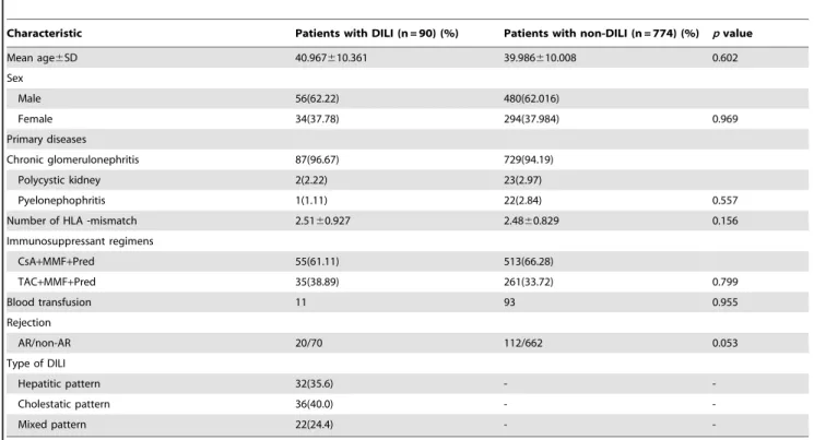
Table 2.Comparison of clinical characteristics between patients with DILI and non-DILI.
Characteristic Patients with DILI (n = 90) (%) Patients with non-DILI (n = 774) (%) pvalue
Mean age6SD 40.967610.361 39.986610.008 0.602
Sex
Male 56(62.22) 480(62.016)
Female 34(37.78) 294(37.984) 0.969
Primary diseases
Chronic glomerulonephritis 87(96.67) 729(94.19)
Polycystic kidney 2(2.22) 23(2.97)
Pyelonephophritis 1(1.11) 22(2.84) 0.557
Number of HLA -mismatch 2.5160.927 2.4860.829 0.156
Immunosuppressant regimens
CsA+MMF+Pred 55(61.11) 513(66.28)
TAC+MMF+Pred 35(38.89) 261(33.72) 0.799
Blood transfusion 11 93 0.955
Rejection
AR/non-AR 20/70 112/662 0.053
Type of DILI
Hepatitic pattern 32(35.6) -
-Cholestatic pattern 36(40.0) -
-Mixed pattern 22(24.4) -
-CsA: cyclosporine, MMF: mycophenolate mofetil, Pred: prednisone, TAC: tacolimous, AR: acute rejection, non-AR: non-acute rejection, DILI: drug induced liver injury. doi:10.1371/journal.pone.0051723.t002
Figure 1. Diagnosis time of drug-induced liver injury after renal transplantation.Twenty-three patients were diagnosed drug-induced liver injury within the first month after operation; 26, 22 and 19 cases presented with DILI between 2 and 6 months, 7 and 12 months and 13 and 24 months after operation, respectively.
version 11.5 software (SPSS Inc., Chicago IL, USA). All statistical tests were two-sided, and statistical significance was set atp,0.05.
Results
Baseline characteristics of 864 renal transplant recipients
The total number of patients was 864, with 536 male and 328 female cases. A total of 10.42% recipients (90/864) had DILI during the first 24 months post-transplantation. Baseline charac-teristics of 864 renal transplant recipients and types of DILI were listed in Table 2. No significant differences in age, sex, primary
diseases, human leukocyte antigen mismatches, blood transfusion, renal transplantation or immunosuppressant regimen were found between patients with DILI and those without (Table 2). The incidence of acute rejection (AR) following renal transplantation was not different between the two groups (p= 0.053).
Twenty-three patients were diagnosed as having drug-induced liver injury within the first month after operation; 26, 22 and 19 cases presented with DILI between 2 and 6 months, 7 and 12 months and 13 and 24 months after operation, respectively. The
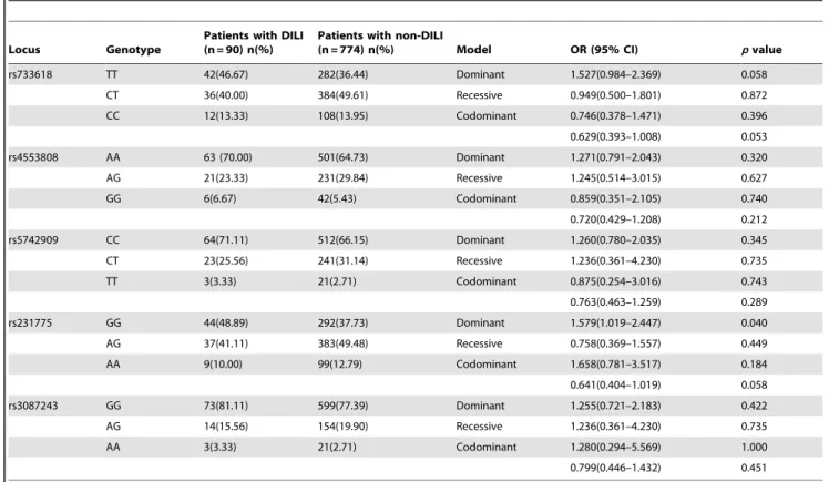
Table 3.The genotype distribution of theCTLA4polymorphisms in patients with DILI and non-DILI.
Locus Genotype
Patients with DILI (n = 90) n(%)
Patients with non-DILI
(n = 774) n(%) Model OR (95% CI) pvalue
rs733618 TT 42(46.67) 282(36.44) Dominant 1.527(0.984–2.369) 0.058
CT 36(40.00) 384(49.61) Recessive 0.949(0.500–1.801) 0.872
CC 12(13.33) 108(13.95) Codominant 0.746(0.378–1.471) 0.396
0.629(0.393–1.008) 0.053
rs4553808 AA 63 (70.00) 501(64.73) Dominant 1.271(0.791–2.043) 0.320
AG 21(23.33) 231(29.84) Recessive 1.245(0.514–3.015) 0.627
GG 6(6.67) 42(5.43) Codominant 0.859(0.351–2.105) 0.740
0.720(0.429–1.208) 0.212
rs5742909 CC 64(71.11) 512(66.15) Dominant 1.260(0.780–2.035) 0.345
CT 23(25.56) 241(31.14) Recessive 1.236(0.361–4.230) 0.735
TT 3(3.33) 21(2.71) Codominant 0.875(0.254–3.016) 0.743
0.763(0.463–1.259) 0.289
rs231775 GG 44(48.89) 292(37.73) Dominant 1.579(1.019–2.447) 0.040
AG 37(41.11) 383(49.48) Recessive 0.758(0.369–1.557) 0.449
AA 9(10.00) 99(12.79) Codominant 1.658(0.781–3.517) 0.184
0.641(0.404–1.019) 0.058
rs3087243 GG 73(81.11) 599(77.39) Dominant 1.255(0.721–2.183) 0.422
AG 14(15.56) 154(19.90) Recessive 1.236(0.361–4.230) 0.735
AA 3(3.33) 21(2.71) Codominant 1.280(0.294–5.569) 1.000
0.799(0.446–1.432) 0.451
DILI: drug induced liver injury, OR: odds ratio, CI: confidence intervals. doi:10.1371/journal.pone.0051723.t003
Table 4.The allele distribution ofCTLA4polymorphisms in patients with DILI and non-DILI.
Locus Allele
Patients with DILI (n = 180) n(%)
patients with non-DILI
(n = 1548) n(%) OR (95% CI) pvalue
rs733618 T 120(66.67) 948(61.24) 0.790(0.570–1.095) 0.156
C 60(33.33) 600 (38.76)
rs4553808 A 147(81.67) 1233(79.65) 0.879 (0.591–1.307) 0.523
G 33(18.33) 315(20.35)
rs5742909 C 151(83.89) 1265(81.72) 1.165(0.767–1.769) 0.474
T 29(16.11) 283(18.28)
rs231775 G 125(69.44) 967(62.47) 1.366(0.978–1.906) 0.066
A 55(30.56) 581(37.53)
rs3087243 G 160(88.89) 1352(87.34) 1.160 (0.712–1.890) 0.552
A 20(11.11) 196(123.66)
scatter plot in Figure 1 showed the distribution of the patients with DILI throughout the entire observation period.
Associations between theCTLA4SNPS and DILI
All polymorphisms were in Hardy-Weinberg equilibrium. Using Haploview version 4.2 software, the five loci were found to be in linkage disequilibrium (LD) (D’ = 0.900–1.000). Regarding the genotype distribution of theCTLA4polymorphisms, no statistical differences for rs733618, rs4553808, rs5742909 or rs3087243 were found between patients with DILI and those without. However, the frequency of the rs231775 GG genotype in recipients with DILI was significantly higher (48.89%) than in those recipients without DILI (37.73%) (p= 0.040, OR = 1.579, 95% CI = 1.019– 2.447, Bonferroni-adjustedp= 0.20) (Table 3).
No differences in the determined allelic frequencies of rs733618, rs4553808, rs5742909 or rs3087243 were found between DILI and non-DILI recipients (Table 4). The allelic distribution of the locus rs231775 was not different between recipients with DILI and those without DILI (p= 0.066, OR = 1.366, 95% CI = 0.978– 1.906).
In subanalysis, in which correction for multiple testing was carried out using the Bonferroni method, no statistical differences in genotype distribution and allelic frequencies of the CTLA4
polymorphisms were found yet in AR group and non-AR group (Table S1, S2, S3 and S4).
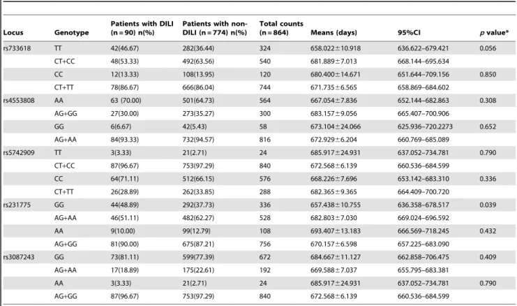
Kaplan-Meier analysis was used to examine the relationships betweenCTLA4SNPs and an early onset of DILI (Table 5); no statistical differences for rs733618, rs4553808, rs5742909 or rs3087243 existed between DILI and non-DILI recipients. A significant difference (p= 0.039) was found between patients bearing the rs231775 GG genotype and those with the AA+AG genotypes using the log-rank test (Figure 2). Values of mean and 95% CI for the GG and AG+AA groups were 657.438610.755 (95%CI: 636.358–678.517) days and 682.80367.030 (95%CI: 669.024–696.592) days respectively. A significant association was found between the rs231775 genotype and an early onset of DILI in recipients.
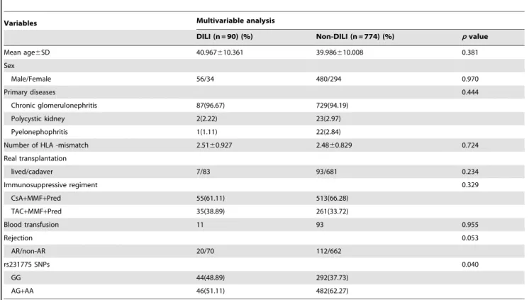
To further examine the associations of DILI with these variables, univariate and multivariate analyses were carried out with the variables age, gender, primary disease, immunosuppres-sive regimen, blood transfusion, HLA mismatch and rs231775 genotype (Table 6).
Multivariate analyses revealed that age, gender, primary disease, immunosuppressive regimen, blood transfusion, HLA mismatch and renal transplantation were independent of DILI; however, the analyses showed that a risk factor, recipient rs231775 genotype (p= 0.040) was associated with DILI.
Figure 2. Association betweenCTLA4 SNPs and early onset of drug-induced liver injury in renal transplantation. No statistical differences for rs733618 (Figure 2a), rs4553808 (Figure 2b), rs5742909 (Figure 2c) or rs3087243(Figure 2e) was found between DILI and non-DILI recipients. A significant difference (p = 0.039) was found between patients bearing the rs231775 GG genotype and those with the AA+AG genotypes using the log-rank test (Figure 2d).
doi:10.1371/journal.pone.0051723.g002
Table 5.Correlation between early onset of DILI andCTLA4genotypes in recipients.
Locus Genotype
Patients with DILI (n = 90) n(%)
Patients with non-DILI (n = 774) n(%)
Total counts
(n = 864) Means (days) 95%CI pvalue*
rs733618 TT 42(46.67) 282(36.44) 324 658.022610.918 636.622–679.421 0.056 CT+CC 48(53.33) 492(63.56) 540 681.88967.013 668.144–695.634
CC 12(13.33) 108(13.95) 120 680.400614.671 651.644–709.156 0.850 CT+TT 78(86.67) 666(86.04) 744 671.73566.565 658.869–684.602
rs4553808 AA 63 (70.00) 501(64.73) 564 667.05467.836 652.144–682.863 0.308 AG+GG 27(30.00) 273(35.27) 300 683.15769.056 665.407–700.906
GG 6(6.67) 42(5.43) 58 673.104624.066 625.936–720.2273 0.652 AG+AA 84(93.33) 732(94.57) 816 672.92966.204 660.769–685.089
rs5742909 TT 3(3.33) 21(2.71) 24 685.917624.931 637.052–734.781 0.790 CT+CC 87(96.67) 753(97.29) 840 672.56866.139 660.536–684.599
CC 64(71.11) 512(66.15) 576 668.22667.696 653.142–683.310 0.336 CT+TT 26(28.89) 262(33.85) 288 682.36569.365 664.409–700.720
rs231775 GG 44(48.89) 292(37.73) 336 657.438610.755 636.358–678.517 0.039 AG+AA 46(51.11) 482(62.27) 528 682.80367.030 669.024–696.592
AA 9(10.00) 99(12.79) 108 693.407613.183 666.569–718.245 0.432 AG+GG 81(90.00) 675(87.21) 756 670.15766.598 657.225–683.090
rs3087243 GG 73(81.11) 599(77.39) 672 684.667611.127 662.858–706.475 0.409 AG+AA 17(18.89) 175(22.61) 192 669.58867.037 655.795–683.381
AA 3(3.33) 21(2.71) 24 685.917624.931 637.052–734.781 0.790 AG+GG 87(96.67) 753(97.29) 840 672.56866.139 660.536–684.599
DILI: drug induced liver injury, CI: confidence intervals. *log-rank test.
The association ofCTLA4haplotype and DILI
No differences in the frequencies of seven haplotypes covering the 5 SNPs existed between the DILI and non-DILI recipients (Table 7). Five haplotypes were estimated for 4 of the SNPs
(excluding rs733618); the frequency of haplotype ACGG was significantly higher in the DILI group (68.9%) than in the non-DILI group (61.1%) (p= 0.041). No statistically significant differences were found between the DILI and non-DILI groups for the rest of the haplotypes (p.0.05) (Table 7).
Discussion
In kidney transplant recipients, immunosuppressive therapy is usually administered as a triple regimen and typically includes cyclosporine A (CsA)/tacrolimus (TAC)+mycophenolate mofetil (MMF)+prednisone (Pred). The mechanism of DILI has not been completely elucidated, although drugs such as CsA and steroids may induce liver cholestasis and/or hepatocyte lesions [2], causing a direct toxic effect and immune-mediated damage that may contribute to the pathogenesis of DILI. Our study revealed that the frequency of recipients carrying the rs231775 GG genotype in the DILI cohort was higher than that in the non-DILI group (p= 0.040). These results were consistent with the previously reported finding that the rs231775 G allele could mitigate the negative effect ofCTLA4 on T cell-mediated immune responses [23]. However, the statistical significance between groups did not hold after correction for multiple testing. This may simply be due to the sample size and, hence, lack of power to detect an association. The frequency of haplotype ACGG, including the rs231775G allele, was significantly higher in the DILI group (68.9%) than in the non-DILI group (61.1%) (p= 0.0409).
The fact thatCTLA4SNPs influenced DILI may not imply that this gene product had a direct toxic effect in liver damage. From our clinical experience, while elevation of serum creatinine and AR may be diagnosed by allograft biopsy, high-dose steroids (for instance, three-day therapy with 240–500 mg/d of intravenous
Table 6.Association between DILI and several risk factors.
Variables Multivariable analysis
DILI (n = 90) (%) Non-DILI (n = 774) (%) pvalue
Mean age6SD 40.967610.361 39.986610.008 0.381
Sex
Male/Female 56/34 480/294 0.970
Primary diseases 0.444
Chronic glomerulonephritis 87(96.67) 729(94.19)
Polycystic kidney 2(2.22) 23(2.97)
Pyelonephophritis 1(1.11) 22(2.84)
Number of HLA -mismatch 2.5160.927 2.4860.829 0.724
Real transplantation
lived/cadaver 7/83 93/681 0.234
Immunosuppressive regiment 0.329
CsA+MMF+Pred 55(61.11) 513(66.28)
TAC+MMF+Pred 35(38.89) 261(33.72)
Blood transfusion 11 93 0.955
Rejection 0.053
AR/non-AR 20/70 112/662
rs231775 SNPs 0.040
GG 44(48.89) 292(37.73)
AG+AA 46(51.11) 482(62.27)
CsA: cyclosporine, MMF: mycophenolate mofetil, Pred: prednisone, TAC: tacolimous, AR: acute rejection, non-AR: non-acute rejection, DILI: drug induced liver injury. doi:10.1371/journal.pone.0051723.t006
Table 7.The distribution of haplotypes in 5 locus of CTLA-4 between DILI and non- DILI.
Haplotype Frequency (%) x2 pvalue
DILI Non-DILI
5 locus
TACGG 65.0 59.1 2.270 0.132
CACAG 11.1 16.5 3.481 0.062
CGTAA 10.6 12.5 0.592 0.442
CGTAG 5.5 4.8 0.168 0.682
CACGG 3.9 1.9 3.103 0.078
CGCAG 1.7 2.1 0.167 0.683
TACAG 1.7 1.5 0.059 0.808
4 locus*
ACGG 68.9 61.1 4.181 0.041
ACAG 12.8 17.9 2.966 0.085
GTAA 10.6 12.5 0.592 0.442
GTAG 5.5 4.8 0.168 0.682
GCAG 1.7 2.1 0.167 0.683
methylprednisolone) or increasing doses of CNI were usually administered to these patients and, to some extent, precipitated the development of DILI. Thex2test showed no correlation between
DILI and acute rejection (AR) (p= 0.053) (Table 2). Percentage of AR recipient with DILI(20/112, 17.86%) was higher than no-AR (70/662, 10.57%), but in subanalysis, no statistical differences in genotype distribution and allelic frequencies of the CTLA4
polymorphisms were found yet in AR group and no-AR group. In our previous study [24], correlation betweenCTLA4SNPs and AR was found, whether AR is a risk of DILI or not need further discover. A larger sample size is necessary to confirm or reject the significance of this correlation.CTLA4SNPs influencing to DILI was possibly an internal factor. The association betweenCTLA4
SNPs and liver damage has been studied in two papers. Kanno et al [17] identified that the CTLA4+49 (rs231775) genotype was positively associated with liver damage in primary biliary cirrhosis (PBC) in Japanese populations and that PBC patients with the G/ G genotype had significantly higher serum levels of ALT, GGT, and IgM than did patients with the A/A or A/G genotype. Valenti et al [18] observed a significantly higher prevalence of the susceptibleCTLA4allele (both in the heterozygous [OR 2.5] and in the homozygous [OR 4.6] state) in patients with alcoholic liver disease (ALD) compared to healthy subjects; this relationship was independent of age, sex and geographical origin. The CTLA4
polymorphic G allele may confer susceptibility to ALD and, in the homozygous state, to alcoholic cirrhosis by interfering with the immune response. Ethnicity may influence a person’s susceptibility to DILI [25]. For instance, African-Americans were predisposed to anticonvulsant-induced DILI, while Caucasians are particularly prone to abacavir- and flucloxacillin-induced DILI [25]. The frequency of the G allele at theCTLA4+49 (rs231775) locus was much higher in the Chinese population than in other populations [26]. This may indicate an even more significant role of this genetic bias. In addition, using log-rank analysis, we discovered that the rs231775 GG genotype was associated with an early onset of DILI (p= 0.039) (Table 5 and Figure 2).
Susceptibility factors such as age and gender were thought to confer an increased risk for the development of DILI [25]. We have discovered that age and gender are not, in fact, susceptibility factors for the development of DILI using multivariate analysis (p= 0.381 and 0.970, respectively), which contradicts some previous studies. In general, increased age was a risk factor for DILI (for example, an age .49 increases the risk of isoniazid hepatotoxicity) [27]. Excessive use of sodium valproate and erythromycin resulted in hepatotoxicity predominantly in children [28]. Women are widely viewed as being more likely to develop DILI, and the ALFSG has reported a female preponderance in ALF due to both paracetamol and idiosyncratic drug reactions (74% and 67%, respectively) [29]. However, a recent examination
of a Spanish registry showed no overall gender difference. Rather, men, who were the predominant gender over age 60, were more likely to have a cholestatic injury, whereas women, who were the predominant gender under age 60, were more susceptible to a hepatitis-like injury [30].
Recent data suggested that DILI was associated with some immune responses, which may be influenced by human leukocyte antigen (HLA) polymorphisms. Polymorphisms of HLA-B*5701 were associated with flucloxacillin- and abacavir-induced DILI [31] and mutations in mitochondrial polymerase 1 with reactions to sodium valproate [32]. In our study, multivariate analysis demonstrated that the number of HLA mismatches was not independently associated with DILI (p= 0.724).
Multivariate analysis showed that a risk factor, recipient rs231775 genotype (p= 0.040), was associated with DILI.
In conclusion, the CTLA4 haplotype ACGG was partially associated with the development of DILI in Chinese kidney transplant recipients. The rs231775 GG genotype may be a risk factor for immunosuppressive drug-induced liver damage in kidney transplantation. The association in the paper was statistically weak, withpvalue close to the threshold. It is possible that different populations and other statistic bias. It is need to further study on different and larger patient cohorts. The mechanism of immunosuppressant-induced DILI in renal trans-plantation has not been completely elucidated and needs to be studied in depth.
Supporting Information
Table S1 The genotype distribution of the CTLA4
polymorphisms in AR patients with DILI and non-DILI.
(DOC)
Table S2 The genotype distribution of the CTLA4
polymorphisms in AR patients with DILI and non-DILI.
(DOC)
Table S3 The allele distribution of CTLA4
polymor-phisms in AR patients with DILI and non-DILI.
(DOC)
Table S4 The allele distribution of CTLA4
polymor-phisms in no-AR patients with DILI and non-DILI.
(DOC)
Author Contributions
Conceived and designed the experiments: YG FG. Performed the experiments: JG. Analyzed the data: YL. Contributed reagents/materi-als/analysis tools: YF JQ. Wrote the paper: YG.
References
1. Hurst FP, Belur P, Nee R, Agodoa LY, Patel P, et al. (2011) Poor outcomes associated with neutropenia after kidney transplantation: analysis of United States Renal Data System. Transplantation 92:36–40.
2. Rita Rezzani (2004) Cyclosporine A and adverse effects on organs: histochemical studies. Progress in Histochemistry and Cytochemistry 39: 85– 128.
3. Kulak CA, Borba VZ, Ju´nior JK, Custo´dio MR (2012) Osteoporosis After Transplantation Curr Osteoporos Rep 10:48–55.
4. Kupeli E, Ulubay G, Colak T, Ozdemirel TS, Ozyurek BA, et al. (2011) Pulmonary complications in renal recipients after transplantation. Transplant Proc 43:551–553.
5. Engels EA, Pfeiffer RM, Fraumeni JF, Kasiske BL, Israni AK, et al. (2011) Spectrum of cancer risk among US solid organ transplant recipients. JAMA 306:1891–901.
6. Alegre ML, Frauwirth KA, Thompson CB (2001) T-cell regulation by CD28 and CTLA-4. Nat Rev Immunol 1:220–228.
7. Grohmann U, Orabona C, Fallarino F, Vacca C, Calcinaro F, et al. (2002) CTLA-4-Ig regulates tryptophan catabolism in vivo. Nat Immunol 3:1097–1101. 8. Wing K, Onishi Y, Prieto-Martin P, Yamaguchi T, Miyara M, et al. (2008) CTLA-4 control over Foxp3+regulatory T cell function. Science 322:271–275.
9. Bouqbis L, Izaabel H, Akhayat O, Pe´rez-Lezaun A, Calafell F, et al. (2003)Association of the CTLA4 promoter region (21661G allele) with type 1 diabetes in the South Moroccan population. Genes Immun 4:132–7. 10. Hudson LL, Rocca K, Song YW, Pandey JP (2002) CTLA-4 gene
polymorphisms in systemic lupus erythematosus: a highly significant association with a determinant in the promoter region. Hum Genet 111:452–5. 11. Ueda H, Howson JM, Esposito L, Heward J, Snook H, et al. (2003) Association
of the T-cell regulatory gene CTLA4 with susceptibility to autoimmune disease. Nature 423:506–511.
13. Wu J, Tang JL, Wu SJ, Lio HY, Yang YC (2009) Functional polymorphism of CTLA-4 and ICOS genes in allogeneic hematopoietic stem cell transplantation. Clin Chim Acta 403:229–233.
14. Tapirdamaz O, Pravica V, Metselaar HJ, Hansen B, Moons L, et al. (2006) Polymorphisms in the T cell regulatory gene cytotoxic T lymphocyte antigen 4 influence the rate of acute rejection after liver transplantation. Gut 55:863–868. 15. De Reuver P, Pravica V, Hop W, Boor P, Metselaar HJ, et al. (2003) Recipient ctla-4+49 G/G genotype is associated with reduced incidence of acute rejection after liver transplantation. Am J Transplant 3:1587–1594.
16. Kusztal M, Koscielska-Kasprzak K, Drulis-Fajdasz D, Magott-Procelewska M, Patrzałek D, et al. (2010) The influence of CTLA-4 gene polymorphism on long-term kidney allograft function in Caucasian recipients. Transpl Immunol 23:121–124.
17. Kanno Y, Rai T, Monoe K, Saito H, Takahashi A, et al. (2006) Possible association of cytotoxic T lymphocyte antigen-4 genetic polymorphism with liver damage of primary biliary cirrhosis in Japan. Fukushima J Med Sci 52:79–85. 18. Valenti L, De Feo T, Fracanzani AL, Fatta E, Salvagnini M, et al. (2004)
Cytotoxic T lymphocyte associated antigen 4 A49G polymorphism is associated with susceptibility to and severity of alcoholic liver disease in Italian patients. Alcohol & Alcoholism 39: 276–280.
19. Maria VA, Victorino RM (1997) Development and validation of a clinical scale for the diagnosis of drug-induced hepatitis. Hepatology 26:664–669. 20. Navarro V (2010) Hepatic Adverse Event Nomenclature document. Available:
h t t p : / / w w w . f d a . g o v / D r u g s / S c i e n c e R es ea r c h / R e s e a r c h A r e a s / ucm080336htm. Accessed 2010 Jan 12.
21. Racusen LC, Colvin RB, Solez K, Mihatsch MJ, Halloran PF, et al. (2003) Antibody-mediated rejection - an addition to the Banff classification of renal allograft rejection. Am J Transplant 3:708–714.
22. Barrett JC, Fry B, Maller J, Daly MJ (2005) Haploview: analysis and visualization of LD and haplotype maps. Bioinformatics 21:263–265.
23. Kouki T, Sawai Y, Gardine CA, Fisfalen ME, Alegre ML (2000) CTLA-4 gene polymorphism at position 49 in exon 1 reduces the inhibitory function of CTLA-4 and contributes to the pathogenesis of Graves’ disease. J Immunol 165:6606– 6611.
24. Gao JW, Guo YF, Fan Y, Qiu JX, Bao ED, et al. (2012) Polymorphisms in cytotoxic T lymphocyte associated antigen-4 influence the rate of acute rejection after renal transplantation in 167 Chinese recipients. Transplant Immunology 26: 207–211.
25. Pauli-Magnus C, Meier PJ (2006) Hepatobiliary transporters and drug-induced cholestasis. Hepatology 44: 778–787.
26. Lee CS, Lee YJ, Liu HF, Su CH, Chang SC, et al. (2003) Association of CTLA-4 gene A-G polymorphism with rheumatoid arthritis in Chinese. Clin Rheumatol 22: 221–224.
27. Fountain FF, Tolley E, Chrisman CR, Self TH (2005) Isoniazid hepatotoxicity associated with treatment of latent tuberculosis infection: a 7-year evaluation from a public health tuberculosis clinic. Chest 128:116–123.
28. Maddrey WC (2005) Drug-induced hepatotoxicity: J Clin Gastroenterol; 39:S83–89.
29. Lee WM, Squires RH Jr, Nyberg SL, Doo E, Hoofnagle JH (2008) Acute liver failure: Summary of a workshop. Hepatology 47:1401–1415.
30. Lucena MI, Andrade RJ, Kaplowitz N, Garcı´a-Cortes M, Ferna´ndez MC, et al. (2009) Phenotypic characterization of idiosyncratic drug-induced liver injury: The influence of age and sex. Hepatology 49:2001–2009.
31. Daly AK, Donaldson PT, Bhatnagar P, Shen Y, Pe’er I, et al. (2009) HLA-B*5701 genotype is a major determinant of drug-induced liver injury due to flucloxacillin. Nat Genet 41: 816–819.