R E V I E W
Molecular, Neurochemical, and Behavioral Hallmarks of
Reserpine as a Model for Parkinson’s Disease: New
Perspectives to a Long-Standing Model
Anderson H.F.F. Leão
1*; Aldair J. Sarmento-Silva
1*; José R. Santos
2; Alessandra M. Ribeiro
1,3;
Regina H. Silva
1,41Memory Studies Laboratory, Department of Physiology, Universidade Federal do Rio Grande do Norte, Natal, RN, 2Biology Department, Universidade Federal de Sergipe, São Cristóvão, SE,
3Department of Biosciences, Universidade Federal de São Paulo, Santos, SP, Brazil,
4Behavioral Neuroscience Laboratory, Department of Pharmacology, Universidade Federal de São Paulo, São Paulo, SP, Brazil.
Keywords
animal model, dopamine, Parkinson’s disease, reserpine, rodent.
Corresponding author:
Regina H. Silva, PhD, Departamento de Farmacologia, UNIFESP, Rua Botucatu, 862, Edifício Leal Prado, 1° andar, São Paulo, SP CEP 04023062, Brazil (E-mail:
reginahsilva@gmail.com)
Received 18 November 2014 Accepted 23 February 2015
Published Online Article Accepted 2 March 2015
* These authors contributed equally to this work.
doi:10.1111/bpa.12253
Abstract
The administration of reserpine to rodents was one of the first models used to investigate
the pathophysiology and screening for potential treatments of Parkinson’s disease (PD).
The reserpine model was critical to the understanding of the role of monoamine system in
the regulation of motor and affective disorders, as well as the efficacy of current PD
treatments, such as L-DOPA and dopamine agonists. Nevertheless, with the introduction of
toxin-induced and genetic models of PD, reserpine became underused. The main rationale
to this drawback was the supposed absence of reserpine construct validity with PD. Here,
we highlight classical and recent experimental findings that support the face,
pharmaco-logical, and construct validity of reserpine PD model and reason against the current
rationale for its underuse. We also aim to shed a new perspective upon the model by
discussing the main challenges and potentials for the reserpine model of PD.
INTRODUCTION
Parkinson’s disease (PD) is the second most common
neuro-degenerative disorder after Alzheimer’s disease. Its onset is rarely
before the age of 50 years and a sharp increase of the incidence
occurs after the age of 60 years (19). PD affects approximately
1%–2% of the population over the age of 60 (63), with a higher
prevalence in men than in women (19, 62). Most importantly,
it is a disorder with progressive onset and escalating deterioration
of quality of life (28). Therefore, PD is a social and economic
burden to countries with increasing life expectancy, and for this
reason, the scientific interest in the disorder is continuously
emphasized.
PD diagnosis is based on its cardinal motor symptoms, which
include bradykinesia, rigidity, resting tremor, and postural
insta-bility (108). However, even though PD is essentially a motor
dis-order, patients present equally incapacitating nonmotor symptoms.
Furthermore, those symptoms may appear previously or
concomi-tantly to motor symptoms (126) and include sleep disorders (83,
134, 152), anxiety (154), depression (15, 97), neuropathic pain and
nociceptive sensitization (27, 72, 196), impulsivity (160, 203,
204), dementia and executive function impairment (1, 7, 49, 123),
olfactory dysfunction (7, 60), and constipation (48, 152).
The motor alterations are a consequence of dopaminergic
neuronal loss in the substantia nigra (SN) (92, 108), where the
main dopaminergic projection to the motor-regulating nucleus in
the basal ganglia originates (52, 120). Nonetheless, loss of
dopaminergic neurons in the ventral tegmental area (VTA)—
projecting to limbic areas and to prefrontal cortex—is also
reported in PD (192, 197). This loss results in emotional and
cognitive deficits (154, 165). Furthermore, other
neurotrans-mission disturbances are described, as revealed by
histopatho-logical markers in serotonergic (101, 194), noradrenergic (28, 211,
213), and cholinergic (197, 211) neurons.
stress response (
DJ-1
) (59, 163, 202, 206). Hence, impairment of
these pathways leads to oxidative stress and defective protein
folding, signaling, and degradation (47, 104, 114, 184). Finally, the
accumulation of defective protein aggregates—mainly constituted
by
α-synuclein, parkin, and ubiquitin, known as Lewy’s bodies
(200)—is followed by cell death. Thus, the pathogenesis of PD
primarily relates to the generation of oxidative stress and
accumu-lation of defective proteins.
The genetic alterations are in accordance with epidemiological
associations to PD. These associations comprise exposure to
envi-ronmental toxins that act on the respiratory chain (42, 143, 195)—
such as pesticides, heavy metals, and carbon monoxide—and
neuroinflammation (88, 200). Both events result in the generation
of toxic reactive oxygen (ROS) and reactive nitrogen species,
giving rise to cell damage and eventually cell death. In brief, PD
harbors the oxidative imbalance as a common molecular pathway
to cellular stress and neurodegeneration. Thus, animal models of
PD aim to reproduce the aforementioned cellular and molecular
damages (44, 61, 129), while clinical and preclinical therapeutic
strategies target different candidate steps of these pathways to slow
PD progression (34, 91).
ANIMAL MODELS OF PD
Current studies use genetic and neurotoxic approaches to
repro-duce pathophysiological hallmarks in animal models of PD. In
genetic studies, some strategies focus on the overexpression of
normal or truncated autosomal dominant genes, such as
SNCA
(23,
105, 137, 205) and
LRRK2
(117, 118), and knockout or
knock-down of autosomal recessive genes, as
Parkin
,
PINK1
, or
DJ-1
(106, 107, 157, 191). Nevertheless, none of these strategies
reca-pitulates the key clinical and neuropathological features of PD and
they only account for 5%–10% of PD cases (206). As a result, the
most frequently used strategy is to induce oxidative imbalance and
dopamine (DA) depletion by the administration of toxins or drugs
that act upon dopaminergic neurons (37, 44, 61, 71, 129, 136, 167,
177, 210).
1-Methyl-4-phenyl-1,2,3,6-tetrahydropyridine
(MPTP)
and
6-hydroxydopamine (6-OHDA) are the most used toxins in animal
models of PD because of their rather selective actions upon
dopaminergic neurons (9, 18, 61, 129). Both enter the dopaminergic
neuron by the DA transporter (DAT) and inhibit the complex I in the
respiratory chain, causing adenosine triphosphate (ATP) reduction,
oxidative damage, protein aggregation, cell death, and DA
deple-tion (61, 94, 129, 181). MPTP is a highly lipophilic protoxin that
readily crosses the blood–brain barrier when peripherally
adminis-tered (161). Once in the brain, MPTP is converted by glial
monoamine oxidase (MAO)-B into its intermediate
1-methyl-4-phenyl-2,3,dihydropyridinium, which is rapidly oxidized into
1-methyl-4-phenylpyridinium
and
then
reabsorbed
by
the
dopaminergic neuron through the DAT (45). A disadvantage of this
model is that rodents are more resilient to cell damage induced by
MPTP compared with primates. This results in the need for higher
dosages and increased variability in neurodegeneration within
treated animals (43, 61, 170). In addition, there is a high risk of
contamination to researchers because of the handling of large doses
of MPTP and the respective biological waste (155).
6-OHDA, on the other hand, does not cross the blood–brain
barrier and is directly administered into the brain (18, 26, 61, 170).
Contrastingly from MPTP, 6-OHDA enters noradrenergic neurons
as well, through the noradrenaline (NA) transporter (NAT) (29).
This lack of specificity is usually resolved by the coadministration
of inhibitors of NA and serotonin (5-HT) reuptake, such as
nortriptyline or desipramine (27, 56, 188). Although safer
regard-ing contamination risk compared to MPTP, bilateral administration
of 6-OHDA results in extensive neuronal loss and severe motor
impairment followed by death. After administration, animals need
tube-feeding because of aphagia and adipsia (55, 198). In order to
avoid these issues, most studies perform the unilateral lesion with
6-OHDA and assess motor deficit by inducing unilateral rotating
behavior with dopaminergic agonists (171, 188). Although
rota-tional behavior lacks face validity with PD (55), some studies
evaluate forelimb akinesia (evaluated by adjusted stepping and
limb-use asymmetry tests) after unilateral 6-OHDA administration
(145, 169, 183). Nevertheless, even though the forelimb akinesia
provides face validity, the unilateral lesion is still a weak approach
to mimic PD pathology and symptomatology.
Alternatively, studies have employed environmental toxins such
as rotenone, paraquat and maneb to model PD in rodents (9). Of
those, rotenone is the most used because of its lipophilic structure,
easiness to cross biological membranes, ability to inhibit complex I,
and generate ROS (16, 93, 172). However, despite its close
relation-ship to epidemiological risk factors of PD, rotenone’s lack
of selective action results in systemic and peripheral toxicity (74,
151, 158) and highly variable dopaminergic lesions (22, 43, 172,
212).
Finally, the administration of reserpine—an inhibitor of the
vesicular transporter of monoamines in the central nervous system
(VMAT2)—was one of the earliest animal models of PD. Reserpine
is an alkaloid extracted from
Rauwolfia serpentine
and was first
used as a potent antihypertensive drug because of its capacity to
deplete cellular monoamine content (76, 125, 150). The clinical use
of reserpine led to the observation that patients chronically treated
with reserpine developed lethargy, depression, and motor
dyskine-sia, implicating the monoamine system in the pathophysiology of
affective and motor disorders (76, 102). Readily after, reserpine was
used in rodents to mimic parkinsonian motor and nonmotor
impair-ments (17, 38, 39, 51, 69, 164, 175). Although considered outdated
in comparison with the aforementioned models, the reserpine
model mimics key features of PD symptomatology,
neuroche-mistry, and pharmacology. For this reason, the model was useful to
elucidate the relevance of dopaminergic neurotransmission to
motor control as well as to screen for candidate drugs for treatment
of PD. This review will highlight a new perspective upon the model
and reason against the current rationale for the undervaluation of
the reserpine-induced parkinsonism model.
MOTOR AND NONMOTOR BEHAVIORAL
IMPAIRMENT IN THE RESERPINE
MODEL
VMAT2 (201), leading to total monoamine depletion, including
DA, NA, and 5-HT.
Besides the typical motor impairment, reserpine is also able to
produce aversive (70, 174) and recognition (167) memory deficits,
anxiety-like behavior (25, 112), depressive and anhedonic-like
behaviors (10, 11, 175), and nociceptive sensitization (10, 11, 119,
144). Moreover, the memory impairment and the anxiety-like
behavior were described in a dose range (0.1–0.5 mg/kg) that did
not produce motor impairment (25, 70, 167, 174). This outcome
allowed the dissociation of an important confounding factor in
behavioral analyses.
More recently, the repeated treatment with low doses of
reser-pine (0.1 mg/kg) has been suggested as a progressive model of PD
(71, 167). Under this treatment regimen, animals progressively
developed motor impairment in the open field, catalepsy bar, and
oral movement tests after repeated injections of a low dose
(0.1 mg/kg) of reserpine. Deficits in these motor tests recapitulate
main motor symptoms of PD, such as hypokinesia and
bradykinesia, in the open field and catalepsy bar test (ie, slowness
and difficulty to initiate movements) and resting tremor in the oral
movement test.
In the aforementioned study (167), the motor impairments were
preceded by cognitive impairment in the novel object recognition
task. This impairment was also accompanied by neuronal
altera-tions compatible with the pathophysiology of PD such as reduction
in tyrosine hydroxylase (TH) immunostaining (167) and increased
lipid peroxidation in the striatum (71). Furthermore, the object
recognition index positively correlated with VTA immunostaining
for TH, suggesting neuronal pathways disruption other than the
nigrostriatal pathway playing an important role in nonmotor
symp-toms of PD. In addition, the object recognition deficit occurred
after a 1-h interval between training and test sessions (167), but not
when the two sessions were 24-h apart (71). In other words,
reserpine-treated rats presented short-term, but not for long-term,
memory deficit previously to motor deficits. Thus, performance in
the task requires recognition and executive functions. These
find-ings are in accordance with early PD symptomatic description, as
executive function, attention deficit and episodic and procedural
memory impairment have been described (20, 64, 115, 160, 162,
204). Furthermore, acute administration of low dose of reserpine
resulted in emotional processing deficits in aversive memory tasks,
such as context conditioning (70) and discriminative avoidance
(40) task, but not motor impairment. In parallel, immobility in the
forced swim test correlated with pain indexes, indicating a
comorbid
relationship
between
different
reserpine-induced
nonmotor symptoms (10). Similarly, PD nonmotor impairments
comprise anxiety (154), depression (15, 97), and nociceptive
sensitization (30, 72, 196). Thus, nonmotor findings induced by
reserpine resemble nonmotor PD symptoms, reinforcing
reser-pine’s face validity as a PD model.
PHARMACOLOGICAL AND PREDICTIVE
QUALITY OF THE RESERPINE MODEL
The use of reserpine was critical to the first demonstration of the
therapeutic efficacy of L-DOPA (38, 178). This effect was shortly
after observed in humans (54) and the reserpine model was
estab-lished for screening of potential symptomatic treatment efficacy of
new drugs for PD. Indeed, besides L-DOPA, the reserpine model
predicted other current symptomatic anti-Parkinson treatments:
apomorphine (85), pramipexole (68, 122), ropinirole (77),
rotigotine (199), pergolide (51, 98), bromocriptine (98, 99), and
cabergoline (133). Likewise, reserpine-induced motor impairment
is also reversed by agents that are used in association with
L-DOPA,
for
example:
muscarinic
antagonists,
such
as
benztropine and trihexyphenidyl (85); MAO-B or
catechol-O-methyltransferase (COMT) inhibitors, such as selegiline (51, 176),
rasagiline (73), and tolcapone (121); and amantadine (51, 53, 85,
100, 176). Table 1 summarizes different types of motor
impair-ment induced by reserpine that are reversed by these drugs. In fact,
reserpine is still currently used to assess anti-parkinsonian efficacy
of novel agents, such as D3 receptor agonists (80), inhibitors of
glutamate release (103), group III metabotropic glutamate receptor
agonists or positive allosteric modulators (14, 32, 142), group I
muscarinic metabotropic receptor antagonists or allosteric
modu-lator (207), and mixed adenosine A2A/A1 antagonists (13, 173).
Reserpine is also employed in the screening for antioxidant and
anti-inflammatory treatments to prevent motor impairments such as
dyskinesia (5, 10, 24, 66, 139, 147, 148). Current literature on oral
dyskinesia implicates oxidative stress on the pathophysiology of the
disorder (3, 4, 136, 186, 187). Accordingly, monoamine depletion in
reserpine-treated rats is followed by increase of reactive oxygen and
nitrogen species and cell damage (179). The metabolism of
catecholamine (CA) intrinsically results in ROS formation, which is
increased as a consequence of free CA in the cytoplasm of
reserpine-treated rats (127, 156). Thus, oxidative stress and cell
damage sums up to the monoamine depletion to impair motor
performance. For this reason, treatment with antioxidants is able to
revert reserpine-induced oxidative stress and oral dyskinesia (3,
147). Finally, the treatment with 40 mg/kg vitamin E concomitant to
the repeated treatment with 0.1 mg/kg reserpine (71, 167)
pre-vented cognitive and motor impairments (168), as well as the
reduction of TH immunostaining in rats (unpublished data).
These neurochemical imbalances resemble features of PD, as
oxidative stress and DA depletion, which are keystones of the
pathophysiology of the disease (33, 79). Thus, the
pharmacologi-cal mechanism of reserpine comprises important qualities of PD
pathophysiology and constitutes a good model for screening for
candidate drugs to both symptomatic treatment and possible
slowing of PD symptom progression. This advantage is reinforced
by its low toxicity to researchers, low cost, and reproducibility
among laboratories, which points out the reserpine model of PD as
a suitable model for drug screening.
MOLECULAR AND NEUROCHEMICAL
FEATURES OF THE RESERPINE MODEL
Despite the robust face and pharmacological validities, the current
literature does not recognize reserpine as a useful PD model,
arguing the lack of construct validity (61). This drawback is due to
the experimental observations that (i) reserpine do not induce
neurodegeneration and protein aggregation (61, 208); (ii) motor
performance, monoamine content, and TH staining are partially
restored after treatment interruption (144, 167); and (iii) reserpine
lacks specificity regarding dopaminergic neurotransmission (10,
11, 119, 141, 144).
variance across studies. Reserpine peripherally administered in the
dose range of 1–10 mg/kg is known to produce a robust
(70%–95%) depletion of monoamine content in several brain areas
(10, 11, 58, 65, 86, 90, 119, 141, 144, 189; for a summary, see
Table 2). This monoamine depletion starts 30 minutes after
reser-pine injection and may endure up to 14 days, finally returning to
normal levels after 21 days of retrieval (90, 144). At first, the
absence of specificity was considered a disadvantage regarding
accurate modeling of PD neurochemistry. However, there is
evi-dence of relevant alterations in 5-HT and NA imbalances in PD as
well (28, 101, 194, 211, 213). This argues in favor of the
resem-blance of the neurochemical disruptions in the reserpine model
with those in PD. Moreover, this characteristic is especially
impor-tant to the aforementioned nonmotor deficits of PD. For instance,
NA and 5-HT transmissions are related to cognitive and emotional
function (130, 175). Accordingly, reserpine treatment results in
monoamine depletion in areas involved in emotional processing—
as the amygdala (119)—and cognition—as the hippocampus,
cortex (9, 10), and prefrontal cortex (144). Furthermore, repeated
reserpine treatment reduces TH staining in the hippocampus,
prefrontal cortex, dorsal striatum, VTA, SN pars compacta (SNpc),
and locus coeruleus (167).
Finally, acute or short-term DA depletion by reserpine treatment
results in upregulation of D1, but not D2 (46, 132, 189).
Never-theless, long-term treatment also leads to D2 upregulation (140,
193). These neurochemical modifications also occur because of
dopaminergic denervation in untreated PD patients. Functional
imaging techniques report upregulation of D2 receptor, whereas
upregulation of D1 is not yet clearly defined (87, 95).
Another highly reproducible biochemical alteration in the
reser-pine model is the induction of oxidative stress. Reserreser-pine, in the
dose range of 1–10 mg/kg, is able to induce decreases in catalase,
superoxide dismutase, total content of reduced glutathione, and
ATP. Similarly, it increases glutathione peroxidase activity,
oxi-dized glutathione, lipid peroxidation, nitric oxide (NO), and iron
(2–4, 10, 11, 24, 35, 36, 65, 66, 71, 119, 138, 139, 147, 149, 159,
166, 174, 179, 186, 187; for a summary, see Table 3). Overall,
there is an increase in oxidative damage. Nevertheless, some
studies report contradicting results. Those differences seem to
emerge from different dosage, treatment regimen, and brain area
studied. For example, repeated treatment with low doses of
reser-pine (0.1 mg/kg) produced cumulative effects upon lipid
peroxidation in the striatum, but not hippocampus, of rats (71). As
well, catalase activity is generally reduced in all brain areas—
except for the striatum in which some studies found increased
activity (186, 187) or no significant differences (4, 66). This
oppo-site outcome may be due to a differential fine-tuning of catalase
activity regulation in the striatum, as catecholaminergic
metabo-lism intrinsically leads to oxidative stress (127, 156). In fact,
hydrogen peroxide (H2O2) is one of the main products of CA
metabolism by MAO-A (127, 156), and naturally one may
specu-late that catalase in catecholaminergic neurotransmission is
differ-entially modulated by increases in H2O2
in order to provide
antioxidant protection. Indeed, this is endorsed by the observation
Table 1.Predictive validity of reserpine Parkinson’s disease (PD) model effectiveness for symptomatic treatment of different motor disturbances in PD. The table was constructed and updated according to the table presented by Duty and Jenner (61). The drug list was compiled from the Parkinson’s UK website: parkinsons.org.uk/content/drug-treatments-parkinsons (accessed 6 October 2014). Abbreviations: COMT = catechol-O-methyltransferase; DA = dopamine; MAO = monoamine oxidase.Treatment Rigidity Hypokinesia Catalepsy Tremor Oral dyskinesia References
L-DOPA±Carbidopa + + + + − (51, 85, 99, 133, 176)
DA agonists
Bromocriptine + + + − − (98, 99, 133, 176)
Cabergoline + + + − − (133)
Pergoline + + + + − (51, 98, 122)
Pramipexole − + + − − (68, 122)
Ropinirole − − + − − (77)
Apomorphine + + + − − (85, 98, 99)
Glutamate antagonists
Amantadine + + − + − (51, 85, 176)
Anticholinergics − − − − − −
Orphenadrine − − − − − −
Procyclidine − − − − − −
Trihexyphenidyl + − − − − (85)
Benztropine + − − − − (85)
COMT inhibitors
Entacapone − − − − − −
Tolcapone − − − − − −
MAO-B inhibitors
Rasagiline − + − − − (73)
Selegiline + + − − + (51, 176)
Antioxidative and Dietary therapy
Vitamin E − − − − + (3, 66)
Co-enzyme Q10 − − − − − −
that catecholaminergic neurons are relatively abundant in
popula-tions of catalase-positive microperoxisomes (124). Thus, it seems
that treatment duration and brain area studied define the extent of
oxidative damage induced by reserpine.
The oxidative stress induced by reserpine is related to increased
DA metabolism as a result of the reduction on the number of DA
molecules in the vesicle (146) and increased DA turnover (67, 141,
179). Accordingly, MAO-A inhibitor reverts L-DOPA and reserpine
induced increase in oxidized glutathione (179, 180). In addition,
free DA and metabolites in the cytoplasm results in auto-oxidation
of DA and DOPAC to their corresponding reactive quinones—
DA-Q and DOPAC-Q, respectively—(12, 127, 156), which
contrib-utes to cell apoptosis and synuclein dimerization (84).
The generation of highly reactive molecules results in early
cell damage—as consistently evidenced by lipid peroxidation
(Table 3)—initiating proinflammatory signaling by tumor necrosis
factor (TNF)-α
and interleukin (IL)-1β
(10, 11). Subsequently, the
increase in proinflammatory cytokines activates microglia, which
leads to a vicious circle of adhesion, inflammation, and release of
more cytokines. Activated microglia upon dopaminergic neurons
also results in increased NO (10, 11, 24). Afterwards, NO—in the
presence of superoxide (O2
−)—produces peroxynitrite (NO3
−)
(127, 156), which is highly reactive and has been shown to
inac-tivate TH via S-thiolation on cysteine residues (8, 96, 110, 111). In
this context, repeated treatment with a low dose of reserpine
(0.1 mg/kg) resulted in reduced TH immunostaining in several
brain areas—that is hippocampus, prefrontal cortex, dorsal
striatum, SNpc, and VTA (167).
Ultimately, these events may terminate in the commitment with
apoptotic pathways. In other words, there is a reduction in
anti-apoptotic molecules, as Bcl-2 (65, 119), and an increase in
proapoptotic molecules, as caspase-3 (10, 11, 119).
Nevertheless, whether reserpine leads to permanent cell damage
or neurodegeneration is not clear yet. In this respect, repeated
treatment with 0.1 mg/kg of reserpine every other day for 20 days
resulted in a reduction of TH immunostaining that was partially
reversed after 30 days of treatment withdrawal (167). Likewise, the
same protocol increased
α-synuclein immunostaining in SN and
dorsal striatum and these effects were reversed after treatment
interruption (data not published). Of notice, such increase did not
result in protein inclusions and studies addressing if actual
neuronal loss occurs are currently being held. Thus, in light of the
current evidence (extent of TH reduction and
α-synuclein
increase, restauration of motor performance, and reversion of
reduction in TH and
α-synuclein immunostaining after
interrup-tion of treatment), data regarding the repeated low-dose reserpine
treatment should be interpreted in terms of TH expression
reduc-tion rather than neurodegenerareduc-tion.
On the other hand, some evidence support long-lasting or
per-manent cellular and behavioral changes within a high dose chronic
reserpine treatment. Treatment with 1 mg/kg of reserpine every
other day for 6 weeks resulted in persistent behavioral and
neurochemical changes (oral dyskinesia, DA depletion and D1 and
D2 receptor upregulation) up to 60 days after treatment withdrawal
(140). Thus, we do not discard the possibility of some extent of
permanent cell damage or cell death after reserpine treatment,
depending on dose and/or length of treatment.
In this context, untreated VMAT2 genetically deficient mice—
which express only 5% of functional VMAT2—presents
age-associated neurodegeneration in SNpc, locus coeruleus, and dorsal
raphe, followed by
α-synuclein accumulation and TH and
tyramine transporter immunostaining reduction (41, 185). This
VMAT2-deficient mice also presents L-DOPA responsive motor
impairment, twofold increase in DA concentration in cytosol,
Table 2.Monoamine content depletioninduced by different reserpine treatment regi-mens in rodents. Abbreviations: 5-HT = seroto-nin; BLA = basolateral amygdala; CTX = cortex; DA = dopamine; HPC = hippocampus; NA = noradrenaline; N/A = not applicable; PFC = prefrontal cortex; SN = substantia nigra; STR = striatum; THA = thalamus.
Dose (mg/kg) Structure Time window
DA NA 5-HT References
(50×) 0.01 STR 24 h 0% ∼45% 0% (141)
(50×) 0.1 STR 24 h ∼90% ∼90% ∼65%
(50×) 1.0 STR 24 h ∼95% ∼90% ∼90%
5.0 SN 2 h ∼85% N/A N/A (90)
24 h ∼70%
STR 2 h >95%
24 h >95%
1.0 STR 6 h ∼80% N/A ∼50% Unpublished data
24 h ∼90% ∼80%
96 h ∼75% ∼80%
5.0 STR 24 h ∼95% N/A N/A (65)
5.0 STR 24 h ∼70% N/A N/A (189)
10.0 STR 18 h ∼95% N/A N/A (86)
STR* 18 h >95%
1.0 STR 24 h ∼55% N/A N/A (58)
(3×) 1.0 BLA 24 h ∼75% ∼80% ∼70% (119)
(3×) 1.0 CTX 48 h ∼75% ∼60% ∼70% (10)
(3×) 1.0 CTX 48 h ∼80% ∼70% ∼80% (11)
HPC 48 h ∼70% ∼60% ∼85%
3.0 THA* 24 h ∼75% >95% >95% (144)
PFC* 24 h ∼90% >95% ∼90%
*Microdialysis studies.
reduction in TH phosphorylation associated with catechol
feed-back, 95% of DA depletion, and increased DA turnover (50, 135,
185). Moreover, these alterations are accompanied by nonmotor
impairments, such as deficit in olfactory discrimination, delayed
gastric emptying, altered sleep latency, anxiety-like behavior, and
age-dependent depressive behavior (185). In short, all behavioral
and neurochemical alterations in VMAT2-deficient mice resemble
the effects of reserpine treatment. As both reserpine and
VMAT2-deficient mice models are similar in terms of functional construct,
we speculate that neurodegeneration is a plausible outcome in
long-term VMAT2 functional blockade by reserpine treatment. As
mentioned earlier, this issue is currently under investigation.
In conclusion, reserpine treatment is able to induce (i)
monoamine depletion, (ii) oxidative stress, (iii) inflammation, (iv)
proapoptotic commitment, (v) reduction in tyrosine hydroxylase
and increase in
α-synuclein immunostaining, and (vi) DA
recep-tors upregulation (for summary of neurochemical events after
reserpine administration, see Figure 1). Despite that there is still
no evidence of some important pathological features of PD—such
as
protein
aggregation,
permanent
cellular
damage,
and
neurodegeneration—most of the reserpine-induced neurochemical
alterations are clearly reminiscent of PD pathophysiology and thus
holds a satisfactory resemblance to PD phenomenology.
There-fore, the lack of construct validity should not be an argument
against the use of the reserpine model to study PD.
It should be noted that the aforementioned toxin-based animal
models do not account for all pathophysiological features of PD as
well. 6-OHDA leads to neurodegeneration and motor impairment,
but studies have not shown protein inclusions, while MPTP
admin-istration resulted in Lewy’s body-like inclusions specifically in
particular mice lineages. Likewise, rotenone treatment induces
Lewy’s body-like inclusions and neurodegeneration in rats, but the
extent of neurodegeneration is highly variable (78, 81, 109, 113,
128, 190).
FINAL CONSIDERATIONS
In addition to the aforementioned features, one might question if
the reserpine model mimics risk factors of PD, such as age and sex,
for example. Neurochemical studies regarding age-related effects
Table 3.Molecular changes related to oxidative stress induced by different reserpine treatment regimens in rodents. Abbreviations: CAT = catalase; GPX = glutathione peroxidase; GSH = reduced glutathione; GSSG = oxidized glutathione; GST = glutathione-S-transferase; LPO = lipid peroxide; NO = nitric oxide; NS = not significant; SOD = superoxide dismutase.Structure Dose
(mg/kg)
Time window
CAT SOD GPX GST GSH GSSG GSSG/GSH LPO NO References
Total brain 5.0 24 h ↓ ↓ ↑ (65)
(3×) 1.0 3 h ↓ ↓ ↓ ↑ (147)
(3×) 1.0 24 h ↓ ↓ ↓ ↑ (138)
(3×) 1.0 24 h ↓ ↓ ↓ ↑ (139)
(3×) 1.0 17 days ↓ ↓ ↓ ↑ (166)
Cortex (2×) 1.0 24 h NS (149)
(3×) 1.0 24 h NS (35)
(3×) 1.0 48 h ↓ ↓ ↓ ↑ ↑ (11)
(3×) 1.0 48 h ↓ ↓ ↑ ↑ (10)
(3×) 1.0 96 h NS (159)
10 2 h NS (180)
Striatum (10×) 0.1 24 h ↑ (2)
(10×) 0.1 48 h ↑ (71)
(2×) 0.5 24 h NS (66)
(2×) 1.0 24 h NS NS (4)
(2×) 1.0 24 h ↑ ↑ (187)
(2×) 1.0 24 h ↑ (3)
(2×) 1.0 24 h ↑ (36)
(2×) 1.0 24 h ↑ ↓ (186)
(2×) 1.0 24 h NS (149)
(3×) 1.0 24 h ↑ (35)
(3×) 1.0 96 h NS (159)
5.0 90 minutes ↑ NS ↑ ↑ (24)
10 2 h ↑ (180)
Hippocampus (10×) 0.1 48 h NS (71)
(2×) 1.0 24 h NS (149)
(3×) 1.0 48 h ↓ ↓ ↓ ↑ ↑ (11)
(3×) 1.0 48 h ↓ ↓ ↑ ↑ (10)
5.0 90 minutes NS NS ↑ ↑ (24)
Substantia nigra (2×) 1.0 24 h NS (149)
Basolateral amygdala (3×) 1.0 24 h ↓ ↑ (119)
of reserpine treatment found that older rats presents reduced DA
turnover (6) and a tendency to reduced DA recovery (153)
com-pared with younger animals. Furthermore, oral dyskinesia is
increased in older rats (2, 4, 35) and reserpine treatment results in
cumulative (182) and persistent (21) oral dyskinesia in older
animals. However, current literatures have not directly addressed
the influence of age on other reserpine-induced motor deficits. Up
to date, the low-dose repeated reserpine treatment has been
con-ducted with 6-month-old rats (unlike studies with other
parkinsonism-inducing drugs, which are usually conducted with
3-month-old animals), but the studies did not include other age
groups (71, 167).
Moreover, regarding sex differences, we have recently
con-ducted the low-dose repeated reserpine treatment (0.1 mg/kg) in
male and female Swiss mice and found that female mice took
longer
to
develop
motor
impairment
in
the
catalepsy
(Figure 2A,B) and oral dyskinesia (Figure 2C) tests (refer to
Figure 2 legend for methods and statistical analysis). Conversely,
DOPALH2O2
DOPAC ALDH Fe+2
OH-O-2
oxidative stress
L-DOPA L-Tyrosine
MAO AADC TH
DA
DA
lipid peroxide DA-Q DOPAC-Q
cell damage TNF-α
IL-1β Caspase-3
NO NO
NO
Activate
microglia
chemokines
Adhesion
phagocytosis
inflammation
+
O -2
O -2 O
-2 NO
-3
reserpine
Apoptosis
?
70%–90% monoamine depletion
Bcl-2
upregulation
D1
D2
Cell damage
(1) (2)
(3) (4)
(5)
(6)
(7)
(8) (9)
(10) (11)
(12)
(13)
(14) (13)
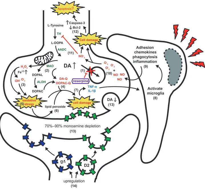
Figure 1. Neurochemical and molecular events after reserpine treat-ment. (1) Reserpine precludes dopamine (DA) storage. (2) Increased DA is metabolized in the cytoplasm (3) generating reactive oxygen species (ROS) and (4) highly reactive quinones (DA-Q and DOPAC-Q) (5) result-ing in oxidative stress and (6) lipid peroxidation.(7) Accumulation of ROS and reactive quinones leads to cell damage and proinflammatory signalization. (8) Activation of microglia by tumor necrosis factor (TNF)-α
and interleukin (IL)-1β(9) amplify proinflammatory signalization resulting
other study reported increased oral dyskinesia in female mice that
was inconsistent at different time points (174). Contradicting
results regarding oral dyskinesia might be explained by differences
in protocol—that is length of treatment, dosage, and type of motor
parameter (vacuous chewing vs. jaw twitching). Nevertheless,
studies with CD-1 mice have suggested that female animals
present a more efficient VMAT2 function (57, 58), which could
explain the need of a longer treatment for female mice to develop
the motor alterations (data displayed in Figure 2). Importantly, this
result is in accordance with the lower incidence of PD in women
(19, 62) and adds to the similarities between the reserpine model
and the clinical condition.
The exposed prospect of reserpine-induced behavioral,
pharma-cological, and neurochemical effects restates the use of reserpine as
a valuable and promising model for PD study. Thus, the current
underuse of reserpine to investigate PD features should be
recon-sidered. Of notice, the use of reserpine could be important to the
relevance of VMAT2 functionality to PD in humans. Indeed,
polymorphisms in promoter regions that increases transcription of
VMAT2 are protective against PD (31, 82) and reduction in VMAT2
and its mRNA in nigrostriatal neurons have been reported in PD
patients (89, 131). Furthermore, VMAT2 is present in Lewy’s
bodies in the SN of PD patients (209) and VTA dopaminergic
neurons that are spared in PD harbors higher levels of VMAT2
(131). Finally, increased cytoplasmic DA influences the
confor-mational state of
α-synuclein, promoting stabilization of its
patho-genic form (75, 116). Thus, because functional VMAT2 expression
is protective against dopaminergic neurodegeneration, its
long-term blockage might represent an interesting approach to model PD.
In conclusion, we believe that the scientific effort on reserpine
PD model validation should focus in answering whether
neurodegeneration and cell death occur after chronic reserpine
treatment, as well as the exploitation of the model to investigate
progression of symptoms and neurochemical features of PD
patho-physiology. We recently presented a low-dose reserpine-induced
progressive model of PD that could be useful to investigate such
inquiry (71, 167). Therefore, in view of the presented experimental
evidence, the reserpine-induced PD model in rodents reaches
robust face and pharmacological validity criteria, besides
present-ing a significant number of neurochemical and molecular features
that closely resemble the pathophysiology of the disease. Taken
together, these characteristics render the reserpine model a useful
tool for PD basic research.
REFERENCES
1. Aarsland D, Andersen K, Larsen JP, Lolk A (2003) Prevalence and characteristics of dementia in Parkinson disease: an 8-year prospective study.Arch Neurol60:387–392.
2. Abílio V, Vera J, Ferreira L, Duarte C, Carvalho R, Grassl Cet al (2002) Effects of melatonin on orofacial movements in rats. Psychopharmacology (Berl)161:340–347.
3. Abílio VC, Araujo CCS, Bergamo M, Calvente PRV, D’Almeida V, Ribeiro RA, Frussa-Filho R (2003) Vitamin E attenuates reserpine-induced oral dyskinesia and striatal oxidized glutathione/reduced glutathione ratio (GSSG/GSH) enhancement in rats.Prog Neuropsychopharmacol Biol Psychiatry
27:109–114.
4. Abílio VC, Silva RH, Carvalho RC, Grassl C, Calzavara MB, Registro Set al(2004) Important role of striatal catalase in
aging- and reserpine-induced oral dyskinesia.Neuropharmacology 47:263–272.
5. Al-Bloushi S, Safer A-M, Afzal M, Mousa SA (2009) Green tea modulates reserpine toxicity in animal models.J Toxicol Sci 34:77–87.
6. Algeri S, Achilli G, Calderini G, Perego C, Ponzio F, Toffano G (1987) Age-related changes in metabolic responses to chronic monoamine depletion in central dopaminergic and serotonergic systems of rats treated with reserpine.Neurobiol Aging8:61–66. 7. Anang JBM, Gagnon J-F, Bertrand J-A, Romenets SR, Latreille V,
Panisset Met al(2014) Predictors of dementia in Parkinson disease: a prospective cohort study.Neurology83:1253–1260. 8. Ara J, Przedborski S, Naini AB, Jackson-Lewis V, Trifiletti RR,
Horwitz J, Ischiropoulos H (1998) Inactivation of tyrosine hydroxylase by nitration following exposure to peroxynitrite and 1-methyl-4-phenyl-1,2,3,6-tetrahydropyridine (MPTP).Proc Natl Acad Sci U S A95:7659–7663.
9. Arif IA, Khan HA (2010) Environmental toxins and Parkinson’s disease: putative roles of impaired electron transport chain and oxidative stress.Toxicol Ind Health26:121–128.
10. Arora V, Chopra K (2013) Possible involvement of
oxido-nitrosative stress induced neuro-inflammatory cascade and monoaminergic pathway: underpinning the correlation between nociceptive and depressive behaviour in a rodent model.J Affect Disord151:1041–1052.
11. Arora V, Kuhad A, Tiwari V, Chopra K (2011) Curcumin ameliorates reserpine-induced pain-depression dyad: behavioural, biochemical, neurochemical and molecular evidences.
Psychoneuroendocrinology36:1570–1581.
12. Asanuma M, Miyazaki I, Ogawa N (2003) Dopamine- or L-DOPA-induced neurotoxicity: the role of dopamine quinone formation and tyrosinase in a model of Parkinson’s disease. Neurotox Res5:165–176.
13. Atack JR, Shook BC, Rassnick S, Jackson PF, Rhodes K, Drinkenburg WHet al(2014) JNJ-40255293, a novel adenosine A2A/A1 antagonist with efficacy in preclinical models of Parkinson’s disease.ACS Chem Neurosci5:1005–1019. 14. Austin PJ, Betts MJ, Broadstock M, O’Neill MJ, Mitchell SN,
Duty S (2010) Symptomatic and neuroprotective effects following activation of nigral group III metabotropic glutamate receptors in rodent models of Parkinson’s disease.Br J Pharmacol
160:1741–1753.
15. Barone P (2011) Treatment of depressive symptoms in Parkinson’s disease.Eur J Neurol18:11–15.
16. Bashkatova V, Alam M, Vanin A, Schmidt WJ (2004) Chronic administration of rotenone increases levels of nitric oxide and lipid peroxidation products in rat brain.Exp Neurol186:235–241. 17. Baskin P, Salamone J (1993) Vacuous jaw movements in rats
induced by acute reserpine administration: interactions with different doses of apomorphine.Pharmacol Biochem Behav 46:793–797.
18. Beal MF (2001) Experimental models of Parkinson’s disease.Nat Rev Neurosci2:325–332.
19. Benito-León J, Bermejo-Pareja F, Morales-González JM, Porta-Etessam J, Trincado R, Vega S, Louis ED (2004) Incidence of Parkinson disease and parkinsonism in three elderly populations of central Spain (NEDICES).Neurology62:734–741.
20. Benito-León J, Louis ED, Posada IJ, Sánchez-Ferro Á, Trincado R, Villarejo Aet al(2011) Population-based case-control study of cognitive function in early Parkinson’s disease (NEDICES).J Neurol Sci310:176–182.
21. Bergamo M, Abílio VC, Queiroz CMT, Barbosa-Júnior HN, Abdanur LRA, Frussa-Filho R (1997) Effects of age on a new animal model of tardive dyskinesia.Neurobiol Aging18:623–629.
22. Betarbet R, Sherer TB, Mackenzie G, Garcia-Osuna M, Panov AV, Greenamyre JT (2000) Chronic systemic pesticide exposure reproduces features of Parkinson’s disease.Nat Neurosci 3:1301–1306.
23. Lo Bianco C, Ridet J-L, Schneider BL, Déglon N, Aebischer P (2002) Alpha-synucleinopathy and selective dopaminergic neuron loss in a rat lentiviral-based model of Parkinson’s disease.Proc Natl Acad Sci U S A99:10813–10818.
24. Bilska A, Dubiel M, Sokołowska-Jezewicz M, Lorenc-Kocib E, Włodek L (2007) Alpha-lipoic acid differently affects the reserpine-induced oxidative stress in the striatum and prefrontal cortex of rat brain.Neuroscience146:1758–1771.
25. Bisong SA, Brown R, Osim EE (2010) Comparative effects of Rauwolfia vomitoria and chlorpromazine on locomotor behaviour and anxiety in mice.J Ethnopharmacol132:334–339.
26. Blesa J, Phani S, Jackson-Lewis V, Przedborski S (2012) Classic and new animal models of Parkinson’s disease.J Biomed Biotechnol2012:1–10.
27. Bonito-Oliva A, Masini D, Fisone G (2014) A mouse model of non-motor symptoms in Parkinson’s disease: focus on pharmacological interventions targeting affective dysfunctions. Front Behav Neurosci8:290.
28. Braak H, Del K, Rüb U, de Vos RAI, Jansen ENH, Braak E (2003) Staging of brain pathology related to sporadic Parkinson’s disease. Neurobiol Aging24:197–211.
29. Breese GR, Traylor TD (1971) Depletion of brain noradrenaline and dopamine by 6-hydroxydopamine.Br J Pharmacol42:88–99. 30. Brefel-Courbon C, Ory-Magne F, Thalamas C, Payoux P, Rascol O
(2013) Nociceptive brain activation in patients with neuropathic pain related to Parkinson’s disease.Parkinsonism Relat Disord 19:548–552.
31. Brighina L, Riva C, Bertola F, Saracchi E, Fermi S, Goldwurm S, Ferrarese C (2013) Analysis of vesicular monoamine transporter 2 polymorphisms in Parkinson’s disease.Neurobiol Aging 34:1712.e9–1712.e13.
32. Broadstock M, Austin PJ, Betts MJ, Duty S (2012)
Antiparkinsonian potential of targeting group III metabotropic glutamate receptor subtypes in the rodent substantia nigra pars reticulata.Br J Pharmacol165(4b):1034–1045.
33. Brundin P, Li J-Y, Holton JL, Lindvall O, Revesz T (2008) Research in motion: the enigma of Parkinson’s disease pathology spread.Nat Rev Neurosci9:741–745.
34. Brundin P, Barker RA, Conn PJ, Dawson TM, Kieburtz K, Lees AJet al(2013) Linked clinical trials—the development of new clinical learning studies in Parkinson’s disease using screening of multiple prospective new treatments.J Parkinsons Dis3:231–239. 35. Burger M, Fachinetto R, Calegari L, Paixão MW, Braga AL,
Rocha JBT (2004) Effects of age on reserpine-induced orofacial dyskinesia and possible protection of diphenyl diselenide.Brain Res Bull64:339–345.
36. Burger ME, Alves A, Callegari L, Athayde FR, Nogueira CW, Zeni G, Rocha JBT (2003) Ebselen attenuates reserpine-induced orofacial dyskinesia and oxidative stress in rat striatum.Prog Neuropsychopharmacol Biol Psychiatry27:135–140. 37. Cannon JR, Tapias VM, Na HM, Honick AS, Drolet RE,
Greenamyre JT (2009) A highly reproducible rotenone model of Parkinson’s disease.Neurobiol Dis34:279–290.
38. Carlsson A, Lindqvist M, Magnusson T (1957)
3,4-dihydroxyphenylalanine and 5-hydroxytryptophan as reserpine antagonists.Nature180:1200.
40. Carvalho RC, Patti CC, Takatsu-Coleman AL, Kameda SR, Souza CF, Garcez-do-Carmo Let al(2006) Effects of reserpine on the plus-maze discriminative avoidance task: dissociation between memory and motor impairments.Brain Res1122:179–183. 41. Caudle WM, Richardson JR, Wang MZ, Taylor TN, Guillot TS,
McCormack ALet al(2007) Reduced vesicular storage of dopamine causes progressive nigrostriatal neurodegeneration. J Neurosci27:8138–8148.
42. Caudle WM, Guillot TS, Lazo CR, Miller GW (2012) Industrial toxicants and Parkinson’s disease.Neurotoxicology33:178–188. 43. Cenci MA, Whishaw IQ, Schallert T (2002) Animal models of
neurological deficits: how relevant is the rat?Nat Rev Neurosci 3:574–579.
44. Chesselet M-F, Richter F (2011) Modelling of Parkinson’s disease in mice.Lancet Neurol10:1108–1118.
45. Chiba K, Trevor A, Castagnoli N (1984) Metabolism of the neurotoxic tertiary amine, MPTP, by brain monoamine oxidase. Biochem Biophys Res Commun120:574–578.
46. Chipkin RE, McQuade RD, Iorio LC (1987) D1 and D2 dopamine binding site up-regulation and apomorphine-induced stereotypy. Pharmacol Biochem Behav28:477–482.
47. Chuang RS, Gitler AD (2013) Parallel PARKing: Parkinson’s genes function in common pathway.Neuron77:377–379. 48. Clairembault T, Leclair-Visonneau L, Neunlist M, Derkinderen P
(2014) Enteric glial cells: New players in Parkinson’s disease?Mov Disorddoi: 10.1002/mds.25979
49. Coelho M, Marti MJ, Sampaio C, Ferreira JJ, Valldeoriola F, Rosa MM, Tolosa E (2015) Dementia and severity of parkinsonism determines the handicap of patients in late-stage Parkinson’s disease: the Barcelona-Lisbon cohort.Eur J Neurol 22(2):305–312.
50. Colebrooke RE, Humby T, Lynch PJ, McGowan DP, Xia J, Emson PC (2006) Age-related decline in striatal dopamine content and motor performance occurs in the absence of nigral cell loss in a genetic mouse model of Parkinson’s disease.Eur J Neurosci 24:2622–2630.
51. Colpaert FC (1987) Pharmacological characteristics of tremor, rigidity and hypokinesia induced by reserpine in rat. Neuropharmacology26:1431–1440.
52. Da Cunha C, Wietzikoski EC, Dombrowski P, Bortolanza M, Santos LM, Boschen SL, Miyoshi E (2009) Learning processing in the basal ganglia: a mosaic of broken mirrors.Behav Brain Res 199:157–170.
53. Danysz W, Gossel M, Zajaczkowski W (1994) Are NMDA antagonistic properties relevant for antiparkinsonian-like activity in rats? Case of amantadine and memantine.J Neural Transm 7:155–166.
54. Degkwitz R, Frowein R, Kulenkampff C, Mohs U (1960) On the effects of L-dopa in man and their modification by reserpine, chlorpromazine, iproniazid and vitamin B6.Klin Wochenschr 38:120–123.
55. Deumens R, Blokland A, Prickaerts J (2002) Modeling Parkinson’s disease in rats: an evaluation of 6-OHDA lesions of the
nigrostriatal pathway.Exp Neurol175:303–317.
56. Didonet JJ, Cavalcante JC, Souza LDS, Costa MSMO, André E, Soares-Rachetti VDPet al(2014) Neuropeptide S counteracts 6-OHDA-induced motor deficits in mice.Behav Brain Res 266:29–36.
57. Dluzen DE, McDermott JL (2008) Sex differences in dopamine-and vesicular monoamine-transporter functions: Implications for methamphetamine use and neurotoxicity.Ann N Y Acad Sci 1139:140–150.
58. Dluzen DE, Bhatt S, McDermott JL (2008) Differences in reserpine-induced striatal dopamine output and content between
female and male mice: implications for sex differences in vesicular monoamine transporter 2 function.Neuroscience154:1488–1496. 59. Dodson MW, Guo M (2007) Pink1, Parkin, DJ-1 and
mitochondrial dysfunction in Parkinson’s disease.Curr Opin Neurobiol17:331–337.
60. Driver-Dunckley E, Adler CH, Hentz JG, Dugger BN, Shill HA, Caviness JNet al(2014) Olfactory dysfunction in incidental Lewy body disease and Parkinson’s disease.Parkinsonism Relat Disord 20:1260–1262.
61. Duty S, Jenner P (2011) Animal models of Parkinson’s disease: a source of novel treatments and clues to the cause of the disease.Br J Pharmacol164:1357–1391.
62. Eeden SKVD, Tanner CM, Bernstein AL, Fross RD, Leimpeter A, Bloch DA, Nelson LM (2003) Incidence of Parkinson’s disease: variation by age, gender, and race/ethnicity.Am J Epidemiol 157:1015–1022.
63. Elbaz A, Bower JH, Maraganore DM, McDonnell SK, Peterson BJ, Ahlskog JEet al(2002) Risk tables for parkinsonism and Parkinson’s disease.J Clin Epidemiol55:25–31.
64. Elgh E, Domellöf M, Linder J, Edström M, Stenlund H, Forsgren L (2009) Cognitive function in early Parkinson’s disease: a population-based study.Eur J Neurol16:1278–1284. 65. El-Ghazaly MA, Sadik NAH, Rashed ER, Abd-El-Fattah AA
(2013) Neuroprotective effect of EGb761(R) and low-dose whole-bodyγ-irradiationin a rat model of Parkinson’s disease. Toxicol Ind Health21:1–17.
66. Faria RR, Abílio VC, Grassl C, Chinen CC, Negrão LTR, de Castro JPMVet al(2005) Beneficial effects of vitamin C and vitamin E on reserpine-induced oral dyskinesia in rats: critical role of striatal catalase activity.Neuropharmacology48:993–1001. 67. Fekete MI, Szentendrei T, Herman JP, Kanyicska B (1980) Effects
of reserpine and antidepressants on dopamine and DOPAC (3,4-dihydroxyphenylacetic acid) concentrations in the striatum, olfactory tubercle and median eminence of rats.Eur J Pharmacol 64:231–238.
68. Ferger B, Buck K, Shimasaki M, Koros E, Voehringer P, Buerger E (2010) Continuous dopaminergic stimulation by pramipexole is effective to treat early morning akinesia in animal models of Parkinson’s disease: a pharmacokinetic-pharmacodynamic study usingin vivomicrodialysis in rats.Synapse64:533–541. 69. Fernagut PO, Diguet E, Labattu B, Tison F (2002) A simple
method to measure stride length as an index of nigrostriatal dysfunction in mice.J Neurosci Methods113:123–130. 70. Fernandes VS, Ribeiro AM, Melo TG, Godinho M, Barbosa FF,
Medeiros DSet al(2008) Memory impairment induced by low doses of reserpine in rats: possible relationship with emotional processing deficits in Parkinson disease.Prog
Neuropsychopharmacol Biol Psychiatry32:1479–1483. 71. Fernandes VS, Santos JR, Leão AHFF, Medeiros AM, Melo TG,
Izídio GSet al(2012) Repeated treatment with a low dose of reserpine as a progressive model of Parkinson’s disease.Behav Brain Res231:154–163.
72. Fil A, Cano-de-la-Cuerda R, Muñoz-Hellín E, Vela L, Ramiro-González M, Fernández-de-Las-Peñas C (2013) Pain in Parkinson disease: a review of the literature.Parkinsonism Relat Disord19:285–294.
73. Finberg JPM, Youdim MBH (2002) Pharmacological properties of the anti-Parkinson drug rasagiline; modification of endogenous brain amines, reserpine reversal, serotonergic and dopaminergic behaviours.Neuropharmacology43:1110–1118.
75. Follmer C, Roma L, Einsiedler CM, Lara A, Moncores M, Weissmu Get al(2007) Dopamine affects the stability, hydration, and packing of protofibrils and fibrils of the wild type and variants ofα-synuclein.Biochemistry46:472–482.
76. Freis ED (1954) Mental depression in hypertensive patients treated for long periods with large doses of reserpine.N Engl J Med 251:1006–1008.
77. Fukuzaki K, Kamenosono T, Nagata R (2000) Effects of ropinirole on various parkinsonian models in mice, rats, and cynomolgus monkeys.Pharmacol Biochem Behav 65:503–508.
78. Galte D, Terzioglu M (2008) Parkinson’s disease: genetic vs. toxin-induced rodent models.FEBS J275:1384–1391.
79. Gao HM, Hong JS (2011) Gene-environment interactions: key to unraveling the mystery of Parkinson’s disease.Prog Neurobiol 94:1–19.
80. Ghosh B, Antonio T, Reith M, Dutta A (2010)
(4-(2-((5-Hydroxy-1, 2, 3, 4-tetrahydronaphthalen-2-yl)(propyl) amino) ethyl) piperazin-1-yl) quinolin-8-ol and its analogues as highly potent dopamine D2/D3 agonists.J Med Chem 53:2114–2125.
81. Giráldez-Pérez RM, Antolín-Vallespín M, Muñoz MD, Sánchez-Capelo A (2014) Models ofα-synuclein aggregation in Parkinson’s disease.Acta Neuropathol Commun2:176. 82. Glatt CE, Wahner AD, White DJ, Ruiz-Linares A, Ritz B (2006)
Gain-of-function haplotypes in the vesicular monoamine transporter promoter are protective for Parkinson disease in women.Hum Mol Genet15:299–305.
83. Gómez-Esteban JC, Tijero B, Somme J, Ciordia R, Berganzo K, Rouco Iet al(2011) Impact of psychiatric symptoms and sleep disorders on the quality of life of patients with Parkinson’s disease. J Neurol258:494–499.
84. Goldstein DS, Sullivan P, Cooney A, Jinsmaa Y, Sullivan R, Gross DJet al(2012) Vesicular uptake blockade generates the toxic dopamine metabolite 3,4-dihydroxyphenylacetaldehyde in PC12 cells: relevance to the pathogenesis of Parkinson’s disease.J Neurochem123:932–943.
85. Goldstein J, Barnett A, Malick J (1975) The evaluation of anti-parkinson drugs on reserpine-induced rigidity in rats.Eur J Pharmacol33:183–188.
86. Gołembiowska K, Dziubina A (2012) The effect of adenosine A(2A) receptor antagonists on hydroxyl radical, dopamine, and glutamate in the striatum of rats with altered function of VMAT2. Neurotox Res22:150–157.
87. Guttman M (1992) Dopamine receptors in Parkinson’s disease. Neurol Clin10:377–386.
88. Hald A, Lotharius J (2005) Oxidative stress and inflammation in Parkinson’s disease: is there a causal link?Exp Neurol 193:279–290.
89. Harrington KA, Augood SJ, Kingsbury AE, Foster OJF, Emson PC (1996) Dopamine transporter (DAT) and synaptic vesicle amine transporter (VMAT2) gene expression in the substantia nigra of control and Parkinson’s disease.Brain Res Mol Brain Res 36:157–162.
90. Heeringa MJ, Abercrombie ED (1995) Biochemistry of somatodendritic dopamine release in substantia nigra: anin vivo comparison with striatal dopamine release.J Neurochem 65:192–200.
91. Henchcliffe C, Severt WL (2011) Disease modification in Parkinson’s disease.Drugs Aging28:605–615.
92. Henderson JM, Stanic D, Tomas D, Patch J, Horne MK, Bourke D, Finkelstein DI (2005) Postural changes after lesions of the substantia nigra pars reticulata in hemiparkinsonian monkeys. Behav Brain Res160:267–276.
93. Höglinger GU, Féger J, Prigent A, Michel PP, Parain K, Champy Pet al(2003) Chronic systemic complex I inhibition induces a hypokinetic multisystem degeneration in rats.J Neurochem 84:491–502.
94. Hsieh Y, Mounsey RB, Teismann P (2011) MPP+-induced toxicity in the presence of dopamine is mediated by COX-2 through oxidative stress.Naunyn Schmiedebergs Arch Pharmacol 384:157–167.
95. Hurley MJ, Jenner P (2006) What has been learnt from study of dopamine receptors in Parkinson’s disease?Pharmacol Ther 111:715–728.
96. Ischiropoulos H (2003) Biological selectivity and functional aspects of protein tyrosine nitration.Biochem Biophys Res Commun305:776–783.
97. Ishihara L, Brayne C (2006) A systematic review of depression and mental illness preceding Parkinson’s disease.Acta Neurol Scand 113:211–220.
98. Johnels B (1982) Locomotor hypokinesia in the reserpine-treated rat: drug effects from the corpus striatum and nucleus accumbens. Pharmacol Biochem Behav17:283–289.
99. Johnson A, Loew D, Vigouret J (1976) Stimulant properties of bromocriptine on central dopamine receptors in comparison to apomorphine, (+)-amphetamine and L-dopa.Br J Pharmacol 56:59–68.
100. Jurna I, Grossmann W, Nell T (1973) Depression by amantadine of tremor induced by reserpine and oxotremorine in the rat.Naunyn Schmiedebergs Arch Pharmacol152:141–152.
101. Kalaitzakis ME, Gentleman SM, Pearce RKB (2013) Disturbed sleep in Parkinson’s disease: anatomical and pathological correlates.Neuropathol Appl Neurobiol39:644–653.
102. Kane JM, Smith JM (1982) Tardive dyskinesia; prevalence and risk factors, 1959 to 1979.Arch Gen Psychiatry39:473–481.
103. Kaur S, Starr M (1996) Motor effects of lamotrigine in naive and dopamine-depleted mice.Eur J Pharmacol304:1–6.
104. Kim SW, Ko HS, Dawson VL, Dawson TM (2005) Recent advances in our understanding of Parkinson’s disease.Drug Discov Today Dis Mech2:427–433.
105. Kirik D, Annett LE, Burger C, Muzyczka N, Mandel RJ, Bjo A (2003) Nigrostriatal alpha-synucleinopathy induced by viral vector-mediated overexpression of human alpha-synuclein: a new primate model of Parkinson’s disease.Proc Natl Acad Sci U S A 100:2884–2889.
106. Kitada T, Asakawa S, Hattori N, Matsumine H, Yamamura Y, Minoshima Set al(1998) Mutations in the parkin gene cause autosomal recessive juvenile parkinsonism.Nature392:605–608. 107. Kitada T, Pisani A, Porter DR, Yamaguchi H, Tscherter A,
Martella Get al(2007) Impaired dopamine release and synaptic plasticity in the striatum of PINK1-deficient mice.Proc Natl Acad Sci U S A104:11441–11446.
108. Klockgether T (2004) Parkinson’s disease: clinical aspects.Cell Tissue Res318:115–120.
109. Korecka JA, Eggers R, Swaab DF, Bossers K, Verhaagen J (2013) Modeling early Parkinson’s disease pathology with chronic low dose MPTP treatment.Restor Neurol Neurosci31:155–167. 110. Kuhn DM, Aretha CW, Geddes TJ (1999) Peroxynitrite
inactivation of tyrosine hydroxylase: mediation by sulfhydryl oxidation, not tyrosine nitration.J Neurosci19:10289–10294. 111. Kuhn DM, Sakowski SA, Sadidi M, Geddes TJ (2004)
Nitrotyrosine as a marker for peroxynitrite-induced neurotoxicity: the beginning or the end of the end of dopamine neurons?J Neurochem89:529–536.
113. Lane E, Dunnett S (2008) Animal models of Parkinson’s disease and L-dopa induced dyskinesia: how close are we to the clinic? Psychopharmacology (Berl)199:303–312.
114. Lee FJS, Liu F (2008) Genetic factors involved in the pathogenesis of Parkinson’s disease.Brain Res Rev58:354–364.
115. Lees AJ, Smith E (1983) Cognitive deficits in the early stages of Parkinson’s disease.Brain106:257–270.
116. Li HT, Lin DH, Luo XY, Zhang F, Ji LN, Du HNet al(2005) Inhibition of alpha-synuclein fibrillization by dopamine analogs via reaction with the amino groups of alpha-synuclein. Implication for dopaminergic neurodegeneration.FEBS J272:3661–3672. 117. Li X, Patel JC, Wang J, Avshalumov M V, Nicholson C, Buxbaum
JDet al(2010) Enhanced striatal dopamine transmission and motor performance with LRRK2 overexpression in mice is eliminated by familial Parkinson’s disease mutation G2019S.J Neurosci 30:1788–1797.
118. Lin X, Parisiadou L, Gu X-L, Wang L, Shim H, Sun Let al(2009) Leucine-rich repeat kinase 2 regulates the progression of
neuropathology induced by Parkinson’s-disease-related mutant alpha-synuclein.Neuron64:807–827.
119. Liu S, Zhao R, Li X, Guo H, Tian Z (2014) Attenuation of reserpine-induced pain/depression dyad by gentiopicroside through downregulation of GluN2B receptors in the amygdala of mice. Neuromolecular Med16:350–359.
120. Lotharius J, Brundin P (2002) Pathogenesis of Parkinson’s disease: dopamine, vesicles and alpha-synuclein.Nat Rev Neurosci 3:932–942.
121. Maj J, Rog Z, Skuza G, Sowifiska H, Superata J (1990) Behavioural and neurochemical effects of Ro 40–7592, a new COMT inhibitor with a potential therapeutic activity in Parkinson’s disease.J Neural Transm2:101–112.
122. Maj J, Rogóz Z, Skuza G, Kołodziejczyk K (1997) The behavioural effects of pramipexole, a novel dopamine receptor agonist.Eur J Pharmacol324:31–37.
123. Mata IF, Leverenz JB, Weintraub D, Trojanowski JQ, Hurtig HI, Van Deerlin VMet al(2014) APOE, MAPT, and SNCA genes and cognitive performance in Parkinson disease.JAMA Neurol 71:1405–1412.
124. McKenna O, Arnold G, Holtzman E (1976) Microperoxisome distribution in the central nervous system of the rat.Brain Res 117:181–194.
125. McQueen EG, Doyle AE, Smirk FH (1954) Mechanism of hypotensive action of reserpine, an alkaloid of Rauwolfia serpentina.Nature174:1015.
126. Mehndiratta M, Garg RK, Pandey S (2011) Nonmotor symptom complex of Parkinson’s disease—an under-recognized entity.J Assoc Physicians India59:302–308.
127. Meiser J, Weindl D, Hiller K (2013) Complexity of dopamine metabolism.Cell Commun Signal11:34.
128. Meredith GE, Halliday GM, Totterdell S (2004) A critical review of the development and importance of proteinaceous aggregates in animal models of Parkinson’s disease: new insights into Lewy body formation.Parkinsonism Relat Disord10:191–202. 129. Meredith GE, Sonsalla PK, Chesselet MF (2008) Animal models
of Parkinson’s disease progression.Acta Neuropathol 115:385–398.
130. Millan MJ, Agid Y, Brüne M, Bullmore ET, Carter CS, Clayton NSet al(2012) Cognitive dysfunction in psychiatric disorders: characteristics, causes and the quest for improved therapy.Nat Rev Drug Discov11:141–168.
131. Miller GW, Erickson JD, Perez JT, Penland SN, Mash DC, Rye DB, Levey AI (1999) Immunochemical analysis of vesicular monoamine transporter (VMAT2) protein in Parkinson’s disease. Exp Neurol148:138–148.
132. Missale C, Nisoli E, Liberini P, Rizzonelli P, Memo M, Buonamici Met al(1989) Repeated reserpine administration up-regulates the transduction mechanisms of D1 receptors without changing the density of [3H]SCH 23390 binding.Brain Res483:117–122. 133. Miyagi M, Arai N, Taya F, Itoh F, Komatsu Y, Kojima M, Isaji M
(1996) Effect of cabergoline, a long-acting dopamine D2 agonist, on reserpine-treated rodents.Biol Pharm Bull11:1499–1502. 134. Monderer R, Thorpy M (2009) Sleep disorders and daytime
sleepiness in Parkinson’s disease.Curr Neurol Neurosci Rep 9:173–180.
135. Mooslehner KA, Chan PM, Xu W, Liu L, Smadja C, Humby T et al(2001) Mice with very low expression of the vesicular monoamine transporter 2 gene survive into adulthood: potential mouse model for parkinsonism.Mol Cell Biol21:5321–5331. 136. Morin N, Jourdain VA, Di Paolo T (2014) Modeling dyskinesia in
animal models of Parkinson disease.Exp Neurol256:105–116. 137. Mulcahy P, O’Doherty A, Paucard A, O’Brien T, Kirik D, Dowd E
(2013) The behavioural and neuropathological impact of intranigral AAV-α-synuclein is exacerbated by systemic infusion of the Parkinson’s disease-associated pesticide, rotenone, in rats.Behav Brain Res243:6–15.
138. Nade VS, Shendye NV, Kawale LA, Patil NR, Khatri ML (2013) Protective effect of nebivolol on reserpine-induced neurobehavioral and biochemical alterations in rats.Neurochem Int63:316–321. 139. Naidu PS, Singh A, Kulkarni SK (2006) Effect of Withania
somnifera root extract on reserpine-induced orofacial dyskinesia and cognitive dysfunction.Phytother Res20:140–146. 140. Neisewander JL, Lucki I, McGonigle P (1991) Neurochemical
changes associated with the persistence of spontaneous oral dyskinesia in rats following chronic reserpine treatment.Brain Res 558:27–35.
141. Neisewander JL, Castañeda E, Davis DA (1994) Dose-dependent differences in the development of reserpine-induced oral dyskinesia in rats: support for a model of tardive dyskinesia. Psychopharmacology (Berl)116:79–84.
142. Niswender C, Johnson K, Weaver C, Jones C, Xiang Z, Luo Q et al(2008) Discovery, characterization, and antiparkinsonian effect of novel positive allosteric modulators of metabotropic glutamate receptor 4.Mol Pharmacol74:1345–1358. 143. Noyce AJ, Bestwick JP, Silveira-Moriyama L, Hawkes CH,
Giovannoni G, Lees AJ, Schrag A (2012) Meta-analysis of early nonmotor features and risk factors for Parkinson disease.Ann Neurol72:893–901.
144. Oe T, Tsukamoto M, Nagakura Y (2010) Reserpine causes biphasic nociceptive sensitivity alteration in conjunction with brain biogenic amine tones in rats.Neuroscience169:1860–1871. 145. Olsson M, Nikkhah G (1995) Forelimb akinesia in the rat
Parkinson model: differential effects of dopamine agonists and nigral transplants as assessed by a new stepping test.J Neurosci 15:3863–3875.
146. Omiatek DM, Bressler AJ, Cans A-S, Andrews AM, Heien ML, Ewing AG (2013) The real catecholamine content of secretory vesicles in the CNS revealed by electrochemical cytometry.Sci Rep3:1447.
147. Patil R, Kasture S (2012) Protective effect ofRubia cordifoliaon reserpine-induced orofacial dyskinesia.Nat Prod Res
26:2159–2161.
148. Patil R, Dhawale K, Gound H, Gadakh R (2012) Protective effect of leaves ofMurraya koenigiion reserpine-induced orofacial dyskinesia.Iran J Pharm Res11:635–641.