Contents lists available atScienceDirect
Journal of Ethnopharmacology
journal homepage:www.elsevier.com/locate/jep
Acute and sub chronic toxicity study of aqueous extract from the leaves and
branches of Campomanesia velutina (Cambess) O. Berg
Marcela Carolina de Paula Michel Araújo
a, Neila Márcia Silva Barcellos
b, Paula Melo de
Abreu Vieira
c, Thiago Magalhães Gouveia
a, Martha Oliveira Guerra
d, Vera Maria Peters
d, Dênia
Antunes Saúde-Guimarães
a,⁎aLaboratory of Medicinal Plants, School of Pharmacy, Federal University of Ouro Preto,, Minas Gerais state, Brazil
bLaboratory of Experimental Pharmacology, School of Pharmacy, Federal University of Ouro Preto, Minas Gerais state, Brazil
cLaboratory of Morfopathology, Department of Biological Sciences, Federal University of Ouro Preto, Minas Gerais state, Brazil
dCentro de Biologia da Reprodução. Universidade Federal de Juiz de Fora, Minas Gerais state, Brazil
A R T I C L E I N F O
Keywords:
Campomanesia velutina in vivoacute toxicity
in vivosub chronic toxicity
A B S T R A C T
Ethnopharmacological relevance: Campomanesia velutinaleaves and branches infusions are used in Brazilian
folk medicine to treat diarrhea and to ameliorate intestinal cramps, respectively.
Aim of the study:Carry out the acute and sub chronic pre-clinical evaluation and thus assess the safety and
toxicological potential of the specie.
Materials and methods: In vivotoxicity was evaluated by acute and sub chronic toxicity assays conducted
according to the guidelines of the Brazilian Agency of National Health Surveillance (Agência Nacional de Vigilância Sanitária–ANVISA). For acute toxicity evaluation, a single dose of aqueous extracts from the leaves (AEL) and branches (AEB) ofCampomanesia velutinawere orally administered to mice at doses of 300, 600 and 1200 mg/kg. Then, the animals were observed for 14 days. In the sub chronic study, the extracts were orally administered to mice for 14 days at doses of 300, 600 and 1200 mg/kg. To assess the toxicological effects, animals were closely observed on general behavior, clinical signs of toxicity, body weight, food and water intake. At the end of the experiment, it was performed biochemical and hematological evaluations, as well as histopathological analysis from the following organs: brain, heart, lungs, liver, stomach, small intestine (section) and left kidney. Preliminary phytochemical analysis was performed using thin layer chromatography (TLC) and colorimetric pharmacognostic tests.
Results:In oral acute assay, treatment with AEB at the major dose (1200 mg/kg) caused diarrhea, abdominal
cramps and tremors in females. These effects were reversed at 4th hour. Normochromic normocytic anemia was observed in males treated with AEL 300 mg/kg and AEB 600 and 1200 mg/kg as well as in females treated with AEB 300 and 1200 mg/kg. The kidney of all treated animals showed moderate inflammation and a few hemorrhagic points. In sub chronic assay, treatment with AEL 600 mg/kg, AEL 1200 mg/kg and AEB 1200 mg/kg caused hyper excitability in females that was not reversed. Treatments also had impact on weight gain and the relative weight of males’ brain was increased on group treated with AEL 300 mg/kg, AEB 300 and AEB 1200 mg/kg. Although changes in hematological parameters were not observed, serum creatinine levels were significantly higher in males treated with AEB 300 mg/kg. Besides, the heart of all treated animals showed intense hyperemia. Preliminary phytochemical analysis revealed the presence offlavonoids, tannins and phenolic compounds.
Conclusions:Toxicity signs were mainly observed after treatment with AEL and AEB at the two highest tested
doses (600 and 1200 mg/kg), suggesting that the extracts are relatively safe at its effective dose (300 mg/kg). However, alterations on hematological and biochemical parameters and on the kidney and heart of the animals were not closely related with the dose, implying caution on its use.
http://dx.doi.org/10.1016/j.jep.2017.02.043
Received 25 October 2016; Accepted 25 February 2017
⁎Corresponding author.
E-mail address:[email protected](D.A. Saúde-Guimarães).
Abbreviations:AEB, Aqueous extracts from the branches; AEL, Aqueous extracts from the leaves; ALT, Alanine aminotransferase; ANVISA, Agência Nacional de Vigilância Sanitária; AST, Aspartate aminotransferase; EDTA, Ethylenediaminetetraacetic acid; HCT, Hematocrit; HGB, Hemoglobin concentration; i.p, Intraperitoneal; MCH, Mean corpuscular hemoglobin; MCHC, Hemoglobin concentration; MCV, Mean corpuscular volume; NHI, National Institute of Health; PLT, platelet count; RBC, Red blood cell count; RDW, Red cell volume distribution; TLC, Thin layer chromatography; UFOP, Universidade Federal de Ouro Preto; WBC, White blood cell count; WHO, World Health Organization
Available online 28 February 2017
0378-8741/ © 2017 Elsevier B.V. All rights reserved.
1. Introduction
The use of medicinal plants has increased and gained popularity in recent years. The World Health Organization (WHO) estimates that 80% of Africa and Asia population use traditional medicine for primary health care. The scenario is similar in developed countries, where 70– 80% of the population use some form of complementary or alternative medicine (WHO, 2008). With the global explosion of phythotherapy, the safety of medicinal plants has become a public health problem (Neergheen-Bhujun, 2013). Medicinal species do not receive adequate attention in global discussions related to health and the potential toxicity of the majority is not well stablished or simply unknown (Tilburt and Kaptchuk, 2008). Extremely toxic substances like strych-nine, the digitoxines, cyanogenic glycosides, among others, are ex-tracted from plants. Therefore, we can only assure that the use of particular specie is secure after a careful investigation (Lapa et al., 2004).
Campomanesia velutina (Cambess) O. Berg is one of the thirty species from the Campomanesia genus (Myrtaceae). It is endemic specie in Brazil and occurs at three different Brazilian biomas: cerrado, caatinga and atlantic forest (Landrum, 1986). The specie has arboreal size and can reach 8 m high. It is exclusive from the interior of gallery forests. Theflowering occurs from August to October and fruiting from October to November. Its fruits arefleshy, 12–15 mm wide, have an orange color and are very appreciated for human consumption “in natura”or as homemade pastries, ice cream, liqueurs and soft drinks (Carrara, 1997; Arantes and Monteiro, 2002).Campomanesiaspecies differentiation is difficult due to the similarity between them. Therefore, in Brazil, Campomanesia species, including Campomanesia velutina, are popularly known as“gabiroba”, “ gua-vira”,“cambuci”, among others.
In folk medicine, ethnopharmacological studies identified the use of the leaves infusion to treat diarrhea. In addition, branches infusion is used to ameliorate intestinal cramps (Oliveira et al., 2010). The specie was also quoted as a medicinal plant in two surveys conducted in different regions of Brazil, one at Sertão do Ribeirão, Santa Catarina state (Giraldi and Hanazaki, 2010) and another at cerrado areas (Dias and Laureano, 2009). Reported pharmacological activities of Campomanesia velutinademonstrated the anti-inflammatory, antino-ciceptive, anti-hyperuricemic and XO inhibitory acitivity of its extracts and fractions (Michel et al., 2013; Araújo et al., 2016).
The evidences describing the biological effects ofCampomanesia velutinaare increasing. However, despite the traditional use and the reported biological effects of Campomanesia velutina, there are no reports regarding its safety or toxicity. Therefore, this study aimed to carry out the acute and sub chronic pre-clinical evaluation of the specie considering its use in folk medicine and potential use in phytotherapy.
2. Materials and methods
2.1. Plant material
Leaves from Campomanesia velutina (Cambess.) O. Berg were collected in Lagoa Santa city, Minas Gerais state, Brazil, in December of 2012, with permission of Chico Mendes Institute of Biodiversity Conservation (Instituto Chico Mendes de Conservação da Biodiversidade –ICMBio/ Sistema de Autorização e Informação em Biodiversidade–SISBIO), license number 17021-5. The plant botani-cal identification was realized by Dr. Marcos E. Guerra Sobral, Department of Natural Sciences, Federal University of São João Del-Rei (Universidade Federal de São João Del-Del-Rei - UFSJ), Minas Gerais state, Brazil. A voucher specimen (HUFSJ 4637) was deposited at the herbarium of UFSJ.
2.2. Preparation of extracts
The leaves and branches were air-dried and ground. In order to obtain the aqueous extracts, 450.0 g of leaves powder and 900.0 g of branches powder were extracted, separately, with 4.5 L and 9.0 L of water, respectively. The water was removed by lyophilization, yielding 19.0 g of the aqueous extract of leaves (AEL) and 21.0 g of the aqueous extract of branches (AEB). The material was solubilized in distilled water at different doses immediately before administration.
2.3. Preliminary phytochemical analysis
The presence of alkaloids, flavonoids, anthocyanines, tannins, saponins, triterpenes, steroids, coumarins, quinones, anthraquinones and cardiac glycosides in the aqueous extracts from leaves and branches of Campomanesia velutina was assayed using thin layer chromatography (TLC) and colorimetric pharmacognostic tests (Wagner et al., 1984; Matos, 1997).
2.4. Animals
The animal experiments were conducted in accordance with the Guide for the Care and Use of Laboratory Animals of the National Institute of Health (NIH). Male (30 ± 5 g) and female (25 ± 5 g) albino Swiss mice were supplied by Animal Science Center of Federal University of Ouro Preto (Universidade Federal de Ouro Preto – UFOP). Animals were divided into experimental groups, housed in plastic cages and maintained on a 12-h light/12-h dark cycle. They were given standard chow and water ad libitum. The Ethical Committee on Animal Experimentation of UFOP (no. 2012/69) approved all experimental procedures.
2.5. Preparation of extract samples
Campomanesia velutinaextracts AEL and AEB were solubilized in distilled water in order to obtain solutions of 30, 60 and 120 mg/ml. The doses evaluated in this experiment were 300, 600 and 1200 mg/kg. Thus, for every gram of animal weight, was administered 10 uL of the solutions prepared above. Dose of 300 mg/kg is a therapeutic dose found in a previous study (Michel et al., 2013). The remaining doses were a progression of this dose.
2.6. Toxicity assays
To evaluate the pre-clinical toxicity ofCampomanesia velutina, the aqueous extract of the leaves (AEL) and the aqueous extract of the branches (AEB) had its safety parameters assessed by conducting the acute and sub chronic toxicity study. The studies were realized in accordance with the guidelines established by National Health Surveillance Agency (Agência Nacional de Vigilância Sanitária – ANVISA) in the Resolution number 90 (Brasil, 2004) and in the Guide for conducting non-clinical safety studies required for drug development (Brasil, 2010).
2.6.1. Acute toxicity assay
(100/20 mg/kg i.p.). After the anesthesia has reached depth, cardiac puncture was performed to collect blood for biochemical and hemato-logical evaluations. Then, the animals were euthanized by cervical dislocation and the following organs were removed for histopathologi-cal analysis: brain, heart, lungs, liver, stomach, small intestine (section) and left kidney.
2.6.2. Sub chronic toxicity assay
The animals were divided into seven experimental groups of 20 animals each (10 males and 10 females). Group 1 received 10 µL/g of distilled water and served as control. Groups 2, 3 and 4 were treated with AEL at the doses of 300, 600 and 1200 mg/kg, respectively. Groups 5, 6 and 7 received AEB at the doses of 300, 600 and 1200 mg/ kg, respectively. All treatments were administered once a day for 14 days by oral gavage. During the treatment period, animals were observed on general behavior, clinical signs of toxicity, mortality, food and water intake. Body weights were measured before and after administration on days 4, 7, 10 and 14. At the end of the experiment (day 15), animals were anesthetized with ketamine/xylasin (100/ 20 mg/kg i.p.). After the anesthesia has reached depth, cardiac puncture was performed to collect blood for biochemical and hemato-logical evaluations. Then, the animals were euthanized by cervical dislocation and the following organs were removed for histopathologi-cal analysis: brain, heart, lungs, liver, stomach, small intestine (section) and left kidney.
2.7. Hematological analysis
The hematological evaluation was performed in all surviving animals at the end of the experiment. A volume of 100 µL of blood was transferred to tubes containing 20 µL of EDTA. The complete blood count was performed using an automated hematology analyzer VET 2800 (Mindray–Brazil). Hematological evaluations included red blood cell count (RBC), hemoglobin concentration (HGB), hematocrit (HCT), mean corpuscular volume (MCV), mean corpuscular hemoglo-bin (MCH), hemoglohemoglo-bin concentration (MCHC), red cell volume distribution (RDW), platelet count (PLT) and white blood cell count (WBC).
2.8. Serum biochemistry analysis
The biochemical evaluation was performed in all surviving animals at the end of the experiment. The remaining collected blood was transferred to tubes without anticoagulant and allowed to stand for 45 min at room temperature before being centrifuged at 3500 rpm for 10 min. The serum from each sample was recovered and stored in cryogenic tubes at −80 °C freezer until assayed. Creatinine, urea, aspartate aminotransferase (AST), alanine aminotransferase (ALT) and total proteins were evaluated using the LabMax Progress device (Labtest–Brazil) and kits from Labtest.
2.9. Histopathology
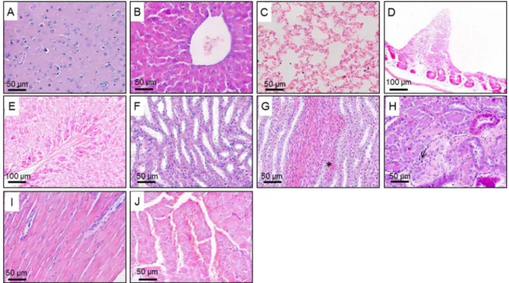
For histological analysis, the organs were collected from all surviv-ing animals, washed with saline solution 0.9% (w/v), weighed andfixed in formaldehyde-calcium solution. The organs were cleaved and processed for paraffin embedding. Sections (5 µm thick) were prepared and stained with hematoxylin and eosin (HE). The tissues were analyzed in optical microscope for their general structure, degenerative changes, necrosis evidence and signs of inflammation. The images were obtained with the microscope Leica DM5000B, scanned through micro camera Leica DFC340 FX using the Leica Application Suite software (version 2.4.0 R1).
2.10. Statistical analysis
The relative weight of organs, hematological and biochemical data were expressed as mean ± standard error of the mean (SEM). Data were submitted to analysis of variance (One-Way ANOVA) followed by Newman-Keuls multiple comparison test.
The animal's weight was transformed in percentage of weight relative to the initial weight. The results were expressed as mean ± SEM. Data were submitted to analysis of variance (two-way ANOVA) followed by Bonferroni test.
Data from individual average daily food and water intake were expressed as mean ± SEM. Because the data did not follow a normal distribution, it was used the Kruskal-Wallis test followed by Dunn's post-test.
The software GraphPad Prism 5.0 (GraphPad Software, USA) was used for statistical analysis. P values < 0.05 were considered statisti-cally significant.
3. Results
3.1. Preliminary phytochemical screening
The preliminary phytochemical analysis of AEL and AEB revealed the presence of phenolic compounds includingflavonoids and tannins.
3.2. Acute toxicity
3.2.1. General signs and mortality
No signs of toxicity or behavioral changes were observed after the treatment with AEL. Only AEB at the major dose (1200 mg/kg) caused diarrhea, abdominal cramps and tremors. Each sign was observed in one female. However, these signs were transient and disappeared mostly in the fourth hour after the administration of the extracts.
No deaths were recorded within 72 h after administration of the extracts and the control in animals of both sexes. After this period, one male died on 4th day after the treatment with AEL 300 mg/kg and one female died on 10th day after the treatment with AEB 300 mg/kg. There are no differences between the survival curves of these treat-ments and the control group.
3.2.2. Body weight, relative organ weight, food and water intake Male and female mice treated with AEL and AEB at the three evaluated doses showed weight gain throughout the entire experimen-tal period. The statistical analysis revealed that time, not the treat-ments, was responsible for the increasing on animals’ weight. The increase was the same in treated and control groups (Fig. 1). The treatments did not affect relative organ weights, food and water intake (Data not shown).
3.2.3. Hematological parameters
Treatment with AEL at the three doses did not produce any changes on female hematological parameters. However, on males treated with AEL 300 mg/kg was observed a reduction on RBC, HGB, HCT and WBC. Moreover, males treated with AEL 600 and 1200 mg/kg showed a reduction on RBC and HCT. Treatment with AEB produced altera-tions on male and females hematological parameters. Females treated with AEB 300 mg/kg showed reduction on HGB. In addition, AEB 1200 mg/kg produced reduction on HGB and PLT. On males, AEB 600 and 1200 mg/kg reduced HGB (Table 1).
3.2.4. Biochemical parameters
3.2.5. Histopathological analysis
The acute administration of AEL and AEB did not produce significant dose-dependent histopathological alterations. At the three evaluated doses, it were not observed tissue changes on brain, heart, lungs, liver, stomach and small intestine of male and female mice. However, the kidney of all treated animals showed moderate infl am-mation and a few hemorrhagic points. The histological findings are shown inFig. 2.
3.3. Sub chronic toxicity
3.3.1. General signs and mortality
No signs of toxicity or behavioral changes were observed on males during the treatment with AEL and AEB at the three doses. However, on female mice treated with AEL 600 and 1200 mg/kg was observed hyper excitability in 3rd and 6th day, respectively, which persisted throughout the treatment. Nevertheless, this effect was only seen in one animal of each group. The same effect was observed after
administra-Table 1
Hematological parameters of Swiss mice treated with a single dose (300, 600 or 1200 mg/kg) of aqueous extracts fromCampomanesia velutinaleaves (AEL) and branches (AEB) and observed for 14 days.
Gender Parameter Treatment
Control AEL AEL AEL AEB AEB AEB
(Water) 300 mg/kg 600 mg/kg 1200 mg/kg 300 mg/kg 600 mg/kg 1200 mg/kg
Male RBC (106/µL) 7.48 ± 0.23 3.72 ± 0.95*** 6.17 ± 0.14** 5.66 ± 0.31*** 8.17 ± 0.25 7.25 ± 0.08 7.15 ± 0.29 HGB (g/dL) 14.00 ± 0.93 7.70 ± 1.85*** 13.05 ± 0.23 11.33 ± 0.67 11.74 ± 1.25 10.48 ± 0.41* 10.58 ± 0.49*
HCT (%) 41.68 ± 1.24 18.20 ± 4.25*** 34.90 ± 0.53* 31.52 ± 1.75*** 45.45 ± 1.39 39.53 ± 0.92 38.57 ± 1.58
VCM (fL) 54.70 ± 1.31 54.40 ± 0.89 56.65 ± 0.79 55.70 ± 0.36 55.20 ± 0.65 54.50 ± 0.82 53.95 ± 0.78
HCM (pg) 18.79 ± 1.18 20.40 ± 1.04 21.17 ± 0.21 20.03 ± 0.33 17.83 ± 2.47 17.92 ± 3.03 16.97 ± 2.19
CHCM (g/dL) 31.70 ± 1.86 31.76 ± 5.54 33.47 ± 3.91 32.21 ± 3.72 29.68 ± 2.19 29.86 ± 2.56 29.23 ± 1.86
PLT (103/µL) 824.3 ± 75.2 563.5 ± 252.0 609.0 ± 102.7 495.2 ± 109.6 666.2 ± 171.9 814.5 ± 49.2 656.0 ± 60.3 WBC (103/µL) 5.85 ± 0.84 1.67 ± 0.27* 5.33 ± 0.77 4.05 ± 0.56 6.60 ± 1.11 4.48 ± 0.64 4.47 ± 0.85 Female RBC (106/µL) 7.02 ± 0.55 6.23 ± 0.24 6.69 ± 0.23 7.37 ± 0.41 6.37 ± 0.25 7.15 ± 0.47 6.46 ± 0.65 HGB (g/dL) 12.58 ± 0.49 12.02 ± 0.38 13.63 ± 0.27 13.20 ± 1.07 9.77 ± 0.36* 10.95 ± 0.52 9.55 ± 0.79*
HCT (%) 38.04 ± 3.49 34.52 ± 1.19 36.33 ± 1.07 39.85 ± 1.95 35.75 ± 1.25 39.55 ± 2.36 36.73 ± 3.27
VCM (fL) 53.92 ± 0.89 55.46 ± 0.69 54.47 ± 1.17 54.18 ± 0.72 56.18 ± 0.52 55.48 ± 0.52 54.78 ± 0.80
HCM (pg) 16.30 ± 2.30 19.34 ± 0.44 20.45 ± 0.41 17.85 ± 1.00 15.38 ± 0.22 15.43 ± 0.38 14.90 ± 0.49
CHCM (g/dL) 34.08 ± 2.98 34.90 ± 0.87 37.58 ± 0.46 32.93 ± 1.87 29.88 ± 2.54 29.46 ± 1.84 29.46 ± 2.65
PLT (103/µL) 693.0 ± 185.3 515.8 ± 104.4 612.2 ± 110.5 525.2 ± 23.8 531.8 ± 146.1 583.5 ± 47.5 283.3 ± 98.1* WBC (103/µL) 6.82 ± 1.53 4.82 ± 0.49 5.45 ± 0.46 4.35 ± 0.50 5.45 ± 0.47 5.42 ± 0.43 5.47 ± 0.58
n =6 males and 6 females. One-way ANOVA followed by Newman-Keuls multiple comparison test. *p < 0,5; **p < 0,05; ***p < 0,01
Red blood cell count (RBC), hemoglobin concentration (HGB), hematocrit (HCT), mean corpuscular volume (MCV), mean corpuscular hemoglobin (MCH), hemoglobin concentration (MCHC), red cell volume distribution (RDW), platelet count (PLT) and white blood cell count (WBC).
AEL - Males
0 4 7 10 14
90 100 110 120 130 140 150
160 Control (Vehicle)
300 mg/kg 600 mg/kg 1200 mg/kg
Days
R
e
la
tiv
e
w
e
ig
h
t (
%
)
AEL - Females
0 4 7 10 14
90 100 110 120 130 140 150
160 Control (Vehicle)
300 mg/kg 600 mg/kg 1200 mg/kg
Days
R
e
la
tiv
e
w
e
ig
h
t (
%
)
AEB - Males
0 4 7 10 14
90 100 110 120 130 140 150
160 Control (Vehicle)
300 mg/kg 600 mg/kg 1200 mg/kg
Days
R
e
la
tiv
e
w
e
ig
h
t (
%
)
AEB - Females
0 4 7 10 14
90 100 110 120 130 140 150
160 Controle (Veículo)
EAG 300 mg/kg EAG 600 mg/kg EAG 1200 mg/kg
Days
R
e
la
ti
v
e
w
e
ig
h
t (%
)
tion of AEB 1200 mg/kg. In this group, one female was hyperactive from 5th day of treatment through the end of the experiment.
Only one male treated with AEB 600 mg/kg died during the experiment. The statistical analysis showed no differences between the survival curves of this treatment and the control group.
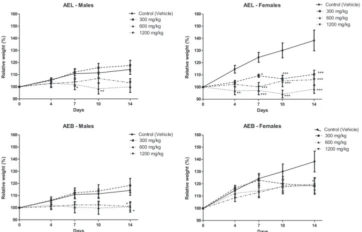
3.3.2. Body weight, relative organ weight, food and water intake Treatment with AEL and AEB for 14 days caused significant alterations on animals´ weight gain. The impact of the treatments over animals’weight can be seen atFig. 3. At the end of the experiment, males treated with AEL 600 mg/kg, AEB 600 and AEB 1200 mg/kg showed relative weight significantly lower than the control group. The effect of AEL over the female weight was very pronounced. This extract
at the three doses caused weight reduction at almost all evaluated times. Females treated with AEB 300 mg/kg also showed relative weight significantly lower than the control group on the 14th day.
None of the treatments affected organ weights. However, the relative weight of the males’brain was increased on groups treated with AEL and AEB 300 mg/kg and AEB 1200 mg/kg (Fig. 4).
The treatments did not affect food and water intake (Data not shown).
3.3.3. Hematological parameters
Treatment for 14 days with AEL and AEB at the three doses did not produce any changes on hematological parameters of males and females Swiss mice (Table 3).
Table 2
Biochemical parameters of Swiss mice treated with a single dose (300, 600 or 1200 mg/kg) of aqueous extracts fromCampomanesia velutinaleaves (AEL) and branches (AEB) and observed for 14 days.
Gender Treatment Parameter
ALT (U/L) AST (U/L) Total Protein (g/dL) Creatinine (mg/dL) Ureia (mg/dL)
Male Vehicle (Water) 58.5 ± 7.6 150.5 ± 18.7 5.19 ± 0.34 0.232 ± 0.044 71.75 ± 7.71
AEL 300 mg/kg 112.8 ± 36.2 237.6 ± 57.0 5.14 ± 0.14 0.177 ± 0.048 62.00 ± 7.83
AEL 600 mg/kg 92.8 ± 33.8 197.8 ± 54.9 5.13 ± 0.29 0.215 ± 0.018 66.00 ± 2.52
AEL 1200 mg/kg 78.0 ± 18.2 218.2 ± 63.8 5.14 ± 0.12 0.218 ± 0.020 66.00 ± 2.45
AEB 300 mg/kg 99.0 ± 38.4 196.5 ± 49.9 5.28 ± 0.22 0.273 ± 0.047 54.20 ± 4.41
AEB 600 mg/kg 51.3 ± 10.0 122.2 ± 8.2 5.70 ± 0.19 0.240 ± 0.035 65.20 ± 1.11
AEB 1200 mg/kg 85.4 ± 19.5 118.5 ± 21.5 5.25 ± 0.27 0.168 ± 0.017 65.33 ± 5.06
Female Vehicle (Water) 63.6 ± 7.1 151.2 ± 16.5 5.04 ± 0.13 0.266 ± 0.047 60.80 ± 8.31
AEL 300 mg/kg 45.2 ± 9.4 107.6 ± 15.6 5.59 ± 0.26 0.248 ± 0.023 53.83 ± 6.18
AEL 600 mg/kg 71.4 ± 4.4 158.0 ± 11.6 5.24 ± 0.58 0.295 ± 0.007 51.67 ± 3.38
AEL 1200 mg/kg 64.2 ± 10.9 153.8 ± 17.0 5.25 ± 0.33 0.232 ± 0.023 58.00 ± 2.35
AEB 300 mg/kg 52.6 ± 4.6 99.4 ± 7.9 5.38 ± 0.36 0.254 ± 0.017 60.40 ± 2.52
AEB 600 mg/kg 56.2 ± 1.2 128.0 ± 5.7 5.38 ± 0.37 0.238 ± 0.035 59.40 ± 4.35
AEB 1200 mg/kg 58.8 ± 13.0 118.5 ± 21.5 5.62 ± 0.26 0.160 ± 0.021 50.80 ± 9.69
n =6 males and 6 females. One-way ANOVA followed Newman-Keuls multiple comparison test.
3.3.4. Biochemical parameters
The only change was found in males treated with the AEB 300 mg/ kg for 14 days. In these animals, serum creatinine levels were significantly higher compared to the control group treated only with water (Table 4).
3.3.5. Histopathological analysis
The sub chronic administration of AEL and AEB did not produce significant dose-dependent histopathological alterations. At the three evaluated doses, it was not observed tissue changes on brain, lungs, liver, kidney, stomach and small intestine of male and female mice. However, the heart of all treated animals showed intense hyperemia. The histologicalfindings are shown inFig. 2.
4. Discussion
The use of plants as medicines, functional food and nutritional supplements have increased worldwide (Kohler and Baghdadi-Sabeti, 2011). The infusion ofCampomanesia velutinaleaves and branches is used in traditional medicine (Dias e Laureano, 2009; Giraldi and Hanazaki, 2010;Oliveira et al., 2010) and previous studies attested the anti-inflammatory, antinociceptive, anti-hyperuricemic and XO inhibi-tory activity of its extracts (Michel et al., 2013; Araújo et al., 2016). Despite the popular use and the biological effects, there are no studies or data about its safety. In addition, the extracts contain phenolic compounds, like flavonoids and tanines. These metabolites have important biological activities but they can also be related to toxic effects. It is known that phenolic compounds can be hematotoxic and hepatotoxic, and may provoke mutagenesis and carcinogenesis (Michalowicz and Duda, 2007). Therefore, the evaluation of Campomanesia velutinatoxicity is indispensable.
Animal models are widely used to assess the preliminary toxicity because the early identification of undesirable effects is usually predictive of the toxicity in humans and can save time, resources and efforts (Kramer et al., 2010). In this study, several parameters were evaluated after the in vivoacute and sub chronic administration of aqueous extracts fromCampomanesia velutinaleaves and branches.
Mortality is an important criterion on toxicological evaluation (Asare et al., 2012) and both acute and sub chronic administration of the extracts did not induce a significant mortality. No deaths were recorded within 72 h after acute administration of the extracts and at the sub chronic toxicity evaluation, only one animal treated with AEB 600 mg/kg died. However, the results showed no dose-response relationship and mortality was not clearly related with extracts AEL - Males
90 100 110 120 130 140 150
160 Control (Vehicle)
300 mg/kg 600 mg/kg 1200 mg/kg
*
**
0 4 7 10 14
Days R e la tiv e w e ig h t ( % )
AEL - Females
90 100 110 120 130 140 150 160 Control (Vehicle) 300 mg/kg 600 mg/kg 1200 mg/kg * *** *** ** *** *** *** *** *** ***
0 4 7 10 14
Days R e la tiv e w e ig h t ( % )
AEB - Males
90 100 110 120 130 140 150
160 Control (Vehicle)
300 mg/kg 600 mg/kg 1200 mg/kg
* *
0 4 7 10 14
Days R e la tiv e w e ig h t ( % )
AEB - Females
90 100 110 120 130 140 150 160 Control (Vehicle) 300 mg/kg 600 mg/kg 1200 mg/kg *
0 4 7 10 14
Days R e la tiv e w e ig h t ( % )
Fig. 3.Relative body weight of male and female Swiss mice treated for 14 days with different doses (300, 600 or 1200 mg/kg) of aqueous extract fromCampomanesia velutinaleaves (AEL) and branches (AEB). Two-way ANOVA followed by Bonferroni test. * p < 0.05, ** p < 0.1 and *** p < 0.01 when compared to control group.
Brain - males
0.0 0.5 1.0 1.5 2.0 Vehicle AEL 300 mg/kg AEL 600 mg/kg AEL 1200 mg/kg AEB 300 mg/kg AEB 600 mg/kg AEB 1200 mg/kg
**
**
***
B rai n r el at ive w e ight ( % )Fig. 4.Relative weight of brain from male Swiss mice treated with aqueous extract from
administration since deaths occurred only with lower doses, indicating that they might have another cause like an improperly gavage.
Clinical signs of toxicity were observed after the acute administra-tion and during the sub chronic evaluaadministra-tion, but only with higher doses. Besides, the number of affected animals was not significant. In acute toxicity, signs of toxicity were observed only in the group treated with AEB 1200 mg/kg and occurred within thefirst hour after administra-tion of the extract, indicating that it is rapidly absorbed from the gastrointestinal tract, and disappeared 4 h after treatment, indicating its transitory nature. In sub chronic toxicity, the toxicity sign observed were hyper excitability in females treated with AEL and AEB, and once expressed, lasted throughout the treatment.
The increase or decrease in body weight can indicate significant physiological changes such as hormonal variations, liver disorders and decreased absorption of proteins, amino acids and other nutrients (Lee et al., 2012). Furthermore, the reduction in body weight can affect the internal organs weight (TEO et al., 2002) and is a simple and sensitive parameter of toxicity after exposure to toxic agents (Thanabhorn et al., 2006). It is known that improperly performed gavage may cause damage to trachea and esophagus negatively affecting food
consump-tion and consequently weight gain. However, both acute and sub chronic treatment did not significantly change the consumption of food and water, indicating that the observed effects on animals' weight and internal organs were the result of physiological changes and not a consequence of a lower food consumption. Acute treatment did not affect weight gain or the relative organ weight. However, sub chronic treatment reduced the weight gain of males and females, indicating that prolonged intake of these extracts produced significant alterations in the metabolism of the animals. Moreover, the sub chronic treatment with AEL and AEB at some doses increased significantly the relative organ weight of the brain when compared to control group. The increase in relative organ weight could be caused by the induction of xenobiotic enzymes leading to increased proteins synthesis (Diallo et al., 2014). However, this change does not seem to be toxicological relevant, since no histopathological alterations were observed in this organ. Thus, the increase of the relative organ weight of the brain can be due to the variability of the weight of this organ.
The hematopoietic system is one of the most sensitive targets for toxic compounds and an important index of physiological and patho-logical state, both in man and in animal (Mukinda and Syce, 2007). In Table 3
Hematological parameters of Swiss mice treated for 14 days with different doses (300, 600 or 1200 mg/kg) of aqueous extracts fromCampomanesia velutinaleaves (AEL) and branches (AEB).
Gender Parameter Treatment
Control (water) AEL AEL AEL AEB AEB AEB
300 mg/kg 600 mg/kg 1200 mg/kg 300 mg/kg 600 mg/kg 1200 mg/kg
Male RBC (106/µL) 6.28 ± 0.18 5.95 ± 0.23 5.94 ± 0.16 6.01 ± 0.19 5.46 ± 0.15 6.67 ± 0.59 5.40 ± 0.25 HGB (g/dL) 12.98 ± 0.30 12.25 ± 0.37 12.16 ± 0.32 12.77 ± 0.37 11.19 ± 0.46 14.29 ± 1.25 11.29 ± 0.47
HCT (%) 35.94 ± 0.89 33.43 ± 1.04 33.51 ± 0.66 35.81 ± 1.06 32.61 ± 1.02 39.30 ± 3.23 30.48 ± 1.64
VCM (fL) 57.40 ± 0.95 56.30 ± 0.59 56.55 ± 0.65 59.67 ± 0.49 59.79 ± 0.93 59.14 ± 0.61 56.37 ± 0.54
HCM (pg) 20.74 ± 0.38 20.64 ± 0.29 20.50 ± 0.31 21.30 ± 0.29 20.48 ± 0.51 21.46 ± 0.12 20.95 ± 0.21
CHCM (g/dL) 36.13 ± 0.21 36.67 ± 0.17 32.26 ± 0.34 35.68 ± 0.37 34.80 ± 0.49 36.29 ± 0.47 37.20 ± 0.47
PLT (103/µL) 751.0 ± 32.9 594.8 ± 20.7 579.7 ± 29.9 839.0 ± 37.9 715.2 ± 47.2 813.4 ± 102.4 725.7 ± 30.4 WBC (103/µL) 5.05 ± 0.65 3.96 ± 0.48 3.71 ± 0.27 5.20 ± 1.47 4.54 ± 0.57 5.41 ± 0.59 4.52 ± 0.55 Female RBC (106/µL) 5.64 ± 0.27 5.83 ± 0.23 5.50 ± 0.26 5.77 ± 0.26 5.77 ± 0.19 5.73 ± 0,73 4.75 ± 0.16 HGB (g/dL) 11.97 ± 0.53 11.75 ± 0.37 12.18 ± 0.64 12.65 ± 0.60 12.26 ± 0.41 11.01 ± 0,63 10.11 ± 0.37
HCT (%) 33.38 ± 1.32 32.54 ± 1.07 31.28 ± 1.55 34.56 ± 1.57 34.72 ± 1.00 29.24 ± 1,77 28.34 ± 0.51
VCM (fL) 59.53 ± 1.34 56.44 ± 0.34 56.82 ± 0.51 59.96 ± 0.78 60.23 ± 0.64 57.21 ± 0,84 57.86 ± 0.76
HCM (pg) 21.32 ± 0.45 20.22 ± 0.19 22.13 ± 0.39 21.91 ± 0.20 21.25 ± 0.21 21.61 ± 0,31 21.27 ± 0.28
CHCM (g/dL) 35.84 ± 0.52 36.12 ± 0.19 37.94 ± 0.61 36.63 ± 0.51 35.28 ± 0.35 37.33 ± 0,24 36.77 ± 0.30
PLT (103/µL) 661.1 ± 27.7 553.8 ± 23.4 826.3 ± 39.2 723.0 ± 51.6 625.1 ± 40.7 839.8 ± 54,9 675.9 ± 53.7 WBC (103/µL) 3.38 ± 0.51 4.41 ± 0.49 4.65 ± 0.41 4.67 ± 0.68 4.74 ± 0.80 4.90 ± 0,76 3.36 ± 0.34
n =10 males and 10 females. One-way ANOVA followed by Newman-Keuls multiple comparison test.
Red blood cell count (RBC), hemoglobin concentration (HGB), hematocrit (HCT), mean corpuscular volume (MCV), mean corpuscular hemoglobin (MCH), hemoglobin concentration (MCHC), red cell volume distribution (RDW), platelet count (PLT) and white blood cell count (WBC).
Table 4
Biochemical parameters of Swiss mice treated for 14 days with different doses (300, 600 or 1200 mg/kg) of aqueous extracts fromCampomanesia velutinaleaves (AEL) and branches (AEB).
Gender Treatment Parameter
ALT (U/L) AST (U/L) Total Proteins (g/dL) Creatinine (mg/dL) Ureia (mg/dL)
Male Control (Water) 97.4 ± 11.9 129.5 ± 7.9 6.67 ± 0.23 0.136 ± 0.018 48.1 ± 3.0
AEL 300 mg/kg 94.9 ± 12.1 184.1 ± 29.8 6.44 ± 0.13 0.166 ± 0.027 47.0 ± 3.4
AEL 600 mg/kg 97.3 ± 19.7 204.3 ± 42.8 6.84 ± 0.14 0.200 ± 0.041 49.6 ± 2.5
AEL 1200 mg/kg 80.8 ± 9.0 166.7 ± 12.2 6.49 ± 0.12 0.193 ± 0.025 52.1 ± 3.2
AEB 300 mg/kg 85.1 ± 10.8 170.0 ± 21.4 6.89 ± 0.27 0.344 ± 0.096** 48.6 ± 3.3
AEB 600 mg/kg 98.9 ± 11.3 199.1 ± 28.2 6.83 ± 0.19 0.108 ± 0.012 38.33 ± 2.4
AEB 1200 mg/kg 74.3 ± 6.6 141.4 ± 12.7 6.82 ± 0.22 0.119 ± 0.008 39.4 ± 2.3
Female Control (Water) 96.7 ± 11.7 161.2 ± 20.9 6.27 ± 0.12 0.152 ± 0.010 54.7 ± 3.1
AEL 300 mg/kg 96.1 ± 17.5 167.1 ± 26.9 6.25 ± 0.18 0.212 ± 0.023 56.3 ± 4.3
AEL 600 mg/kg 69.3 ± 6.8 120.1 ± 24.7 7.08 ± 0.19 0.186 ± 0.031 51.5 ± 5.3
AEL 1200 mg/kg 87.0 ± 10.9 180.4 ± 26.5 6.61 ± 0.12 0.264 ± 0.057 52.8 ± 4.3
AEB 300 mg/kg 110.3 ± 26.9 226.0 ± 85.3 6.35 ± 0.20 0.122 ± 0.007 50.8 ± 3.7
AEB 600 mg/kg 105.7 ± 17.1 202.9 ± 37.5 6.24 ± 0.14 0.123 ± 0.009 44.9 ± 2.9
this study, hematological parameters showed a large variation between the different extracts, dose and gender. The differences between males and females can be due to hormones produce, especially the estrogen and its effect on B and T lymphocytes cells equilibrium and cytochrome P450 enzymes metabolism (Ahmed et al., 1999; Sin et al., 2007). Furthermore, the observed changes did not show a clear dose-response relationship and some has no clinical significance when occurring isolated. In acute toxicity, the main change was the decrease in hemoglobin levels without changes in MCV, MCH and MCHC indices, featuring a normochromic normocytic anemia. This clinical condition was observed in males treated with AEL 300 mg/kg, AEB 600 and 1200 mg/kg as well as in females treated with AEB 300 and 1200 mg/ kg. Besides anemia, AEL 300 mg/kg caused leukopenia in males. Females treated with AEB 1200 mg/kg also showed reduction on platelet levels. Therefore, the occurrence of these effects following acute ingestion of aqueous extracts may be related to the toxicity caused by the extracts. However, in sub chronic toxicity, AEL and AEB had no effect on hematopoietic system. This result was surprising, since it was expected an increase in the occurrence or even an exacerbation of anemia, leukopenia and thrombocytopenia observed after acute treatment. Thus, the absence of toxic effects on hematopoietic system after a 14-day treatment with AEL and AEB indicates that the observed effects on hematological parameters after acute administration are not strictly related with the administration of the extracts. Indeed, anemia, leukopenia and thrombocytopenia are related to several clinical con-ditions, such as infections, autoimmune diseases and cancer.
It is known that several toxic compounds accumulate in liver where occurs the detoxification (Clarke and Clarke, 1977). Liver damage are usually assessed by the determination of serum transaminases (ALT and AST) and also by the measurement of total proteins. It was not observed any significant alterations in serum levels of these three markers of liver function after acute and sub chronic administration of Campomanesia velutinaextracts and histological analysis of the liver did not show tissue changes, confirming that the administration of the extracts did not cause liver damage.
The kidneys receive about 25% of the cardiac bloodflow and any substance that reaches the systemic circulation will reach this organ. Therefore, they are considered frequent targets of toxicity (Dekant and Vamvakas, 1996). Renal function was evaluated by serum levels of urea and creatinine and by histological analysis. The kidney of animals treated acutely with AEL and AEB showed histological alterations such as discrete inflammation and hemorrhage. However, the histological changes did not affect serum levels of renal function markers, what could be explained by the fact that histopathologyfindings were only focal, occurring in small and distinct regions of the kidney. An increase in serum creatinine levels was observed only in males treated for 14 days with the AEB 300 mg/kg. Creatinine is increased in the serum when the cortex and/or the glomeruli are damaged (Burtis and Bruns, 2015) suggesting a chronic kidney disease (Ravel, 1997). However, the histological evaluation did not reveal alterations in this organ of any treated groups of sub-chronic toxicity.
In addition to the tissue alterations found in the kidneys, the heart of animals treated for 14 days with AEB and AEL showed marked hyperemia, which is an increase in blood inside the vessels. The hyperemia may be due to a vasodilation caused by the extracts, since they containflavonoids, which is a class that previously demonstrated vasodilation effects (Pérez-Vizcaíno et al., 2002).
In conclusion, although the effective dose of 300 mg/kg have not caused the appearance of signs of toxicity or caused significant mortality, it was not possible to establish a clear dose-response relation regarding the histopathologicalfindings and the effects on hematolo-gical and biochemical parameters.
In this context, Campomanesia velutina aqueous extracts from leaves and branches demonstrated a toxic potential in acute and sub chronic administration, especially regarding the hematopoietic system. In addition, kidney and heart showed toxicity signs. Since the specie is
already used in Brazilian folk medicine to treat diarrhea and to ameliorate intestinal cramps and also demonstrated promising effects over inflammatory diseases, like gout, these results provided valuable and primary data on the toxicity profile ofCampomanesia velutinaand will serve as a guide to future studies with this medicinal plant. However, further studies are necessary to fully determine the toxic profile of this plant and should include genotoxicity, mutagenicity, and carcinogenicity evaluations. Furthermore, aspects of the effects of this plant on hematopoiesis, heart and kidney function need to be studied over longer periods.
Conflict of interest
The authors declare no conflict of interests.
Acknowledgments
The authors would like to thank the "Fundação de Amparo à Pesquisa do Estado de Minas Gerais'' (FAPEMIG), grant number APQ-01318-12, APQ-00956-13 and APQ-01160-15, “Rede Mineira de Ensaios Toxicológicos e Farmacológicos da FAPEMIG (Rede TOXIFAR - FAPEMIG), grant number CBB – RED-00008–14, Universidade Federal de Ouro Preto (UFOP) and ”Coordenação de Aperfeiçoamento de Pessoal de Nível Superior”(CAPES) forfinancial support. We also would like to thank the “Laboratório Piloto de Análises Clinicas”(LAPAC/UFOP) for the aid in hematological analysis and the“Centro de Biologia da Reprodução da Universidade Federal de Juiz de Fora (CBR/UFJF) for the aid in biochemistry analysis.
References
Ahmed, S.A., Hissong, B.D., Verthelyi, D., Donner, K., Becker, K., Karpuzoglu-Sahin, E., 1999. Gender and risk of autoimmune diseases: possible role of estrogenic compounds. Environ. Health Persp. 107, 681–686.
Arantes, A.A., Monteiro, R., 2002. A família Myrtaceae na estação ecológica do Panga, Uberlândia, Minas Gerais, Brasil. Lundiana, 3, pp. 111–127.
Araújo, M.C.P.M., Ferraz-Filha, Z.S., Ferrari, F.C., Saúde-Guimarães, D.A., 2016. Campomanesia velutinaleaves extracts exert hypouricemic effects through inhibition of xanthine oxidase and ameliorate inflammatory response triggered by MSU crystals. Braz. J. Pharm. 26, 720–727.
Asare, G.A., Gyan, B., Bugyei, K., Adjei, S., Mahama, R., Addo, P., Otu-Nyarko, L., Wiredu, E.K., Nyarko, A., 2012. Toxicity potentials of the nutraceuticalMoringa oleíferaat supra-supplementation levels. J. Ethnopharmacol. 139, 265–272. BRASIL, 2004. Ministério da Saúde, Agência Nacional de Vigilância Sanitária. Resolução
de Diretoria Colegiada (RDC) n° 48 de 16/03/2004. Diário Oficial da União. Brasil, 2010. Ministério da Saúde, Agência Nacional De Vigilância Sanitária. Guia para a
condução de estudos não clínicos de segurança necessários ao desenvolvimento de medicamentos. Diário Oficial da União.
Burtis, C.A., Bruns, D.E., 2015. Tietz Fundamentals of Clinical Chemistry and Molecular Diagnostics seventh ed.. Elsevier Saunders, St Louis.
Carrara, M.R., 1997. Espécies deCampomanesiaRuiz & pavon (Myrtinae, Myrtaceae) ocorrentes no estado do Rio de Janeiro. Dissertação (Mestrado). Universidade Federal do Rio de Janeiro.
Clarke, E.G.C., Clarke, M.L., 1977. Veterinary Toxicology. Cassel and Collier Macmillan Publishers, London.
Dekant, W., Vamvakas, S., 1996. Biotransformation and membrane transport in nephrotoxicity. Crit. Rev. Toxicol. 26, 309–334.
Diallo, A., Eklu-Gadegbeku, K., Amegbor, K., Agbonon, A., Aklikokou, K., Creppy, E., Gbeassor, M., 2014.In vivoandin vitrotoxicological evaluation of the hydroalcoholic leaf extract ofAgeratum conyzoidesL. (Asteraceae). J. Ethnopharmacol. 155, 1214–1218.
Dias, J.E., Laureano, L.C., 2009. Farmacopéia popular do cerrado. Articulação Pacari, Góias.
Giraldi, M., Hanazaki, N., 2010. Uso e conhecimento tradicional de plantas medicinais no Sertão do Ribeirão, Florianópolis, SC, Brasil. Acta Bot. Bras. 24, 395–406.
Kohler, J.C., Baghdadi-Sabeti, G., 2011. The World Medicines Situation 2011 third ed.. World Health Organization, Geneva.
Kramer, J.A., O’Neill, E., Phillips, M.E., Bruce, D., Smith, T., Albright, M.M., Bellum, S., Gopinathan, S., Heydorn, W.E., Liu, X., Nouraldeen, A., Payne, B.J., Read, R., Vogel, P., Yu, X.Q., Wilson, A.G.E., 2010. Early toxicology signal generation in the mouse. Toxicol. Pathol. 38, 452–471.
Landrum, L.R., 1986. Campomanesia, Pimenta, Blepharocalyx, Legrandia, Acca, Myrrhinium and Luma (Myrtaceae). Flora Neotrop. Monogr. 45, 1–179. Lapa, A.J., Soucarr, C., Lima-Landman, M.T.R., Godinho, R.O., Lima, T.C.M., 2004.
planta ao medicamento,fifth ed. Editora da UFRGS/Editora da UFSC, Porto Alegre.
Lee, M.Y., Shin, I.S., Seo, C.S., Kim, J.H., Han, S.R., Shi, H.K., 2012. Sub chronic oral toxicity studies of the traditional herbal formula Bangpunglongseong-an in Crl:CD (SD) rats. J. Ethnopharmacol. 144, 720–725.
Matos, F.J.A., 1997. Introdução afitoquímica experimental. Edições UFC, Foraleza.
Michalowicz, J., Duda, W., 2007. Phenols–source and toxicity. Polym. J. Environ. Stud. 16, 347–362.
Michel, M.C.P., Guimarães, A.G., Paula, C.A., Rezende, S.A., Sobral, M.E.G., Saúde-Guimarães, D.A., 2013. Extracts from the leaves of Campomanesia velutina inhibits production of LPS/inf-γinduced inflammatory mediators in J774A.1 cells and exerts anti-inflammatory and antinociceptive effects in vivo. Rev. Bras. Farmacogn. 23, 927–936.
Mukinda, J.T., Syce, J.A., 2007. Acute and chronic toxicity of the aqueous extract of Artemisia afrain rodents. J. Ethnopharmacol. 122, 138–144.
Neergheen-Bhujun, V.S., 2013. Understanding the toxicological challenges associated with the use of herbal medicinal products in developing countries. BioMed. Res. Int. 2013, 1–9.
Oliveira, F.C.S., Barros, R.F.M., Moita Neto, J.M., 2010. Plantas medicinais utilizadas em comunidades rurais de Oeiras, semiárido piauiense. Rev. Bras. Plantas Med. 12, 282–301.
Pérez-Vizcaíno, F., Ibarra, M., Cogolludo, A.L., Duarte, J., Zaragozá-Arnaez, F., Moreno, L., López-López, G., Tamargo, J., 2002. Endothelium-independent vasodilator effects of theflavonoid quercetin and its methylated metabolites in rat conductance and resistance arteries. J. Pharmacol. Exp. Ther. 302, 66–72.
Ravel, R., 1997. Laboratório clínico–Aplicações clínicas dos dados laboratoriais sixth ed.. Guanabara Koogan, Rio de Janeiro.
Sin, D.D., Cohen, S.B.Z., Day, A., Coxson, H., Paré, P.D., 2007. Understanding the biological differences in susceptibility to chronic obstructive pulmonary disease between men and women. Proc. Am. Thor. Soc. 4, 671–674.
Teo, S., Strlig, D., Thomas, S., Hoberman, A., Kiorpes, A., Khetani, V., 2002. A 90 days oral gavage toxicity study of D-methylphenidate and D,L-methylphenidate in Sprague-Dawley rats. Toxicology 79, 183–196.
Thanabhorn, S., Jaijoy, K., Thamaree, S., Ingkaninan, K., Panthong, A., 2006. Acute and subacute toxicity study of the ethanol extract from Locinera japonica Thunb., 107, pp. 370–373.
Tilburt, J.C., Kaptchuk, T.J., 2008. Herbal medicine research and global health: an ethical analysis. Bull. World Health Organ. 86, 594–599.