The article was published by Academy of Chemistry of Globe Publications www.acgpubs.org/RNP © Published 03/19/2014 EISSN:1307-6167
Rec. Nat. Prod
. 8:2 (2014) 128-135
A Comparative Study of the Biological Activity of Skin and
Granular Gland Secretions of
Leptodactylus latrans
and
Hypsiboas
pulchellus
from Argentina
Alvaro Siano
1, Paula I. Gatti
1, Maria S. Imaz
2, Elsa Zerbini
2, Arturo C.
Simonetta
3, Rafael Lajmanovich
4†and Georgina G. Tonarelli
1*1
Departamento de Química Orgánica, Facultad de Bioquímica y Cs. Biológicas (FBCB), Universidad Nacional del Litoral (U.N.L). Ciudad Universitaria, (3000) Santa Fe, Argentina.
2
Instituto Nacional de Enfermedades Respiratorias “Dr. Emilio Coni”, Administración Nacional de Laboratorios e Institutos de Salud (A.N.L.I.S.), Blas Parera 8260, (3000) Santa Fe, Argentina
3
Cátedras de Microbiología y Biotecnología, Departamento de Ingeniería en Alimentos, Facultad de Ingeniería Química, U.N.L. Santiago del Estero 2829, (3000) Santa Fe, Argentina.
4
Cátedra de Ecotoxicología, Escuela Superior de Sanidad. FBCB, U.N.L. Ciudad Universitaria, (3000) Santa Fe, Argentina.
†
Consejo Nacional de Investigaciones Científicas y Técnicas (CONICET), Argentina (Received November 8, 2012; Revised July 25, 2013; Accepted October 24, 2013)
Abstract: the potential of the skin of anuran amphibians as a new source of bioactive peptides was investigated .
For this purpose, the study collected data in the mid-eastern region of Argentina. Two anuran amphibian species
were studied which belong to the Hylidae and Leptodactylidae families, Leptodatylus latrans (Ll) and Hypsiboas
pulchellus (Hp). Two methods for the extraction of bioactive components were compared in their effectiveness:
solvent extraction (SE) and transcutaneous amphibian stimulation (TAS). Two different approaches were used to study the extracts: i) the direct analysis of the complete samples by MALDI-TOF-MS, and ii) the characterization of bioactive fractions obtained by HPLC and analyzed by MS. The results show that not only the composition of the samples obtained by SE and TAS is different but also their antimicrobial activity. In this
sense, only the extracts obtained from Ll and Hp by SE inhibited the growth of Mycobacterium tuberculosis
H37Rv. The inhibitory activity of the extracts against the butyrylcholinesterase enzyme (BChE) was also investigated. Samples of Ll showed low percentages of inhibition whereas in Hp samples, the inhibition was moderate (40 -44%). The results suggest that the inhibitory activity of Hp is related to the presence of low molecular weight compounds.
Keywords: amphibians; antimicrobial activity; butyrylcholinesterase inhibition; granular glands; Hypsiboas
pulchellus; Leptodactylus latrans; mass spectrometry; peptides; skin. © 2014 ACG Publications. All rights
reserved.
1. Introduction
All kinds of living organisms produce large repertoires of antimicrobial peptides, which play an important role as first line of defense against microbial invasion [1]. In animals, antimicrobial molecules are produced in the skin, simple epithelial tissues and acute inflammatory cells, where they supplement the humoral and cellular immune system of hosts [2,3].
In response to stress or predator attack, amphibians secrete a complex chemical cocktail from highly specialized skin structures, namely the venom or granular glands [3], which are dispersed throughout the dorsal surface (and rarely on the ventral) and often aggregated to form prominent structures called “macroglands” [4]. The molecular diversity of active components in these glands is high and includes alkaloids, biogenic amines, proteins and peptides [5-7]. These chemical defenses can be directed either against predators or against microorganisms and can provide useful taxonomic and phylogenic information to clarify the evolutionary history of frogs [8]. Peptides from amphibians’ skin have
particularlyshown a broad spectrum of antimicrobial activity against Gram (+) and Gram (-) bacteria,
and fungi. It has also been reported that some of these peptides show anti-tumoral activity with little toxicity against non-malignant cells [9-11].
In Argentina, despite the high richness of wild amphibians, knowledge on the antimicrobial
properties and the presence of alkaloids in amphibians’ skin is limited to a few species [6, 12-14]. In
addition to their ability to inhibit certain bacteria strains, peptides may be able to act as both signal peptides [15] and tumoral markers [16], or they can alter the functioning of certain enzymes [17], as in the case of cholinesterase [18], among others.
Two forms of cholinesterase coexist in several organisms, acetylcholinesterase (AChE) and butyrylcholinesterase (BChE), and even though they are highly homologous (> 65%), they are products of different genes, located on chromosomes 7 and 3 in humans [19].
Although the initiation factors underpinning neurological diseases remain to be elucidated, it is well established that Alzheimer's disease (AD) is associated with a reduction in the levels of acetylcholine, which is the major neurotransmitter in the central nervous system. AChE is considered to be the chief enzyme involved in acetylcholine hydrolysis, as well as in the development of AD [20]. Selective and non-selective cholinesterase inhibitors have been used in the treatment of human diseases and in the control of insects [21-22].
In 1986, two polypeptides with potent anticholinesterase activity were reported: fasciculins I and II (FAS) [23]. FAS inhibit both acetylcholinesterase and butyrylcholinesterase from various sources. Several reports have described methodologies for the extraction of peptides/proteins from amphibian skin using organic polar solvents or buffer solutions [24-25]. In 1986, a method for recovering the secretions by injecting noradrenaline was reported [26]. More recently, a method has been described that uses a square-wave stimulator and a bipolar electrode to obtain the secretions without harming the animal [27].
In this work, we have investigated the chemical and biological properties of the skin secretionsof
Leptodactylus latrans (Anura: Leptodactylidae) and Hypsiboas pulchellus (Anura: Hylidae), which are
the two most widespread species in the mid-eastern region of Argentina.
Two methods for obtaining the biologically active samples were analyzed in order to compare their
effectiveness: electrical stimulation (TAS) and solvent extraction (SE). Ouraim was to select the best,
easiest strategy for the extraction of antimicrobial peptides for further purification and characterization.
2. Materials and Methods
2.1. Collection of amphibian specimens
Adult specimens were collected in the Northern Access to the city of Paraná (province of Entre Ríos, Argentina) during the summer months, in the period 2006-2010. The collected species were:
Leptodactylus latrans (Ll) (Anura: Leptodactylidae) and Hypsiboas pulchellus (Hp) (Anura: Hylidae). 2.2. Methods of biological sampling
The following methods were used to obtain the biological samples from each species: i) electric stimulation of the granular glands using the transamphibian membrane stimulator previously described [27], and ii) extraction with organic solvents in acid medium from the skin.
2.2.1. Transcutaneous amphibian stimulation (TAS)
laterally and dorsally from neck to thigh with activated TAS electrodes for 20-30 sec with a maximum voltage of 20V. During the electrical stimulation the animal skin was rinsed with Milli-Q water, and the aqueous solution was collected in a suitable vessel.
2.2.2. Solvent extraction (SE)
Each specimen was euthanized following the recommendations of the American Society of Ichthyologists & Herpetologists [29] and the approval of FBCB’s animal ethics committee.
The skin was removed and triturated; a solution of ethanol /water (60:40) was added. Aliquots of
acetic acid were added to help the extraction and solubilization of the different compounds. The solution was kept in constant agitation during 2 h at 0ºC, followed by centrifugation at 10,000 rpm, at 4°C, and the process was repeated twice. The supernatant was separated and concentrated under reduced pressure.
The samples obtained by both methods were lyophilized and stored at -20°C.
2.3. Partial purification and analysis of the extracts 2.3.1. Dialysis of the extracts
Lyophilized samples obtained by SE were dissolved in Milli-Q water and a few drops of methanol were added, agitated by vortex and sonicated until complete dissolution. The resulting solution was distributed equally into 1kDa and 2kDa cut-off membranes for dialysis and placed in beakers containing approximately one liter of water each. After stirring with a magnetic stirrer for 24 h, the fractions were concentrated in a rotary evaporator, lyophilized and stored at -20 ° C.
2.3.2. High Performance Reverse phase Liquid Chromatography (RP-HPLC)
The samples obtained by TAS and SE were dissolved in Milli-Q Water containing 0.1% TFA, filtered with a 0.45 µm membrane disc (Merk, Millipore) and analyzed by reverse-phase HPLC on a
Gilson equipment (USA) using an Atlantis (Waters) C18 analytical column (3 µm, 4.6 ID x 150mm).
The run was performed using a linear gradient of 5-70% ACN/H2O for 35 min and 0.1 % TFA was
added to each solvent; flow rate: 0.8mL/min. The eluates were monitored by their absorbance at 220nm; 20 µL of a 1mg/mL solution of each sample were injected (0.2 sensitivity).
For purification purposes a Phenomenex C18 semi- preparative column (250 x 10 mm, 10 µm, 90 Å) was used.
2.3.3. Mass Spectrometry analysis
Samples were analyzed in a MALDI-TOF/TOF Ultraflex II, Bruker Daltonics equipment at
CEQUIBIEM Center (UBA, Buenos Aires, Argentina.), in the mass range of 600-3000 m/z.
2.4. Antimicrobial activity assays against Gram (+) and Gram (-) bacteria 2.4.1. Bacterial strains and culture conditions
The following bacterial strains, belonging to the collection of the Food Engineering Department (Microbiology and Biotechnology Sections), FIQ, Universidad Nacional del Litoral, Argentina, were
used as indicators of antimicrobial activity: Escherichia coli (DBFIQ Ec9), Bacillus cereus (DBFIQ
B28), Staphylococcusaureus (DBFIQ S 21) and Pseudomonas sp (DBFIQ P 55).
The different strains were frozen at -20ºC and -80ºC in Nutrient Broth (Merck) supplemented with 15% (v/v) glycerol for cryopreservation. Before their use, bacteria were propagated overnight at 37ºC in Nutrient Broth (Merck).
2.4.2. Detection of antagonistic activity
the clear inhibition zones were subsequently measured. Each assay was done in triplicate and the results were expressed as an average of the measured values.
2.5. Evaluation of antimycobacterial activity
Mycobacterium tuberculosis H37Rv was grown in an egg-based solid medium
Lowenstein-Jensen with incubation at 37ºC for 3 to 4 weeks. Bacterial suspensions were prepared by dispersing the colonies with 6-9 sterile glass beads in a bottle containing 3-4 mL of Middlebrook 7H9 broth [base broth of Middlebrook 7H9 medium (Difco, Sparks, MD, USA; 4.7 g/L) with 0.2% glycerol, supplemented with 10% oleic acid-albumin-dextrose-catalase (OADC; Becton Dickinson, Sparks, MD, USA)] (7H9-S). Tubes were vigorously agitated for 1 min and afterwards left to settle for 30 min. The supernatants were then adjusted with distilled water to a density of 0.5 Mc Farland standard and diluted 1/25 with 7H9-S. Extract samples were dissolved in sterile phosphate buffered saline (PBS), 0.1 M pH 6.8, at a concentration of 3 mg/mL for LI and 2.5 mg/mL for Hp, and then diluted 1:4 with 7H9-S to obtain a final concentration of 750 g/mL and 625 g/mL for LI and Hp extracts,
respectively. Similarly, the dialized samples were dissolvedin sterile phosphate buffered saline (PBS),
0.1 M pH 6.8 at a concentration 2.5 mg/mL, and then diluted 1:4 with 7H9-S to obtain a final concentration of 625 g/mL.
Antimycobacterial activity testing was performed using the micromethod with clear flat-bottomed 96-well polystyrene plates (Cellstar, Greiner Bio-one, Frickenhausen, Germany). The technique was performed as Leite et al. [31] described with minor changes. With the exception of the first well of each lane, each well was filled with 100 L of 7H9-S broth. 100 L of each extract sample dilution were added to wells 1 and 2 of each lane and then serial two-fold dilutions were prepared from well 2 of each lane. Then 100 L of the inoculum were added to each well. The final concentration of each extract sample was from 2.9 to 375 g/mL for LI extracts and 2.4 to 312.5 g/mL for Hp extracts and dialized samples. Extract sample dilution buffer was used for growth control. Plates were sealed, put into translucent plastic bags and incubated at 37ºC. The bacterial growth (turbidity) was daily examined to monitor growth in control wells. Minimal inhibitory concentration (MIC) was defined as the lowest concentration that exhibited no growth by visual reading during the day in which
development was observedin control wells.
2.6. Hemolysis assays
The assay was performed using human red blood cells (hRBC) and following previously described protocols [32-33]. Briefly, human erythrocytes were isolated from heparinized blood by centrifugation (3,000 rpm for 10 min), after washing three times with saline solution. Erythrocyte
solutions were prepared at a concentration of 0.4 % (v/v) in isotonic-saline solution. Test tubes
containing 1 mL of erythrocyte solution were incubated with 1 mL of increasing concentrations of each extract (from 0.4 to 4.4 mg/mL) for 60 min at 37 ºC. After centrifugation at 3,000 rpm for 5 min, the supernatant absorbance was measured at 405 nm. Lysis induced by 1% Triton X-100 was taken as 100%.
2.7. Protein Quantification
The bicinchoninic acid assay (BCA) was used for protein quantification [34]. For the assay, the extracts were dissolved in Milli-Q water, and 100 L of each sample were incubated with 2 mL of the BCA reagent at 60º C for 30 min. After cooling, absorbance was measured at 562 nm in a Metrolab 1700 UV- Vis spectrophotometer.
A standard curve was developed using Bovine Serum Albumin (BSA) in the concentration range between 0.1 and 1.5 g/mL.
2.8. Anticholinesterase activity assessment
The inhibitory activity towards butyrylcholinesterase (BChE) was evaluated using Ellman’s
method [35]. BChE hydrolyzes the thiol ester butyrylthiocholine to thiocholine and acetate. In turn,
thiocholine reduces the 5,5'-dithiobis-2-nitrobenzoic acid (DTNB) liberating
A commercial kit of Wiener Laboratories SAIC, Argentina (http://www.wiener-lab.com.ar) was
used and the manufacturer’s protocols were followed. The lyophilized extracts and dialyzed fractions
were reconstituted in phosphate buffer pH 7.7 and serial dilutions were made. The samples were incubated with 7 µL of human plasma for 30 min at 25-30°C. Each vial containing the substrate was reconstituted in 3 mL of buffer solution and 900 L of this solution were added to each sample, immediately measuring the absorbance at 405 nm in a Metrolab 1700 UV-VIS spectrophotometer. The enzyme kinetics fitted the initial velocity of the enzyme reaction as a function of substrate concentration.
Maloxon (oxidation product of malathion, an organophosphate pesticide) was used as a positive control of inhibition, and phosphate buffer pH 7.7 as the control of basal activity of the enzyme (BEA) (Data not shown).
All samples were analyzed in triplicate.
Enzymatic Activity (U/l) = (∆Absorbance/30sec) x 22710 % Enzymatic Activity (%EA) = (EAS/BEA) x 100 % Inhibition = 1 - %EA
EAS: Enzymatic Activity of the sample; BEA: Basal Enzymatic Activity
3. Results and Discussion
3.1. Collection of amphibian specimens
The species studied are shown in Table 1 together with data concerning the geographic regions from which they were collected, number of specimens used and their sizes, among others.
Table 1. Collection of amphibian specimens
SE: Solvent extraction; TAS: Transcutaneous Amphibian Stimulation . (**) SVL: snout-vent length. (*) total number of specimens used for the extraction of the biological samples by both methods.
3.2. Antagonistic activity against Gram (+) and Gram (-) bacteria
Table 2 shows that all thesamples obtained from Ll inhibited the four tested bacterial strains,
but the SE samples were more active than those obtained by TAS. Moreover, the inhibitory activity of the dialyzed fractions (ES> 1kDa and ES> 2kDa) was greater than that of the complete extract,
particularly against S. aureus and Pseudomona sp.
The inhibitory activity of the complete extracts of Hp (SE and TAS) and dialyzed fractions are presented in Table 2. In this case, TAS and SE samples were particularly active against Gram (-)
bacteria, but fraction ES> 2kDa was the most active against the S. aureus strain.
Studies carried out in Brazil by Castro et al demonstrated that fractions isolated from skin
extracts of H. albopunctatus inhibited the growth of E. coli and S. aureus bacterial strains. From these
extracts, hylin-a1 was isolated showing important antimicrobial activity against both strains [36]. While there are numerous reports about the antimicrobial properties of amphibian skin secretions of the Hylidae and Ranidae families, there is still little information on the Leptodactylidae
family [37]. Six ocellatins have been identified from L. latrans, which showed moderate antimicrobial
activity against the E. coli strain [38-40].
Species Acronym Year of collection
Source Number of specimens (*)
Average SVL (mm)(**)
Average % of proteins
SE TAS
L.latrans Ll 2006/2008 Northern access to
Paraná (Entre Ríos) 31°42´44.83´´S 60°32´50.25´´W
6 80-120 39.22 5.1
H.pulchellus Hp 2008/2010 Northern access to
Paraná (Entre Ríos) 31°42´44.83´´S 60°32´50.25´´W
Table 2. Antagonistic activity of samples obtained from L. latrans and H. pulchellus against Gram (+)
and Gram (-) bacteria.
Indicator strains
Sample E. coli
DBFIQ Ec9 B. cereus DBFIQ B28
S. aureus DBFIQ S21
Pseudomonas sp. DBFIQ P55
Ll
Diameter of inhibition zones (mm) (*)
SE 5 6 6 6
SE >1kDa 6 10 12 12
SE >2kDa 7 9 11 13
TAS 3 3 3 3
Hp
SE 12 7.5 9 10.5
SE >1kDa 9 5 7 8
SE >2kDa 7 4 15 10
TAS 12 6.5 8 11
Diameter of wells: 6mm. Diameter of wells was subtracted from the inhibited zone (*).DBFIQ: collection of the Food Engineering Department (Microbiology and Biotechnology Section), Facultad de Ingeniería Química, Universidad Nacional del Litoral, Santa Fe, Argentina. SE: complete extract obtained by ethanol-water extraction in acid media; SE> 1kDa and SE> 2kDa: fractions obtained by dialysis with membranes of 1 and 2 kDa cut-off, respectively. TAS: sample obtained by transcutaneous electrical stimulation.
3.3. Minimal inhibitory concentration (MIC) against Mycobacterium tuberculosis H37Rv (MT)
As shown in Table 3, the only samples that inhibited the growth of MT were the ones obtained by the SE method.
Table 3.Determination of theMinimal inhibitory concentration (MIC)against M. tuberculosis
H37Rv of L. latrans and H. pulchellus samples
Species MIC Species MIC
Ll
SE 187.5 µg/mL
Hp
SE 156.3 µg/mL
SE >1kDa 78.1 µg /mL SE >1kDa NI
SE >2kDa NI SE >2kDa NI
TAS NI TAS NI
F. III 90.0 µg /mL NI: No Inhibition.
The SE samples of Ll and Hp inhibited the growth of the MT strain with a MIC of 187.5 µg/mL and 156.3 µg/mL, respectively. After dialysis, it was found that fraction ES>1kDa from Ll was the only one that inhibited the growth of this strain (MIC: 78.1 µg/mL). According to these results, the MW of the active components of Ll would be in the range of 1000 to 2000 Da. In this situation, the results of the MIC were similar to those reported by Cole et al who worked with pleurocidin obtained from fish skin secretions [41].
Many natural products have been reported with inhibitory activity against M. tuberculosis or
related organisms. These compounds have been isolated from diverse natural sources, including terrestrial and marine plants and animals, and microorganisms [42]. However, there are no reports about peptides isolated from the skin of anuran amphibians that inhibit the growth of MT.
3.4. RP-HPLC analysis
species are different, according to the extraction method used, with a greater number of hydrophilic components when the SE method is employed.
Figure 1. Comparative HPLC chromatographic profiles of samples of L. latrans (A) and H. pulchellus (B) obtained by SE and TAS methods.
For Hp, both chromatograms show the presence of compounds in a wide range of hydrophobicities, and for Ll it was not possible to compare both chromatograms, due to the presence of a main component that eluted with 30% ACN, which appeared in both samples.
3.5. Chromatographic profiles of the dialyzed fractions
The chromatographic profiles of the dialyzed fractions ofLl and Hpare shown in Figures 2 and
3, respectively.
The analysis of the Ll chromatograms suggests that the MW of the major component of fractions SE<1kDa and SE<2kDa, which elutes with 30% ACN ( rt: 12 min) is approximately less than 1kDa. This peak was isolated by RP- HPLC and did not show inhibitory activity against the tested strains.
Figure 2. Chromatographic profiles of dialyzed fractions of L. latrans. A: fractions SE> 1kDa
and SE<1kDa. B: fractions SE> 2kDa and SE<2kDa.
A comparative analysis of the Ll chromatograms (Figure 2) also indicates the peaks that elute
between 38 to 54% ACN with a rt of 15-20 min approximately, may correspond to the bioactive peptide fraction.
Figure 3 shows that the Hp extract contains a major peak that elutes with 40% ACN and most probably has a MW greater than 2000 Da. This peak may correspond to a tissue protein, consistent with the high content of proteins detected in this sample (Table 1).
minutes minutes
S
ig
n
a
l I
n
te
n
si
ty
S
ig
n
a
l I
n
te
n
si
ty
minutes minutes
S
ig
n
a
l I
n
te
n
si
ty
S
ig
n
a
l I
n
te
n
si
ty
SE
SE
Figure 3. Chromatographic profiles of dialyzed fractions of H. pulchellus. A: fractions SE> 1kDa
and SE<1kDa. B: fractions SE> 2kDa and SE<2kDa.
In fraction ES>1kDa, several peaks eluting between 8-12 min and with 18-28% ACN were detected, which may correspond to the mixture of bioactive peptides. These peaks along with others also appear in fraction ES>2kDa and elute with similar % ACN.
3.6. Mass Spectrometry Analysis of L. latrans and H. pulchellus samples
Figure 4. MALDI-TOF MS of L. latrans samples obtained by SE (A and B) and TAS (C).
minutes minutes
S
ig
n
a
l I
n
te
n
si
ty
S
ig
n
a
l I
n
te
n
si
ty
The MS spectra of the Ll samples (Figure 4) showed that the one obtained by SE (Figures 4.A and B) has a very complex composition. The presence of compounds in a wide range of masses was observed. Ions with m/z: 2558.7, 2250.4, 2201.4 and 2274.0 correspond to ocellatins -1, -2, -3 and -4, according to previously reported data that indicated for them a MW of 2559.19, 2250.34, 2200.89 and 2274.24 Da, respectively, determined by ESI-MS and MALDI-TOF-MS (Table 4) [39]. These ocellatins are caboxamide type. Furthermore, several ions with m/z between 600 and 1450 that may correspond to enzymatically degraded peptides were also detected (Figure 4).
Table 4. Sequences of reported ocellatins
Nr: Number of residues. MW: Molecular weight
Unlike that observed in the SE sample, a reduced number of ions was detected in the Ll sample obtained by TAS (Figure 4C). For this, an ion with m/z: 1605.995 showed the highest relative abundance. Subsequent studies by MALDI-TOF-TOF allowed us to determine that this peak corresponds to an anionic peptide (data not shown). Ions 1739.040 and 1304.759 may correspond to truncated versions of ocellatin-2 (Table 4) which resulted from the analysis of the reported sequences [38, 40]. The reduced number of ions detected in the TAS sample is in agreement with its lower antimicrobial activity.
The presence of several peptidases involved in propeptide processing was particularly studied in
the skin of Xenopus laevis. Among them, several studies have reported the presence of a
dipeptidylaminopeptidase [43], a cysteine endopeptidase [44], a highly specific protease that cleaves after a single arginine of a consensus sequence of peptide hormone precursors [45], and an endopeptidase named magainase that recognizes helical peptides of amphipatic nature with more than 12 residues [46].
Leite et al. reported the isolation of two novel antimicrobial peptides from the skin secretions of
Leptodactylus latrans, ocellatins-5 and -6 (Table 4) and five truncated forms of the same sequences.
They proposed that these truncated forms of ocellatins were enzymatically processed products, cleaved by a skin enzyme. Also, the amino acid sequence of the two novel peptides shows structural similarity with other antimicrobial peptides found in the skin secretion of other leptodactylidae frogs. This observation is consistent with the hypothesis that many frog skin antimicrobial peptides are evolutionarily related, having arisen from multiple duplications of an ancestral gene that existed before the evolutionary divergence of the different species [38]. None of the other ions detected by MALDI-TOF-MS corresponds to other sequences identified from the Leptodactylidae family, such as laticeptins [47], pentadactilins [48], falaxins [49] or leptoglicins [50].
Samples obtained by both methods from Hp (Figure 5) have a predominance of ions with m/z between 700 and 1000. A small number of ions with m/z greater than 1000 and high relative abundance were found in both samples: 1892.998 and 1850.990 in SE, and 1504.080 and 1463.982 in TAS samples.
According to the data shown in Table 1, the TAS sample of Hp contains high percentages of proteins (47.45%). Then, a great number of components of this sample may be peptides and proteins of high molecular weight that were not considered in this study. The same conclusion resulted from the analysis of the % of proteins detected in the dialyzed sample of Hp (69.02%).
Many peptides from frog skin have been isolated and characterized so far on the basis of their biological activity and after several steps of chromatographic purification, which is a very time-consuming approach. The characterization of proteins from venoms with a combination of mass spectrometric techniques appears as a straightforward and efficient alternative method [51-53].
In this sense, the direct MALDI-TOF MS analysis of complete extracts of Ll obtained by SE allowed the identification of known groups of biological active peptides, ocellatins, based on their
Identification Sequence Nr MW
molecular mass. This finding suggests that this kind of studies can be useful as part of chemotaxonomic studies to classify skin components by virtue of their mass fingerprints.
Figure 5. MALDI-TOF MS of H. pulchellus obtained by SE (A and B) and TAS (C y D). 3.7. Purification of fraction SE>1kDa obtained from Ll
The sample was purified by RP-HPLC using a C18 semi-preparative column, and 7 fractions were obtained (Figure 6). The ability of each fraction to inhibit the growth of MT was investigated, and it was found that only fraction number three (F.III) was active against this mycobacterial strain.
Figure 6. Chromatographic fractionation of SE > 1kDa sample of L. latrans by reversed-phase
HPLC.
minutes
S
ig
n
a
l
In
te
n
si
ty
800 1440 2080 2720 3360 4000 Mass (m /z)
3479.4 0 10 20 30 40 50 60 70 80 90 100 % I n te n s it y
4700 Reflector Spec #1 MC=>NF0.7=>BC=>BC=>AdvBC(32,0.5,0.1)[BP = 1578.7, 3479]
mue s tra 1c alib rad a 2405 07
1 5 7 8 .7 5 1 2 7 2 .6 6 8 4 5 .5 7 1 4 6 6 .6 6 9 1 3 .4 8 1 9 2 8 .8 9 1 5 6 1 .9 6 2 2 2 1 .0 7 2 2 8 2 .1 1 2 4 4 4 .2 2 3 2 1 2 .6 8 2 6 8 9 .3 8 3 3 4 0 .8 4
Fraction III (F.III) was analyzed by MALDI-TOF- MS and four major ions with m/z between 1200 and 1900 were detected: 1578.75, 1272.66, 1466.66 and 1928.89 (Figure 7).
Figure 7. MALDI-TOF MS of Fraction III of L. latrans. The presence of four major components
is observed with m/z in the range 1200-1900: 1578.75, 1272.66, 1466.66 and 1928.89
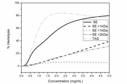
3.8. Hemolytic activity
The hemolytic activity of Ll extracts increased significantly with their concentration, being maximum at 2 mg/mL (82%) for the TAS sample, and at 5 mg/mL (80.7%) for SE ones (Figure 8). The dialyzed fractions were less hemolytic than the complete extract, producing 20% of hemolysis at 3 mg/mL and 30-40% at 5 mg/mL.
Figure 8. Hemolytic activity of L. latrans samples obtained by SE and TAS methods
Leite et al. reported that amphibian protection against pathogens may be related to a synergistic effect of the different compounds present in the granular glands [38]. In this way, and taking into account that SE<1kDa is not hemolytic, the major hemolytic activity of the complete SE extract is related to a synergistic effect of the peptides mixture, and not to the presence of other compounds of low MW, such as alkaloids, amines, steroids and short peptides.
Figure 9. Hemolytic activity of H. pulchellus samples obtained by SE and TAS methods
While the ability to lyse human erythrocytes is not a desired feature in molecules of therapeutic interest, in this study it was considered as a positive indicator of toxic effect and interaction with membranes.
3.9. Inhibition assays of butyrylcholinesterase (BChE)
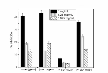
Figures 10 and 11 show the enzyme inhibition variations with the concentration of the tested samples. For Ll (Figure 10), the highest percentage of inhibition (27.5%) was obtained at 1.25 mg/mL with fraction SE>1kDa and the decrease in inhibition observed at higher concentrations could be related to aggregation effects.
Figure 10. Inhibitory effect of L. latrans samples obtained by TAS, SE and dialysis (SE> 1kDa and
SE <1kDa) on BChE enzyme activity.
The inhibitory activity of the TAS sample of Ll was similar to that of SE>1kDa at 5 mg/mL. This was considered an interesting result because a reduced number of ions were detected in the TAS sample by MS, some of which may correspond to truncated fragments of ocellatins (see figure 4C).
Figure 11. Inhibitory effect of H. pulchellus samples obtained by TAS, SE and dialysis (SE> 1kDa
and SE <1kDa) on the BChE enzyme activity.
There are no previous reports that demonstrate antibutyrylcholinesterase activity of biological samples from species of the Hylidae and Leptodactylidae families.
In general, samples obtained from Ll by SE showed higher antimicrobial activity against Gram (+) and Gram (-) bacteria than those obtained by TAS. Also, SE extract of Ll inhibited the growth of
Mycobacterium tuberculosis H37Rv strain.
In the specific case of Hp, both extracts (SE and TAS) showed similar biological properties, although their composition was different. The results suggested the BChE inhibitory activity of Hp is
related to the presence of compounds with low molecular weight.
According to these results, both extraction methods are useful and complementary for the study of the biological properties of new anuran amphibian species.
Abbreviations:
AChE, acetylcholinesterase; ACN, acetonitrile; AD, Alzheimer's disease; BChE,butyrylcholinesterase; FAS, fasciculins; HPLC, high-performance liquid chromatography; Hp, Hypsiboas
pulchellus; Ll, Leptodactylus latrans; MS, mass spectrometry; MALDI, Matrix-assisted laser
desorption/ionization; RP, reverse phase; rt, retention time; SE, solvent extraction; TAS, Transcutaneous amphibian stimulation.; TOF, time of flight.
Acknowledgments
This work was supported by grants from ANPCyT (PICT 2010 Nº 428 Project) and UNL (CAI+D 2009 Project) from Argentina.
The authors thank Paola Peltzer, Andrés Attademo and Agustín Bassó for field assistance.
References
[1] R.I. Lehrer and T. Ganz (1999). Antimicrobial peptides in mammalian and insect host defence, Curr. Opin.
Immunol.11, 23-27.
[2] R.I. Lehrer, T. Ganz and M.E. Selsted (1991). Defensins: endogenous antibiotic peptides of animal cells,
Cell64, 229-230.
[3] C.L Bevins and M. Zasloff (1990). Peptides from frog skin, Annu. Rev. Biochem. 59, 395-414.
[4] W.E. Duellman and L. Trueb (1986). Biology of Amphibians. McGraw-Hill, New York.
[5] J.M. Daly, H.M. Garraffo, T.F. Spande, L.A. Giddings, R.A. Saporito, D.R. Vieites and M. Vences (2008).
Individual and geographic variation of skin alkaloids in three species of Madagascan poison frogs
(Mantella), J. chem. Ecol. 34, 252-279.
[6] J.W. Daly, H.M. Garraffo, T.F. Spande, H.J. Yeh, P.M. Peltzer, P.M. Cacivio, J.D. Baldo and J. Faivovich
(2008). Indolizidine 239 Q and quinolizidine 275I. Major alkaloids in two Argentinian bufonid toads
(Melanophryniscus), Toxicon52, 858-870.
[7] L.H. Lazarus and M. Attila (1993). The toad, ugly and venomous, wears yet a precious jewel in his skin,
[8] J.M. Conlon, J. Kolodziejek and N. Nowotny (2004). Antimicrobial peptides from ranid frogs: taxonomic
and phylogenetic markers and a potential source of new therapeutic agents, Biochim. Biophys. Acta
1696,1-14.
[9] R.A. Cruciani, J.L. Barker, M. Zasloff, H.C. Chen and O. Colamonici (1991). Antibiotic magainins exert
cytolytic activity against transformed cell lines through channel formation, Proc. Natl. Acad. Sci. USA. 88,
3792-3796.
[10] S. Kim, S.S Kim, Y.J. Bang, S.J. Kim and B.J. Lee (2003). In vitro activities of native and designed
peptide antibiotics against drug sensitive and resistant tumor cell lines, Peptides24, 945-953.
[11] M.L. Mangoni (2011). Host-defense peptides: From biology to therapeutic strategies, Cell Mol. Life Sci.
68(13), 2157-5159.
[12] L.G. Gebhard, F.U. Carrizo, A.L. Stern, N.I. Burgardt, J. Faivovich, E. Lavilla and M.R. Ermácora.
(2004). A Kazal prolyl endopeptidase inhibitor isolated from the skin of Phyllomedusa sauvagii, Eur. J.
Biochem.271, 2117-2126.
[13] A. Sánchez Riera, A. Daud, A. Gallo, S. Genta, M. Aybar and S. Sánchez (2003). Antibacterial activity of
lactose-binding lectins from Bufo arenarum skin, Biocell27, 37-46.
[14] R. Zhang, M. Zhou, L. Wang, S. McGrath, T. Chen, X. Chen and C. Shaw (2010). Phylloseptin-1 (PSN-1)
from Phyllomedusa sauvagei skin secretion: a novel broad-spectrum antimicrobial peptide with
antibiofilm activity, Mol. Immunol.47, 2030-2037.
[15] K.N. Pandey (2010). Small peptide recognition sequence for intracellular sorting, Curr. Opin. Biotechnol.
21, 611-620.
[16] G.M. Oremek and N. Sapoutzis (2003). Pro-gastrin-releasing peptide (Pro-GRP), a tumor marker for small
cell lung cancer, Anticancer Res.23, 895-898.
[17] Z. Yu, W. Zhao, J. Liu, J. Lu and F. Chen (2011). QIGLF, a novel angiotensin I-converting
enzyme-inhibitory peptide from egg white protein, J. Sci. Food. Agric.91, 921-926.
[18] R.J. Falkenstein and C. Pena (1999). Interaction of synthetic peptides from fasciculin with
acetylcholinesterase, J. Protein. Chem.18, 233-238.
[19] Q. Yu, H.W. Holloway, T. Utsuki, A. Brossi and N.H. Greig. (1999). Synthesis of novel
phenserine-based-selective inhibitors of butyrylcholinesterase for Alzheimer's disease, J. Med. Chem.42, 1855-1861.
[20] J. Filho, K. Medeiro, M. Diniz, L. Batista, P. Athayde-Filho, M. Silva, E. da-Cunha, J. Silva Almeida and
L. Quintans-Júnior (2006). Natural products inhibitors of the enzyme acetylcholinesterase, Rev. bras.
Farmacogn.16, 258-285.
[21] R.E. Becker, P. Moriearty, L. Unni and S. Vicari (1996). Cholinesterase inhibitors as therapy in Alzheimer
disease: benefit to risk considerations in clinical application, In: Alzheimer Disease from Molecular
Biology to Therapy, eds: Becker R. and Giacobini E.,Birkhäuser Boston, USA, pp. 257-266.
[22] E. Giacobini (1996). Cholinesterase inhibitors do more than inhibit cholinesterase, In: Alzheimer Disease
from Molecular Biology to Therapy, eds: Becker R. and Giacobini E., Birkhäuser Boston, USA, , pp. 187–
204.
[23] D. Rodriguez-Ithurralde, R. Silveira, L. Barbeito and F. Dajas (1983). Fasciculin, a powerful
anticholinesterase polypeptide from Dendroaspis angusticeps venom, Neurochem. Int.5, 267-274.
[24] R. Bolliger and K. Meyer (1957). Isolierung und Identifizierung der Steroide des Giftsekretes der
Berberkröte (Pantherkröte) Bufo mauretanicus, Helvet. Chim. Acta40, 1659-1670.
[25] M. Roseghini, G. Falconieri Erspamer, C. Severini and M. Simmaco (1989). Biogenic amines and active
peptides in extracts of the skin of thirty-two European amphibian species, Comp. Biochem. Physiol. C
Toxicol. Pharmacol. 94, 455-460.
[26] J.F. White and R. Britanisky (1986). Adrenergic agents stimulate and cholinergic agents inhibit H+
secretion by amphibian jejunum, Am. J. Physiol.251, G405-G412.
[27] M.J. Tyler, D.J. Stone and J.H. Bowie (1992). A novel method for the release and collection of dermal,
glandular secretions from the skin of frogs, J. Pharmacol. Toxicol. Methods28, 199-200.
[28] J.B. Grant and B. Land (2002). Transcutaneous Amphibian Stimulator (TAS): A device for the collection
of amphibian skin secretions, Herpetol. Review33, 38-41.
[29] ASIH. (2004). Guidelines for use of live amphibians and reptiles in field and laboratory research.
Herpetological animal care and use committee (HACC) of the American Society of Ichthyologists and Herpetologists. Washington DC, USA.
[30] J.R. Tagg and A.R. McGiven (1971). Assay system for bacteriocins. Appl. Microbiol. 21, 943.
[31] C. Leite, A. Beretta, I. Anno and M. Telles (2000). Standartization of broth microdilution method for
Mycobacterium tuberculosis, Mem. Inst. Oswaldo Cruz95, 127-129.
[32] A. Siano, M.V Húmpola, M.C. Rey, A. Simonetta and G.G. Tonarelli (2011). Interaction of acylated and
substituted antimicrobial peptide analogs with phospholipid-polydiacetylene vesicles. Correlation with
their biological properties, Chem. Biol. Drug Des.78, 85-93.
[33] A. Siano, G. Tonarelli, M.S. Imaz, J.C. Perin, N. Ruggeri, M. Lopez, M.N. Santi and E. Zerbini (2010).
Bactericidal and hemolytic activities of synthetic peptides derived from granulysin, Protein Pept. Lett.17,
517-521.
[34] G.L. Ellman, K.D. Courtney, V. Andres and R.M. Featherstone (1961). A new and rapid colorimetric
determination of acetylcholinesterase activity, Biochem. Pharmacol.7, 88-95.
[35] P.K. Smith, R.I. Krohn, G.T. Hermanson, A.K Mallia, F.H. Gartner, M.D. Provenzano, E.K. Fujimoto,
N.M. Goeke, B.J. Olson and D.C. Klenk (1985). Measurement of protein using bicinchoninic acid, Anal.
[36] M.S. Castro, T.C Ferreira, E.M Cilli, E. Crusca Jr, M.J Mendes-Giannini, A. Sebben, C.A Ricart, M.V Sousa and W. Fontes (2009). Hylin a1, the first cytolytic peptide isolated from the arboreal South
American frog Hypsiboas albopunctatus ("spotted treefrog"), Peptides 30, 291-296.
[37] J.M. Conlon and J. Leprince (2010). Identification and analysis of bioactive peptides in amphibian skin
secretions, Methods Mol. Biol.615, 145-157.
[38] J.M. Leite Jr., L.P. Silva, R.R.. Silva-Leite, A.S Ferrari, S.E. Noronha, H.R. Silva, C. Bloch Jr. and J.R.
Leite. (2010). Leptodactylus ocellatus (Amphibia): mechanism of defense in the skin and molecular
phylogenetic relationships, J. Exp. Zool. A Ecol. Genet. Physiol.313,1-8.
[39] A. Nascimento, L.C. Zanotta, C.M. Kyaw, E.N.F. Schwartz, C.A Schwartz, A. Sebben, M.V. Sousa, W.
Fontes and M.S. Castro (2004). Ocellatins: new antimicrobial peptides from the skin secretion of the
South American frog Leptodactylus ocellatus (Anura: Leptodactylidae), Protein J.23, 501-508.
[40] A. Nascimento, A. Chapeaurouge, J. Perales, A. Sebben, M.V. Sousa, W. Fontes and M.S. Castro (2007).
Purification, characterization and homology analysis of ocellatin 4, a cytolytic peptide from the skin
secretion of the frog Leptodactylus ocellatus, Toxicon 50, 1095-1104.
[41] A.M. Cole, R.O Darouiche, D. Legarda, N. Connell and G. Diamond (2000). Characterization of a fish
antimicrobial peptide: gene expression, subcellular localization, and spectrum of activity, Antimicrob.
Agents Chemother. 44, 2039-2045.
[42] B.R. Copp and A.N. Pearce (2007). Natural product growth inhibitors of Mycobacterium tuberculosis, Nat.
Prod. Rep. 24, 278-297.
[43] C. Mollay, U. Vilas, A. Hutticher and G. Kreil (1986). Isolation of a dipeptidyl aminopeptidase, a putative
processing enzyme, from skin secretion of Xenopus laevis, Eur. J. biochem. 160, 31-35.
[44] N.J. Darby, D.B. Lackey and D.G. Smyth (1991). Purification of a cysteine endopeptidase which is
secreted with bioactive peptides from the epidermal glands of Xenopus laevis, Eur. J. biochem. 195, 65-70.
[45] P.F. Kuks, C. Creminon, A.M. Leseney, J. Bourdais, A. Morel and P. Cohen (1989). Xenopus laevis skin
Arg-Xaa-Val-Arg-Gly-endoprotease. A highly specific protease cleaving after a single arginine of a
consensus sequence of peptide hormone precursors, J. biol. chem. 264, 14609-14612.
[46] N.M. Resnick, W.L. Maloy, H.R. Guy, H.R., Zasloff, M. (1991). A novel endopeptidase from Xenopus that
recognizes alpha-helical secondary structure, Cell66, 541-554.
[47] J.M. Conlon, N. Al-Ghaferi, B. Abraham, A. Sonnevend, J.D. King and P.F. Nielsen (2006). Purification
and properties of laticeptin, an antimicrobial peptide from skin secretions of the south american frog
Leptodactylus laticeps, Protein Pept. Lett.13, 411-415.
[48] J.D. King, N. Al-Ghaferi, B. Abraham, A. Sonnevend, J. Leprince, P.F. Nielsen and J.M. Conlon (2005).
Pentadactylin: an antimicrobial peptide from the skin secretions of the south american bullfrog
Leptodactylus pentadactylus, Comp. Biochem. Physiol. C Toxicol. Pharmacol. 141, 393-397.
[49] L.A. Rollins-Smith, J.D. King, P.F. Nielsen, A. Sonnevend and J.M. Conlon (2005). An antimicrobial
peptide from the skin secretions of the mountain chicken frog Leptodactylus fallax
(Anura:Leptodactylidae), Regul. Pept.124, 173-178.
[50] J.C. Sousa, R.F. Berto, E.A. Gois, N.C. Fontenele-Cardi, J.E. Honorio Jr., K. Konno, M. Richardson, M..F
Rocha, A.A. Camargo, D.C Pimenta, B.A Cardi and K.M. Carvalho (2009). Leptoglycin: a new glycine/leucine-rich antimicrobial peptide isolated from the skin secretion of the south american frog
Leptodactylus pentadactylus (Leptodactylidae), Toxicon54, 23-32.
[51] R.M. Caprioli, T.B Farmer and J. Gile (1997). Molecular imaging of biological samples: localization of
peptides and proteins using MALDI-TOF MS, Anal. Chem. 69, 4751–4760.
[52] P. Favreau, L. Menin, S. Michalet, F. Perret, O. Cheneval, M. Stöcklin, P. Bulet and R. Stöcklin (2006).
Mass spectrometry strategies for venom mapping and peptide sequencing from crude venoms: Case
applications with single arthropod specimen, Toxicon46, 676-687.
[53] B.G. Fry, W. Wuster, S.F. Ryan Ramjan, T. Jackson, P. Martelli and R.M. Kini (2003). Analysis of
Colubroidea snake venoms by liquid chromatography with mass spectrometry: evolutionary and
toxicological implications, Rapid Commun. Mass Spectrom.17, 2047–2062.