Submitted23 October 2015
Accepted 21 January 2016
Published22 February 2016
Corresponding authors
Lihong Ge, [email protected] Shicheng Wei, [email protected]
Academic editor
Alistair Forrest
Additional Information and Declarations can be found on page 13
DOI10.7717/peerj.1684
Copyright
2016 Zheng et al.
Distributed under
Creative Commons CC-BY 4.0
OPEN ACCESS
Insight into the maintenance of
odontogenic potential in mouse
dental mesenchymal cells based on
transcriptomic analysis
Yunfei Zheng1,2,*, Lingfei Jia1,2,3,*, Pengfei Liu4, Dandan Yang4,5, Waner Hu1,
Shubin Chen4, Yuming Zhao6, Jinglei Cai4, Duanqing Pei4, Lihong Ge6and
Shicheng Wei1,3
1Department of Oral and Maxillofacial Surgery, Laboratory of Interdisciplinary Studies, Peking University School and Hospital of Stomatology, Beijing, China
2Department of Orthodontics, Peking University School and Hospital of Stomatology, Beijing, China 3Central Laboratory, Peking University School and Hospital of Stomatology, Beijing, China
4Institute for Stem Cell Biology and Regenerative Medicine, Guangzhou Institute of Biomedicine and Health, Chinese Academy of Sciences, Guangzhou, China
5Experimental Center of Pathogenobiology Immunology, Cytobiology and Genetic, College of Basic Medical Sciences of Jilin University, Jilin, China
6Department of Pediatric Dentistry, Peking University School and Hospital of Stomatology, Beijing, China *These authors contributed equally to this work.
ABSTRACT
Background. Mouse dental mesenchymal cells (mDMCs) from tooth germs of cap or later stages are frequently used in the context of developmental biology or whole-tooth regeneration due to their odontogenic potential.In vitro-expanded mDMCs serve as an alternative cell source considering the difficulty in obtaining primary mDMCs; however, cultured mDMCs fail to support tooth development as a result of functional failures of specific genes or pathways. The goal of this study was to identify the genes that maintain the odontogenic potential of mDMCs in culture.
Methods.We examined the odontogenic potential of freshly isolated versus cultured mDMCs from the lower first molars of embryonic day 14.5 mice. The transcriptome of mDMCs was detected using RNA sequencing and the data were validated by qRT-PCR. Differential expression analysis and pathway analysis were conducted to identify the genes that contribute to the loss of odontogenic potential.
Results. Cultured mDMCs failed to develop into well-structured tooth when they were recombined with dental epithelium. Compared with freshly isolated mDMCs, we found that 1,004 genes were upregulated and 948 were downregulated in cultured mDMCs. The differentially expressed genes were clustered in the biological processes and signaling pathways associated with tooth development. Followingin vitroculture, genes encoding a wide array of components of MAPK, TGF-β/BMP, and Wnt pathways were significantly downregulated. Moreover, the activities ofBdnf,Vegfα,Bmp2, and
Bmp7 were significantly inhibited in cultured mDMCs. Supplementation of VEGFα, BMP2, and BMP7 restored the expression of a subset of downregulated genes and induced mDMCs to form dentin-like structuresin vivo.
SubjectsBioinformatics, Cell Biology, Developmental Biology, Dentistry
Keywords Odontogenesis, Odontogenic potential, RNA-Seq, Transcriptome, Mouse dental mesenchymal cells
INTRODUCTION
In the field of tooth regeneration, the main concept is to mimic the natural tooth development process using stem cells. Cells or tissues with odontogenic potential are required to regenerate a whole tooth (Kollar & Baird, 1970b;Mina & Kollar, 1987). Since the odontogenic potential to instruct tooth organogenesis shifts to the dental mesenchyme at bud stage of odontogenesis (Kollar & Baird, 1970a;Mina & Kollar, 1987), and dental mesenchyme determines the tooth type and size (Kollar & Baird, 1969), it will be preferable to use a mesenchymal cell population with odontogenic potential to provide the inductive signal. Nevertheless, the adult dental stem/progenitor cells appear to lack the potency to regenerate an anatomically correct complete tooth organ (Hu et al., 2014;
Mao & Prockop, 2012), and the odontogenic potential of progenitor cells derived from the
embryonic stem cells or induced pluripotent stem cells was unknown (Otsu et al., 2012;
Ozeki et al., 2013;Seki et al., 2015). Mouse dental mesenchymal cells (mDMCs) isolated
from dental mesenchyme of cap or later stages possess the odontogenic potential and they have frequently been used in the context of developmental biology or whole-tooth regeneration (Honda et al., 2007;Ikeda et al., 2009;Nakao et al., 2007). mDMCs are able to induce tooth formation in a dental or nondental epithelial cell population (Kollar
& Baird, 1969;Kollar & Baird, 1970b;Wang et al., 2010;Angelova Volponi, Kawasaki &
Sharpe, 2013;Cai et al., 2013). As the preparation of mDMCs is time-consuming and
embryos at the right stage are not easily available,in vitro-expanded mDMCs could be an available cell source. However, mDMCs fail to support tooth development when they are grown as a monolayer (Keller et al., 2011), and the genes that contribute to the loss of odontogenic potential in mDMCs were unknown.
RNA sequencing (RNA-seq) technology provides sensitive detection of global gene expression profiles in cells and tissues, and bioinformatics approaches also examine changes in the context of the overall pathway rather than individual genes (Wang, Gerstein
& Snyder, 2009). To identify the genes that maintain the odontogenic potential in mDMCs,
we evaluated the odontogenic potential of freshly isolated versus cultured mDMCs from the lower first molars in embryonic day 14.5 (E14.5) mice, and compared the transcriptome of freshly isolated mDMCs with that of cultured mDMCs. We found a loss of odontogenic potential accompanied by a significant disturbed transcriptome in cultured mDMCs and identified potential genes that contribute to the loss of odontogenic potential, providing new insight into the maintenance of odontogenic potential of mDMCs.
MATERIALS & METHODS
Cell cultureTable 1 Primers for quantitative real-time PCR.
Forward Reverse
Actin 5′-GGC TGT ATT CCC CTC CAT CG-3′ 5′-CCA GTT GGT AAC AAT GCC ATG T-3′ Lhx6 5′-CAT TGA GAG TCA GGT ACA GTG C-3′ 5′-GGG CCG TCC AAA TCA GCT T-3′ Bmp4 5′-TTC CTG GTA ACC GAA TGC TGA-3′ 5′-CCT GAA TCT CGG CGA CTT TTT-3′ Msx1 5′- GCA CAA GAC CAA CCG CAAG-3′ 5′- CGC TCG GCA ATA GAC AGG T-3′ Pax9 5′- CAT TCG GCT TCG CAT CGT G -3′ 5′- CTC CCG GCA AAA TCG AAC C-3′ Fgf10 5′- GCA GGC AAA TGT ATG TGG CAT-3′ 5′- ATG TTT GGA TCG TCA TGG GGA-3′ Bmp2 5′-GGG ACC CGC TGT CTT CTA GT-3′ 5′-TCA ACT CAA ATT CGC TGA GGA C-3′ Fgf3 5′-TGC GCT ACC AAG TAC CAC C-3′ 5′-CAC TTC CAC CGC AGT AAT CTC-3′
University in China (permit Number: CMU-B20100106). The lower first molar tooth germs were dissected from the mandibles of E14.5 mice with fine needles and treated with 0.75 mg/ml dispase (Becton, Dickinson and Co., Franklin Lakes, NJ, USA) for 40 min at 37 ◦C to facilitate separation from dental epithelium. The mesenchymal tissues were then incubated in 0.25% trypsin (Sigma-Aldrich, St. Louis, MO, USA) at 37 ◦C for 1 min and dissociated with gentle pipetting. The cells were then filtered through a 40µm cell
sieve, resuspended, and cultured in Dulbecco’s modified Eagle’s medium (DMEM; Gibco, Grand Island, NY, USA) plus 10% fetal bovine serum (FBS; Gibco) (Duailibi et al., 2004;
Jiang et al., 2014;Yamazaki et al., 2007;Zhao et al., 2014). To maintain the odontogenic
potential of mDMCs, supplementation of VEGFα (Sino Biological Inc, Beijing, China), BMP2 (Sigma-Aldrich), and BMP7 (Sigma-Aldrich) were added into the medium.
Quantitative reverse transcription PCR (qRT-PCR)
Total RNA was extracted with TRIzol (Invitrogen, Carlsbad, CA, USA) and reverse transcribed using an RT-PCR kit (TaKaRa Bio, Otsu, Japan) (Zheng et al., 2014). Quantitative PCR was performed in a Thermal CyclerDice7TM Real Time System with SYBR Green Premix EXTaqTM(TaKaRa Bio). The primers used are listed in Table 1. The
relative expression of genes was calculated using the 2−1Ct method.
Tissue recombination and subrenal culture
The mDMCs were harvested at indicated time points using 0.25% trypsin (Sigma) and spun down to make cell pellets. The cell pellets were cultured at 37 ◦C for 2–3 h and then recombined with freshly isolated E14.5 dental epithelium. The recombinants were further culturedin vitrofor 24 h prior to subrenal culture in adult ICR male mice. The host mice were sacrificed 3 weeks later to harvest the grafted tissue. Grafts were then fixed and subjected to H&E staining for histological analysis.
RNA isolation and sequencing
RNA was extracted using the RNeasy Mini Kit and RNase-Free DNase Set according to the manufacturer’s protocol (Qiagen GmbH, Hilden, Germany). The purity and quantity of RNA were assessed using a spectrophotometer (model 8453; Agilent, Santa Clara, CA, USA). RNA libraries for samples were prepared according to instructions for the Illumina TruSeqTMRNA Sample Prep Kit. Sequencing was performed on an Illumina HiseqTM2000 (Illumina, San Diego, CA, USA) in duplicate.
Bioinformatics analysis
Sequenced reads were mapped to the mouse transcriptome (mm10, Ensembl v73) and then aligned using bowtie (v1.0.1) and RSEM (v1.2.12), as described previously
(Hutchins, Takahashi & Miranda-Saavedra, 2015;Li & Dewey, 2011). EDASeq (v1.11.0)
was used for GC normalization of samples, and differential expression was called using DESeq2 (v1.12.0). The fold change cut-off was set at twofold andp-value < 0.05 were considered to be statistically significant. The Database for Annotation, Visualization and Integrated Discovery (DAVID) was used to determine overrepresented GO categories and KEGG pathways using the entire mouse transcriptome as the background gene set. Protein-protein interaction network of DEGs was constructed using the STRING database
(http://string-db.org/). Ingenuity pathway analysis (Ingenuity Systems, Redwood City, CA,
USA) was used to analyze the upstream regulators of the differential expression between the samples (Razali et al., 2015).
Statistical analysis
Statistical analyses were performed using SPSS PASW version 18. Experiments with cell cultures were performed at least in triplicate and data are expressed as mean±standard deviation (SD). The differences among groups were analyzed using one-way ANOVA. A two-tailedp-value < 0.05 was considered to be statistically significant.
RESULTS
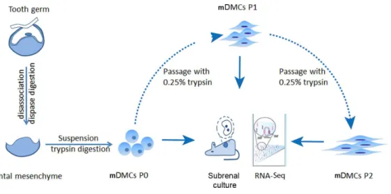
mDMCs were isolated from the developing molars in E14.5 mouse embryos and were designated as P0. One part of the P0 cells was recombined with E14.5 dental epithelium and cultured in kidney; another part was subjected to RNA-seq. The remaining cells were subcultured in standard medium, with the first-passage culture designated as P1 and the second-passage as P2. P1 and P2 cells were also split into two portions to ensure an identical cell state for the recombination assay and the RNA-seq (Fig. 1).
Odontogenic potential of cultured mouse dental mesenchymal cells
mDMCs are typically cultured in Eagle’s minimum essential medium plus fetal bovine serum and antibiotics (Jiang et al., 2014;Keller et al., 2011;Zhao et al., 2014). mDMCs showed an atypical round- or spindle-shaped fibroblast-like morphology with a higher nuclear-to-cytoplasmic ratio, indicating their primitive character. Although the cells retained fibroblastic features up to the second passage, they exhibited a senescent phenotype with increased cytoplasm and augmented volume (Fig. 2A).
Figure 1 Experimental design.Tooth germs from embryonic day 14.5 mice were obtained and digested with dispase to separate the dental mesenchyme from dental epithelium. Freshly isolated dental mesenchy-mal cells were designated as P0 and culturedin vitro. RNA samples from the P0, the first (P1), and second (P2) passages were collected before they were submitted for RNA-seq using an Illumina HiseqTM2000.
when recombined with embryonic dental epithelium, cultured mDMCs failed to support tooth development (Fig. 2B). Moreover, the expression of genes that are essential for tooth development were analyzed using qRT-PCR. The expression ofMsx1,Pax9, and
Lhx6 was significantly reduced in P1 and P2 cells compared with P0 cells (Fig. 2C). The expression ofFgf3andBmp2in cultured mDMCs was reduced compared with the P0 cells, but the expression ofBmp4was not significantly different between the P0 cells and cultured mDMCs (Fig. 2D).
Overview of the mouse dental mesenchymal cells’ transcriptome
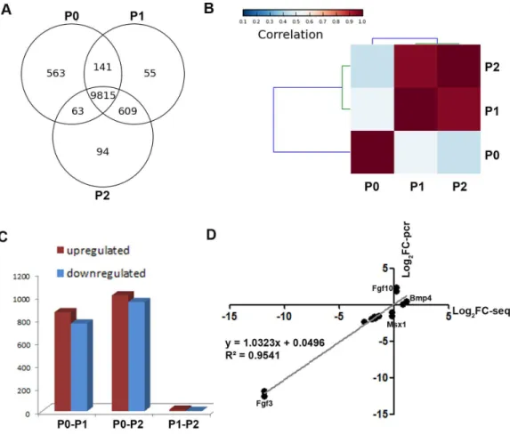
To obtain a global view of genes regulating the loss of odontogenic potential, total mRNA of P0, P1, and P2 cells was extracted and sequenced. After data correction, 11,340 transcripts could be matched exactly to known mouse Ensemble transcripts. A total of 9,815 genes were shared among P0, P1 and P2 cells, whereas 563 genes were expressed exclusively in P0 cells
(Fig. 3A). P0 cells that were not exposed toin vitroculture conditions showed a striking
separation from P1 and P2 cells (Fig. 3B;Fig. S1). The transcriptional disparity between freshly isolated and cultured mDMCs is consistent with their phenotypic differences. Differential expression analysis revealed thatin vitroexpansion of mDMCs promoted the selective overexpression of 859 genes, whereas 763 genes were downregulated in P1 cells
(Fig. 3C). Comparison of the transcriptomes of P0 and P2 cells revealed that 1,004 genes
were upregulated and 948 were downregulated (Fig. 3C). In contrast, 13 genes were upregulated and two genes were downregulated in P1 compared with P2 cells (Fig. 3C). These results suggested that the transcriptome of mDMCs was significantly influenced by
in vitroculture conditions. In addition, the expression levels ofMsx1,Lhx6,Pax9,Bmp4,
Fgf10, Bmp2, and Fgf3were comparable when analyzed with RNA-seq and qRT-PCR
Figure 3 Comparison of the transcriptomic profiles of freshly isolated and cultured mDMCs.(A) The Venn diagram shows the expression profiling of P0, P1 and P2 cells, with each section showing the number of genes. (B) Hierarchical clustering analysis showed that the P1 and P2 were highly correlated, whereas the P0 was independent of them. (C) The number of differentially-expressed genes is illustrated. (D) The expression levels ofMsx1,Lhx6,Pax9,Bmp4,Fgf10,Bmp2, andFgf3were comparable when ana-lyzed with RNA-seq and qRT-PCR. Data are expressed as the mean±standard deviation (SD). *p<0.05.
Gene ontology analysis of differentially expressed genes
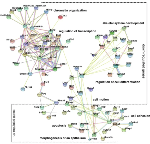
Gene ontology (GO) analysis provides an intuitive and effective approach to understand the function of genes in three domains: biological processes, cellular components, and molecular functions. To understand the function of differentially expressed genes, GO analysis was conducted (Fig. S2) and a network diagram was created to illustrate the communication of differentially expressed genes in the enriched clusters of biological processes (Fig. 4). The network is composed of: (a) genes around the node Dnmt3b, namelyDnmt1,Ezh2,Uhrf1, andRbl1, which participate in chromatin organization and transcriptional regulation; (b) genes around the nodeRunx2, namelyBmp2,Dlx1,Lhx6,
Figure 4 Gene ontology (GO) analysis of the differentially expressed genes.A protein–protein interac-tion network was constructed. DEGs in enriched GO categories are represented in the network.
and their functions are expected because chromatin modification, cell motion, and cell adhesion are basic cellular functions involving the development of multiple organs.
Pathway analysis of the differentially expressed genes
Multiple pathways including fibroblast growth factor (FGF), bone morphogenetic protein (BMP), hedgehog (SHH), and Wnt signaling play critical roles in odontogenesis (Liu et
al., 2013;Thesleff, Vaahtokari & Partanen, 1995); thus, the Kyoto Encyclopedia of Genes
Figure 5 Disturbed pathways and the upstream regulators.(A) Kyoto Encyclopedia of Genes and Genomes (KEGG) pathways involving the differentially expressed genes are listed. (B) A heat map representing the genes within the MAPK, Wnt, and TGFβpathways. (C) The upstream regulators predicted by Ingenuity pathway analysis.
numerous members of these signaling pathways were reduced in cultured mDMCs
(Fig. 5B), and it was difficult to identify the critical regulators that are responsible for
the loss of odontogenic potential in cultured mDMCs.
An upstream analysis predicts which upstream regulators were most likely to be involved based on the current levels of gene expression detected by RNA-seq analysis. Numerous upstream regulators were related to the loss of odontogenic potential in cultured mDMCs. Specifically, the activities ofBdnf,Vegfα,Bmp2, andBmp7 were predicted to be significantly inhibited, whileIgf2,Fgf10, andTgfb1were more active in cultured mDMCs
(Fig. 5C). AlthoughBdnf was slightly increased in cultured mDMCs, its receptorNtrk2
Figure 6 VEGFα, BMP2, and BMP7 play roles in the maintenance of odontogenic potential.The mRNA levels of dental mesenchyme-specific genes in mDMCs cultured in the medium supplemented with 10 ng/ml or 50 ng/ml VEGFα(A), BMP2 (B), and BMP7 (C). (D) Stereoscopic images (upper) and histological images (lower) of the dentin-like structures or the amorphous matrix. Dentin-like structures derived from recombinants with mDMCs cultured in the medium supplemented with VEGFα, BMP2, and BMP7 (VBB). D, dentin-like structures; M, amorphous matrix; NC, negative control. Data are expressed as the mean±standard deviation (SD). *p<0.05. Scale bar: upper left 1 mm, upper right 500µm, lower 50µm.
VEGFα, BMP2, and BMP7 restore the expression of
downregulated genes
Given the pivotal roles of VEGFα, BMP2, and BMP7 in tooth development, we selected these proteins to increase the expression of odontogenic genes. Indeed, tooth mesenchyme-specific genes were upregulated including Msx1 and Pax9 in mDMCs treated with Bmp2 and Bmp7 (Figs. 6Band6C). VEGFαslightly increased the expression of Msx1 and Pax9, but not that of Lhx6 (Fig. 6A). In parallel, VEGFα, BMP2, and BMP7 were adopted to maintain the odontogenic potential of mDMCs. Although well-structured tooth was not observed, dentin-like structures were present (Fig. 6D). Together, VEGFα, BMP2, and BMP7 promoted the expression of dental mesenchyme-specific genes and the formation of dentin-like structures.
DISCUSSION
the present study, we found that cultured mDMCs lost the odontogenic potential and failed to initiate tooth formation. Consistently, the dental mesenchyme-specific transcription factors (Msx1, Pax9, and Lhx6) were decreased in cultured mDMCs, and the mutation of these transcription factors was shown to cause tooth agenesis disorders (Peters et al.,
1998;Satokata & Maas, 1994;Zhao et al., 2013). The loss of odontogenic potential and
decreased expression of dental mesenchyme-specific genes may be due to the absence of inductive signals from the dental epithelium or mesenchyme. Since Bmp and Fgf signaling pathways mediate the odontogenic inductive signals of dental mesenchyme (Liu et al.,
2013;Thesleff, Vaahtokari & Partanen, 1995), the expression of representative components
within these two signaling families was analyzed in mDMCs. The expression of Bmp4 shifts from dental epithelium to mesenchyme, which is concomitant with the shift of instructive potential during tooth development (Vainio et al., 1993), but the expression ofBmp4in the present study was not significantly changed when mDMCs were culturedin vitro. This suggests thatBmp4in cultured mDMCs is not sufficient to substitute for the odontogenic potential and that other factors besidesBmp4are responsible for the inductive potential. Similarly, althoughFgf10stimulates cell proliferation in the dental epithelium during tooth morphogenesis (Kettunen et al., 2000), the mRNA level ofFgf10 was comparable between freshly-isolated and cultured mDMCs. In contrast, the expression of Fgf3andBmp2in cultured mDMCs was significantly reduced. Fgf3regulates the number, position, and interrelation of cusps in molar teeth; mutation of theFgf3gene leads to dosage-dependent morphological changes in teeth of both mice and human patients (Charles et al., 2009).
Bmp2is essential for the differentiation of ameloblasts and odontoblasts (Guo et al., 2015;
Yang et al., 2012). Thus, the downregulation of these genes may account for the loss of
odontogenic potential in cultured mDMCs. However,Bmp2was expressed at a relatively low level in E14.5 mDMCs, andFgf3knockout mice do not exhibit any overt defects in teeth
(Mansour, Goddard & Capecchi, 1993), raising the possibility that the loss of odontogenic
potential in cultured mDMCs may also involve other genes.
Transcriptome analysis, as conducted in this work, has provided insight into the molecular mechanism underlying the loss of odontogenic potential. The transcriptomic profiles reflected the potential of mDMCs, and the mDMCs without odontogenic potential were strikingly separated from those with odontogenic potential. Moreover, the activities of Bdnf,Vegfα,Bmp2, and Bmp7 were predicted to be inhibited. These growth factors were reported to be involved in tooth development.Bdnf and its receptor Ntrk2are involved in the MAPK pathway and play roles in epithelial-mesenchymal interactions in early tooth morphogenetic events (Nosrat et al., 1997).Vegfα is an important factor that induces angiogenesis and related with the MAPK signaling pathway. It is expressed in inner enamel epithelial cells and the basement membrane (Aida et al., 2005;Miwa et
al., 2007), indicating a potential role in the epithelial-mesenchymal interactions.Bmp2
promotes the maturation of odontoblasts andBmp2conditional knockout mice display abnormal tooth phenotypes with a hypomineralization enamel layer, delayed odontoblast differentiation, abnormal dentin tubules, and decreased tooth-related gene expression
(Guo et al., 2015;Yang et al., 2012).Bmp7 is also essential for tooth development and its
pulp cells, representing a potential mediator of the inductive odontogenic potential of dental mesenchyme (Gao et al., 2015). Consistently, we found that VEGFα, BMP2, and BMP7 increased the expression of dental mesenchyme-specific genes. Even though the supplementation with these growth factors did not restore the odontogenic potential of mDMCs, dentin-like structures formed instead of amorphous matrix, suggesting their potential roles in the maintenance of odontogenic potential. Recently, immortalized fetal dental mesenchymal cell lines were established (Huang et al., 2015;Wu et al., 2015), and a medium supplemented with upstream signaling molecules such asVegfα,Bmp2, and
Bmp7 may preserve the biological properties of mDMCs.
Although the effects of Igf2, Fgf10, andTgfb1 were predicted to be enhanced in cultured mDMCs, they are well-known mesenchymal signals that mediate epithelial-mesenchymal interactions during tooth development (Matsumoto et al., 2011;Nakao et
al., 2013;Vaahtokari, Vainio & Thesleff, 1991). Interestingly, the level ofIgf2 in dental
mesenchyme decreased over time during development because of the hypermethylation of CpG islands (Khan et al., 2012); this suggests that the inhibition ofIgf2activity is required for normal tooth development. However, further studies are still needed to investigate whether the overactivation of these factors leads to abnormal phenotype of teeth, and whether inhibitors of these factors play a role in the maintenance of odontogenic potential.
In the present study, supplementation withVegf,Bmp2, andBmp7 did not completely restored the odontogenic potential of mDMCs. However, the GO analysis also provided a clue for the establishment of culture system for mDMCs. GO analysis showed that multiple biological processes involving the development of multiple organs were dysregulated when mDMCs were culturedin vitro. Specifically, Chromatin modifier enzymes play essential roles in the establishment of transcriptional programs accompanying cell differentiation during tooth development (Brook, 2009). Mesenchymal cell migration and compaction are required to induce the expression of critical odontogenic genes and the differentiation of odontoblasts (Hu, Parker & Wright, 2015;Mammoto et al., 2011). The dysregulation of cell motion also contributed to the compromised potential in cultured mDMCs. Since both the mandible and teeth are mineralized tissue derived from the neural crest (Chai
et al., 2000), multiple genes acting in skeletal system development have been identified as
affecting the odontogenic signaling cascades (Komori, 2006;Nakashima et al., 2002). Thus, growth factors or cytokines that related with transcriptional regulation, cell differentiation or mineralization, and cell motion may facilitate the maintenance of odontogenic potential.
ACKNOWLEDGEMENTS
We thank Dr. Andrew Paul Hutchins and Dr. Xiaoshan Wang for their suggestions on data analysis. We thank Prof. Xiaodong Su and Prof. Faming Chen for their suggestions on the revision of the manuscript.
ADDITIONAL INFORMATION AND DECLARATIONS
Funding
This work was supported by the Peking University’s 985 Grants, Open Project of Key Laboratory of Regenerative Biology, Chinese Academy of Sciences (KLRB201401), and National Natural Science Foundation of China (No. 813716977; 81570944). The funders had no role in study design, data collection and analysis, decision to publish, or preparation of the manuscript.
Grant Disclosures
The following grant information was disclosed by the authors: Peking University’s: 985.
Open Project of Key Laboratory of Regenerative Biology, Chinese Academy of Sciences: KLRB201401.
National Natural Science Foundation of China: 813716977, 81570944.
Competing Interests
The authors declare there are no competing interests.
Author Contributions
• Yunfei Zheng conceived and designed the experiments, performed the experiments, analyzed the data, wrote the paper, prepared figures and/or tables.
• Lingfei Jia performed the experiments, analyzed the data, prepared figures and/or tables, reviewed drafts of the paper.
• Pengfei Liu, Dandan Yang and Shubin Chen performed the experiments, reviewed drafts of the paper.
• Waner Hu analyzed the data, prepared figures and/or tables, reviewed drafts of the paper. • Yuming Zhao and Jinglei Cai contributed reagents/materials/analysis tools, reviewed
drafts of the paper.
• Duanqing Pei, Lihong Ge and Shicheng Wei conceived and designed the experiments, reviewed drafts of the paper.
Animal Ethics
The following information was supplied relating to ethical approvals (i.e., approving body and any reference numbers):
Data Availability
The following information was supplied regarding data availability: GEO accession number:GSE65164.
Supplemental Information
Supplemental information for this article can be found online athttp://dx.doi.org/10.7717/
peerj.1684#supplemental-information.
REFERENCES
Aida M, Irie T, Aida T, Tachikawa T. 2005.Expression of protein kinases C betaI, betaII, and VEGF during the differentiation of enamel epithelium in tooth development.
Journal of Dental Research84:234–239DOI 10.1177/154405910508400305.
Angelova Volponi A, Kawasaki M, Sharpe PT. 2013.Adult human gingival epithe-lial cells as a source for whole-tooth bioengineering.Journal of Dental Research
92:329–334DOI 10.1177/0022034513481041.
Brook AH. 2009.Multilevel complex interactions between genetic, epigenetic and environmental factors in the aetiology of anomalies of dental development.Archives of Oral Biology 54(Suppl 1):S3–S17DOI 10.1016/j.archoralbio.2009.09.005.
Cai J, Zhang Y, Liu P, Chen S, Wu X, Sun Y, Li A, Huang K, Luo R, Wang L, Liu Y, Zhou T, Wei S, Pan G, Pei D. 2013.Generation of tooth-like structures from integration-free human urine induced pluripotent stem cells.Cell Regeneration
2(1):6DOI 10.1186/2045-9769-2-6.
Chai Y, Jiang X, Ito Y, Bringas Jr. P, Han J, Rowitch DH, Soriano P, McMahon AP, Sucov HM. 2000.Fate of the mammalian cranial neural crest during tooth and mandibular morphogenesis.Development127:1671–1679.
Charles C, Lazzari V, Tafforeau P, Schimmang T, Tekin M, Klein O, Viriot L. 2009.
Modulation of Fgf3 dosage in mouse and men mirrors evolution of mammalian dentition.Proceedings of the National Academy of Sciences of the United States of America106:22364–22368DOI 10.1073/pnas.0910086106.
Duailibi MT, Duailibi SE, Young CS, Bartlett JD, Vacanti JP, Yelick PC. 2004.
Bioengineered teeth from cultured rat tooth bud cells.Journal of Dental Research
83:523–528DOI 10.1177/154405910408300703.
Gao B, Zhou X, Pi C, Xu R, Wan M, Yang J, Zhou Y, Liu C, Sun J, Zhang Y, Zheng L. 2015.BMP7 and EREG contribute to the inductive potential of dental mesenchyme.
Scientific Reports5:9903DOI 10.1038/srep09903.
Guo F, Feng J, Wang F, Li W, Gao Q, Chen Z, Shoff L, Donly KJ, Gluhak-Heinrich J, Chun YH, Harris SE, MacDougall M, Chen S. 2015.Bmp2 deletion causes an amelogenesis imperfecta phenotype via regulating enamel gene expression.Journal of Cellular Physiology230:1871–1882DOI 10.1002/jcp.24915.
Honda MJ, Tsuchiya S, Sumita Y, Sagara H, Ueda M. 2007.The sequential seeding of epithelial and mesenchymal cells for tissue-engineered tooth regeneration.
Hu X, Lin C, Shen B, Ruan N, Guan Z, Chen Y, Zhang Y. 2014.Conserved odontogenic potential in embryonic dental tissues.Journal of Dental Research93:490–495
DOI 10.1177/0022034514523988.
Hu S, Parker J, Wright JT. 2015.Towards unraveling the human tooth transcriptome: the dentome.PLoS ONE10:e0124801DOI 10.1371/journal.pone.0124801.
Huang Y, Yang Y, Jiang M, Lin M, Li S, Lin Y. 2015.Immortalization and charac-terization of human dental mesenchymal cells.Journal of Dentistry 43:576–582
DOI 10.1016/j.jdent.2015.02.008.
Hutchins AP, Takahashi Y, Miranda-Saavedra D. 2015.Genomic analysis of LPS-stimulated myeloid cells identifies a common pro-inflammatory response but divergent IL-10 anti-inflammatory responses.Scientific Reports5:9100
DOI 10.1038/srep09100.
Ikeda E, Morita R, Nakao K, Ishida K, Nakamura T, Takano-Yamamoto T, Ogawa M, Mizuno M, Kasugai S, Tsuji T. 2009.Fully functional bioengineered tooth replacement as an organ replacement therapy.Proceedings of the National Academy of Sciences of the United States of America106:13475–13480
DOI 10.1073/pnas.0902944106.
Jiang N, Zhou J, Chen M, Schiff MD, Lee CH, Kong K, Embree MC, Zhou Y, Mao JJ. 2014.Postnatal epithelium and mesenchyme stem/progenitor cells in bioengineered amelogenesis and dentinogenesis.Biomaterials35:2172–2180
DOI 10.1016/j.biomaterials.2013.11.061.
Keller L, Kuchler-Bopp S, Mendoza SA, Poliard A, Lesot H. 2011.Tooth engineering: searching for dental mesenchymal cells sources.Frontiers in Physiology2:7
DOI 10.3389/fphys.2011.00007.
Kettunen P, Laurikkala J, Itaranta P, Vainio S, Itoh N, Thesleff I. 2000.Associations of FGF-3 and FGF-10 with signaling networks regulating tooth morphogenesis.
Developmental Dynamics219:322–332
DOI 10.1002/1097-0177(2000)9999:9999<::AID-DVDY1062>3.0.CO;2-J.
Khan QE, Sehic A, Skalleberg N, Landin MA, Khuu C, Risnes S, Osmundsen H. 2012.
Expression of delta-like 1 homologue and insulin-like growth factor 2 through epigenetic regulation of the genes during development of mouse molar.European Journal of Oral Sciences120:292–302DOI 10.1111/j.1600-0722.2012.00976.x.
Kollar EJ, Baird GR. 1969.The influence of the dental papilla on the development of tooth shape in embryonic mouse tooth germs.Journal of Embryology & Experimental Morphology 21:131–148.
Kollar EJ, Baird GR. 1970a.Tissue interactions in embryonic mouse tooth germs. I. Reorganization of the dental epithelium during tooth-germ reconstruction.Journal of Embryology & Experimental Morphology 24:159–171.
Kollar EJ, Baird GR. 1970b.Tissue interactions in embryonic mouse tooth germs. II. The inductive role of the dental papilla.Journal of Embryology & Experimental Morphology 24:173–186.
Li B, Dewey CN. 2011.RSEM: accurate transcript quantification from RNA-Seq data with or without a reference genome.BMC Bioinformatics12:323
DOI 10.1186/1471-2105-12-323.
Liu C, Gu S, Sun C, Ye W, Song Z, Zhang Y, Chen Y. 2013.FGF signaling sustains the odontogenic fate of dental mesenchyme by suppressing beta-catenin signaling.
Development 140:4375–4385DOI 10.1242/dev.097733.
Mammoto T, Mammoto A, Torisawa YS, Tat T, Gibbs A, Derda R, Mannix R, de Bruijn M, Yung CW, Huh D, Ingber DE. 2011.Mechanochemical control of mesenchymal condensation and embryonic tooth organ formation.Developmental Cell21:758–769DOI 10.1016/j.devcel.2011.07.006.
Mansour SL, Goddard JM, Capecchi MR. 1993.Mice homozygous for a targeted disruption of the proto-oncogene int-2 have developmental defects in the tail and inner ear.Development 117:13–28.
Mao JJ, Prockop DJ. 2012.Stem cells in the face: tooth regeneration and beyond.Cell Stem Cell 11:291–301DOI 10.1016/j.stem.2012.08.010.
Matsumoto A, Harada H, Saito M, Taniguchi A. 2011.Induction of insulin-like growth factor 2 expression in a mesenchymal cell line co-cultured with an
ameloblast cell line.In Vitro Cellular & Developmental Biology-Animal 47:675–680
DOI 10.1007/s11626-011-9456-x.
Mina M, Kollar EJ. 1987.The induction of odontogenesis in non-dental mesenchyme combined with early murine mandibular arch epithelium.Archives of Oral Biology
32:123–127DOI 10.1016/0003-9969(87)90055-0.
Miwa Y, Shimada K, Sunohara M, Sato I. 2007.Immunohistochemically localization of vascular endothelial growth factor, vascular endothelial growth factor receptor-2, collagen I and fibronectin in the epithelia-mesenchymal junction of the human tooth germ.Okajimas Folia Anatomica Japonica84:107–110DOI 10.2535/ofaj.84.107.
Nakao K, Morita R, Saji Y, Ishida K, Tomita Y, Ogawa M, Saitoh M, Tomooka Y, Tsuji T. 2007.The development of a bioengineered organ germ method.Nature Methods
4:227–230DOI 10.1038/nmeth1012.
Nakao Y, Mitsuyasu T, Kawano S, Nakamura N, Kanda S, Nakamura S. 2013.Fibroblast growth factors 7 and 10 are involved in ameloblastoma proliferation via the mitogen-activated protein kinase pathway.International Journal of Oncology43:1377–1384
DOI 10.3892/ijo.2013.2081.
Nakashima K, Zhou X, Kunkel G, Zhang Z, Deng JM, Behringer RR, De Crombrugghe B. 2002.The novel zinc finger-containing transcription factor osterix is required for osteoblast differentiation and bone formation.Cell108:17–29
DOI 10.1016/S0092-8674(01)00622-5.
Nosrat CA, Fried K, Lindskog S, Olson L. 1997.Cellular expression of neurotrophin mRNAs during tooth development.Cell and Tissue Research290:569–580
DOI 10.1007/s004410050962.
into dental mesenchymal cells.Stem Cells and Development21:1156–1164
DOI 10.1089/scd.2011.0210.
Ozeki N, Mogi M, Kawai R, Yamaguchi H, Hiyama T, Nakata K, Nakamura H. 2013.
Mouse-induced pluripotent stem cells differentiate into odontoblast-like cells with induction of altered adhesive and migratory phenotype of integrin.PLoS ONE
8:e80026DOI 10.1371/journal.pone.0080026.
Peters H, Neubuser A, Kratochwil K, Balling R. 1998.Pax9-deficient mice lack pharyn-geal pouch derivatives and teeth and exhibit craniofacial and limb abnormalities.
Genes and Development12:2735–2747DOI 10.1101/gad.12.17.2735.
Razali N, Aziz AA, Lim CY, SM J. 2015.Investigation into the effects of antioxidant-rich extract of Tamarindus indica leaf on antioxidant enzyme activities, oxidative stress and gene expression profiles in HepG2 cells.PeeJ 3:e1292.
Satokata I, Maas R. 1994.Msx1 deficient mice exhibit cleft palate and abnormalities of craniofacial and tooth development.Nature Genetics6:348–356
DOI 10.1038/ng0494-348.
Seki D, Takeshita N, Oyanagi T, Sasaki S, Takano I, Hasegawa M, Takano-Yamamoto T. 2015.Differentiation of odontoblast-like cells from mouse induced pluripotent stem cells by Pax9 and Bmp4 transfection.Stem Cells Translational Medicine
4:993–997DOI 10.5966/sctm.2014-0292.
Thesleff I, Vaahtokari A, Partanen AM. 1995.Regulation of organogenesis. Common molecular mechanisms regulating the development of teeth and other organs.
International Journal of Developmental Biology39:35–50.
Vaahtokari A, Vainio S, Thesleff I. 1991.Associations between transforming growth factor beta 1 RNA expression and epithelial-mesenchymal interactions during tooth morphogenesis.Development 113:985–994.
Vainio S, Karavanova I, Jowett A, Thesleff I. 1993.Identification of BMP-4 as a signal mediating secondary induction between epithelial and mesenchymal tissues during early tooth development.Cell75:45–58DOI 10.1016/S0092-8674(05)80083-2.
Wang B, Li L, Du S, Liu C, Lin X, Chen Y, Zhang Y. 2010.Induction of human ker-atinocytes into enamel-secreting ameloblasts.Developmental Biology344:795–799
DOI 10.1016/j.ydbio.2010.05.511.
Wang Z, Gerstein M, Snyder M. 2009.RNA-Seq: a revolutionary tool for transcrip-tomics.Nature Reviews Genetics10:57–63DOI 10.1038/nrg2484.
Wu L, Wang F, Donly KJ, Wan C, Luo D, Harris SE, MacDougall M, Chen S. 2015.
Establishment of immortalized mouse Bmp2 knock-out dental papilla mesenchymal cells necessary for study of odontoblastic differentiation and odontogenesis.Journal of Cellular Physiology230:2588–2595DOI 10.1002/jcp.25061.
Yamazaki H, Tsuneto M, Yoshino M, Yamamura K, Hayashi S. 2007.Potential of dental mesenchymal cells in developing teeth.Stem Cells25:78–87
DOI 10.1634/stemcells.2006-0360.
Zhao H, Feng J, Seidel K, Shi S, Klein O, Sharpe P, Chai Y. 2014.Secretion of shh by a neurovascular bundle niche supports mesenchymal stem cell homeostasis in the adult mouse incisor.Cell Stem Cell14:160–173 DOI 10.1016/j.stem.2013.12.013.
Zhao M, Gupta V, Raj L, Roussel M, Bei M. 2013.A network of transcription fac-tors operates during early tooth morphogenesis.Molecular and Cellular Biology
33:3099–3112DOI 10.1128/MCB.00524-13.