Submitted8 June 2016
Accepted 27 September 2016
Published25 October 2016
Corresponding author
Massimo Ponti, massimo.ponti@unibo.it
Academic editor
James Reimer
Additional Information and Declarations can be found on page 22
DOI10.7717/peerj.2614
Copyright
2016 Ponti et al.
Distributed under
Creative Commons CC-BY 4.0
OPEN ACCESS
Baseline reef health surveys at Bangka
Island (North Sulawesi, Indonesia) reveal
new threats
Massimo Ponti1,2, Francesca Fratangeli2,3, Nicolò Dondi2,3,
Marco Segre Reinach2,3, Clara Serra2,3and Michael J. Sweet4
1Dipartimento di Scienze Biologiche, Geologiche e Ambientali, University of Bologna, Ravenna, Italy 2Reef Check Italia onlus, Ancona, Italy
3Coral Eye, Bangka Island, North Sulawesi, Indonesia
4Environmental Sustainability Research Centre, University of Derby, Derby, United Kingdom
ABSTRACT
Worldwide coral reef decline appears to be accompanied by an increase in the spread of hard coral diseases. However, whether this is the result of increased direct and indirect human disturbances and/or an increase in natural stresses remains poorly understood. The provision of baseline surveys for monitoring coral health status lays the foundations to assess the effects of any such anthropogenic and/or natural effects on reefs. Therefore, the objectives of this present study were to provide a coral health baseline in a poorly studied area, and to investigate possible correlations between coral health and the level of anthropogenic and natural disturbances. During the survey period, we recorded 20 different types of coral diseases and other compromised health statuses. The most abundant were cases of coral bleaching, followed by skeletal deformations caused by pyrgomatid barnacles, damage caused by fish bites, general pigmentation response and galls caused by cryptochirid crabs. Instances of colonies affected by skeletal eroding bands, and sedimentation damage increased in correlation to the level of bio-chemical disturbance and/or proximity to villages. Moreover, galls caused by cryptochirid crabs appeared more abundant at sites affected by blast fishing and close to a newly opened metal mine. Interestingly, in the investigated area the percentage of corals showing signs of ‘common’ diseases such as black band disease, brown band disease, white syndrome and skeletal eroding band disease were relatively low. Nevertheless, the relatively high occurrence of less common signs of compromised coral-related reef health, including the aggressive overgrowth by sponges, deserves further investigation. Although diseases appear relatively low at the current time, this area may be at the tipping point and an increase in activities such as mining may irredeemably compromise reef health.
SubjectsConservation Biology, Ecology, Marine Biology
Keywords Coral diseases, Coral bleaching, Indo-Pacific, Scleractinians, Terpios hoshinota, Chalinula nematifera, Waminoa sp., Cryptochirid crabs, Pyrgomatid barnacles, Skeletal eroding band
INTRODUCTION
resources and global climate change which have all been linked with the onset of mass coral bleaching and a variety of different disease signs (Carpenter et al., 2008;Ban, Graham & Connolly, 2014;Burge et al., 2014;Thompson et al., 2014). Many environmental stressors and anthropogenic disturbances are thought to favour the onset of infectious disease, either on their own or more commonly synergistically (Sutherland, Porter & Torres, 2004; Harvell et al., 2007). For example, anomalous high sea surface temperatures and their increasing frequency have been shown to raise coral susceptibility and pathogen virulence, influencing the severity and rate of spread of infections (Harvell et al., 2002); (Lafferty & Holt, 2003;Randall et al., 2014;Wooldridge, 2014). Furthermore, disease susceptibility has also been linked to high sedimentation rates, water turbidity and eutrophication (Bruckner & Bruckner, 1997;Bruno et al., 2003;Fabricius, 2005;Voss & Richardson, 2006a; Voss & Richardson, 2006b;Haapkylä et al., 2011;Pollock et al., 2014). Interestingly, in controlled experiments the above stressors have proven insufficient to cause the onset of disease without direct physical injury. Such injury has been shown to occur from contact with macroalgae, direct physical damage and predation for example (Nugues et al., 2004; Nicolet et al., 2013;Séré et al., 2015). For this reason, it has been strongly recommended that any coral reef health monitoring undertaken should consider all possible sign of compromised health and not only those of infectious diseases and bleaching (Raymundo, Couch & Harvell, 2008).
Baseline coral health surveys are an important first step in identifying areas of concern where management and mitigation strategies need to be implemented. To date, the majority of published studies on coral diseases have been focused around the Caribbean (for a review seeWeil & Rogers, 2011), and the Australian Great Barrier Reef (e.g.,Willis, Page & Dinsdale, 2004;Haapkylä et al., 2013). More recently, survey effort has increased to cover other areas of the Indo-Pacific such as the Maldives (e.g.,Montano et al., 2012; Montano et al., 2013;Montano et al., 2015;Montano et al., 2016) and certain areas within the ‘Coral Triangle’ (e.g.,Brown & Suharsono, 1990;Hoeksema, 1991;Haapkylä et al., 2007;Haapkylä et al., 2009b;Nugues & Bak, 2009;Burke et al., 2012;Cervino et al., 2012; Sabdono et al., 2014;Haapkylae, Melbourne-Thomas & Flavell, 2015;Johan, Ginanjar & Priyadi, 2015;Miller et al., 2015). Nevertheless, baseline coral health surveys remain sparse in other locations such as Sulawesi, Indonesia, for example (De Vantier & Turak, 2004;Fava et al., 2009). In this study, we therefore aimed to assess the reef health around Bangka Island, within a small archipelago at the northern tip of Sulawesi.
MATERIALS AND METHODS
Study area
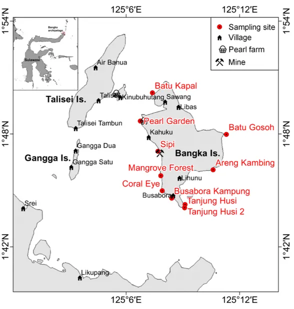
Bangka Island belongs to a small archipelago located at the northern tip of Sulawesi, Indonesia (Fig. 1). These islands are exposed to the main current coming from the western Pacific Ocean and directed toward the Indian Ocean (Tomascik et al., 1997). The central area of the archipelago is shallow, while the outer sides drop rapidly to over 1,000 m depth. The islands are covered by lush vegetation and fringing reefs are alternated with mangroves and volcanic cliffs. The islands are home to some villages and few resorts,
Figure 1 Sketch map of Bangka archipelago (north Sulawesi).Study sites, villages, pearl farms and mine are shown.
of which the oldest was built in 1987. Bangka, the largest of these islands is less than 48 km2. It has a resident population of about 2,500 inhabitants (as of 2013), distributed
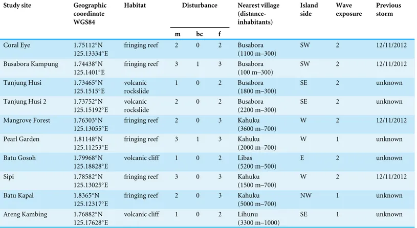
Table 1 Main characteristics of the study sites.Table 1 reports habitat typologies, disturbance levels, nearest village (including distance and inhab-itants), island side, wave exposure, previous storm. Human disturbances were divided into three main groups: m, mechanic (e.g., anchoring, boat strike, SCUBA diving, blastfishing), bc, bio-chemical (e.g., boat engine leaching, villages sewages), f, fishing pressure. Intensities of human distur-bances and wave exposure were classified into four ranked levels (0, none; 1, low; 2, medium; 3, high).
Study site Geographic
coordinate WGS84
Habitat Disturbance Nearest village
(distance-inhabitants) Island side Wave exposure Previous storm
m bc f
Coral Eye 1.75112◦
N 125.13334◦E
fringing reef 2 0 2 Busabora (1100 m–300)
SW 2 12/11/2012
Busabora Kampung 1.74438◦N
125.1401◦E
fringing reef 3 1 3 Busabora (100 m–300)
SW 2 12/11/2012
Tanjung Husi 1.73465◦N
125.1515◦
E
volcanic rockslide
1 0 2 Busabora (1800 m–300)
SE 2 unknown
Tanjung Husi 2 1.73752◦N
125.15192◦E
volcanic rockslide
2 0 2 Busabora (2200 m–300)
SE 2 unknown
Mangrove Forest 1.76303◦
N 125.13055◦E
fringing reef 2 0 3 Kahuku (3600 m–700)
W 2 12/11/2012
Pearl Garden 1.81148◦N
125.11253◦E
fringing reef 3 1 3 Kahuku (2000 m–700)
W 1 unknown
Batu Gosoh 1.79968◦N
125.18828◦
E
volcanic cliff 1 0 2 Libas (5200 m–500)
E 2 unknown
Sipi 1.78582◦N
125.13025◦E
fringing reef 3 0 3 Kahuku (1500 m–700)
W 2 12/11/2012
Batu Kapal 1.8365◦N
125.12317◦E
fringing reef 2 0 3 Kahuku (5000 m–700)
NW 1 unknown
Areng Kambing 1.76882◦
N 125.17628◦E
volcanic cliff 1 0 2 Lihunu (3300 m–1000)
SE 1 unknown
Sampling design and survey method
Benthic assemblages, coral diseases and other signs of compromised health around Bangka Island (North Sulawesi, Indonesia;Fig. 1) were investigated at 10 randomly selected sites at three depths: 3, 6 and 9 m (below the mean lower low water). Surveys were conducted between the months of October and November 2013. Tide levels were calculated by WXTide, a free Windows tide prediction software (http://www.wxtide32. com), using the Manado subordinate station (based on Sungai Kutei reference station, Borneo, Indonesia) and locally calibrated using a depth logger (DST centi-TD from Star-Oddi,http://www.star-oddi.com). Six sites were characterised by fringing reefs, two sites by coral rims growing on volcanic cliffs and the other two by sparse coral heads on volcanic rockslides (seeTable 1). Along fringing reefs, the three chosen depths roughly correspond to reef flat, reef crest and slope (i.e., front reef), which are standard subzones in coral reef monitoring studies (e.g.,Hill & Wilkinson, 2004;Hoeksema, 2012). At each site and depth, five belt transects (10×2 m) were laid randomly along reef contours
(Beeden et al., 2008). A gap of at least 5 m was left between each transect. Overall, 150 belt transects were analysed, covering a total area of 3,000 m2. Scleractinian coral colonies larger than 20 cm in diameter were identified to genus level across all transects.
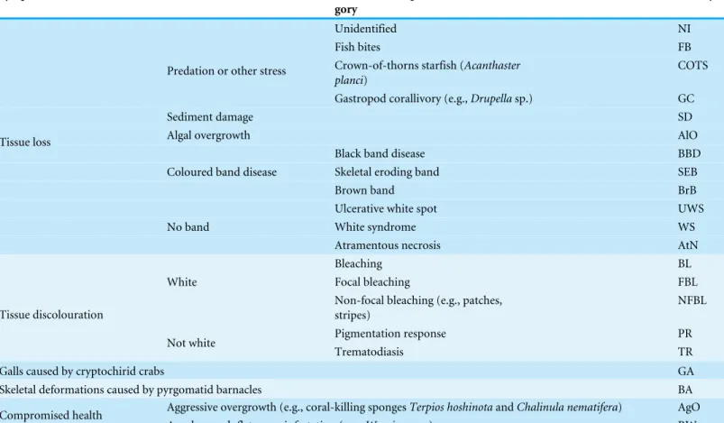
Table 2 Investigated coral diseases and other signs of compromised health.Adopted classification scheme and acronyms (afterBeeden et al., 2008;Raymundo, Couch & Harvell, 2008).
Symptom Disease or compromised health
cate-gory
Acronym
Unidentified NI
Fish bites FB
Crown-of-thorns starfish (Acanthaster planci)
COTS Predation or other stress
Gastropod corallivory (e.g.,Drupellasp.) GC
Sediment damage SD
Algal overgrowth AlO
Black band disease BBD
Skeletal eroding band SEB
Coloured band disease
Brown band BrB
Ulcerative white spot UWS
White syndrome WS
Tissue loss
No band
Atramentous necrosis AtN
Bleaching BL
Focal bleaching FBL
White
Non-focal bleaching (e.g., patches, stripes)
NFBL
Pigmentation response PR
Tissue discolouration
Not white
Trematodiasis TR
Galls caused by cryptochirid crabs GA
Skeletal deformations caused by pyrgomatid barnacles BA
Aggressive overgrowth (e.g., coral-killing spongesTerpios hoshinotaandChalinula nematifera) AgO Compromised health
Acoelomorph flatworm infestation (e.g.,Waminoasp.) RW
Scleractinian taxonomy was mainly based onVeron (2000), with some exceptions taking into account the recent reclassification of certain coral species. For example,Acropora andIsoporagenera were separated according toWallace, Done & Muir (2012). Corals previously from the genusFaviawere reclassified to the genusDipsastraea(Budd et al., 2012).Phymastreawas used to indicate all Indo-Pacific species previously indicated as MontastreainVeron (2000)(according toBudd et al. (2012)) and more recently reassigned toAstrea,ParamontastraeaandFavites(Huang et al., 2014). Similarly we have used the functional group ’Fungia’ to indicate some subgenera recently elevated to genus level (e.g., Danafungia,VerrillofungiaandPleuractis; seeGittenberger, Reijnen & Hoeksema (2011)).
In order to reduce inconsistencies associated with disease identification (Lindop, Hind & Bythell, 2008), each visible sign of coral disease or other compromised health indicators were photographed, identified and assigned to one of the 21 categories defined according to the identification guides byBeeden et al. (2008)andRaymundo, Couch & Harvell (2008). The classification scheme and adopted category acronyms can be seen in
Benthic sessile assemblages, bare rock, sand and coral rubble were quantified by using five photographic samples (50×50 cm), which were randomly located within each
transect. Benthic organisms were assigned to 14 main groups: algae, encrusting-, massive-, erected- and boring- sponges, hydroids, anemones, soft- and hard- corals, gorgonians, giant clams, colonial-, solitary- and social- ascidians. Percent covers were estimated by superimposing a grid of 100 equal-sized cells, using the software PhotoQuad (Trygonis & Sini, 2012). The data of percent cover was averaged across transects. No hard corals were detected at two transects, both of which were at Pearl Garden.
Information on anthropogenic disturbance sources at each site was obtained by interviewing local people, including those working at the resorts, and from data collected in previous surveys carried out applying the Reef Check protocol (Ponti et al., 2012); freely available data athttp://data.reefcheck.us/). Local human disturbances were grouped as mechanic (e.g., anchoring, boat strike, SCUBA diving, blast fishing), bio-chemical (e.g., boat engine leaking, village sewage) and fishing pressure. Storms are the primary natural disturbance in the area. Previous recent storms, such intense as to damage at least the shallow-water corals, were analysed in term of direction, occurrence date and wave exposure at each site. Intensities of human disturbances and wave exposure were classified into four ranked levels according to the maximum impacts recorded in the area (De Vantier & Turak, 2004): 0=almost no human disturbance and/or well sheltered, 1 =low or occasional disturbance and/or sheltered, 2=medium intensity, 3=high and
frequent disturbance and/or very exposed.
A research permit was granted prior to undertaking the survey work by the authority of Lihunu, Likupang eastern districts, Minahasa northern district government (Permit ID: 211/2019/SPP/IX-2013 issued at Lihunu on the 28th September 2013).
Data analyses
In the present study the abundances of each category of coral diseases and other compro-mised health indicators were expressed for each transect and analysed in terms of mean number of affected colonies per square metre of hard corals, based on the extension of hard corals in the transect.
Differences between sites (10 levels, random) and depths (three levels, fixed) were assessed by two-way crossed univariate and multivariate permutational analysis of variance (PERMANOVA,α=0.05;Anderson & Robinson, 2001;Anderson & Ter Braak,
2003).
Univariate tests were performed on Euclidean distances calculated on untransformed data (Anderson & Robinson, 2001). Multivariate tests were based on Bray-Curtis similarity of square root transformed data (Clarke, 1993). Patterns of similarities in diseases and other signs of compromised health among sites and depths were tested on zero-adjusted Bray-Curtis similarity of square root transformed data. An adjustment was applied to solve the indetermination of the Bray-Curtis coefficient, which occurs when it is calculated for pairs of transects without signs of diseases. This was accomplished by adding a dummy variable with a value of 0.22361, that is the square root of the lowest
non-zero value attainable (i.e., 0.05 affected colonies m−hard corals2 ) (Clarke, Somerfield & Chapman, 2006).
Multivariate similarity patterns were displayed by unconstrained ordination plots using the Principal Coordinate Analysis (PCoA, i.e., metric multidimensional scaling; Gower, 1966) based on the centroids of the similarity clouds at each site. Multivariate multiple regressions between similarity patterns and variables were performed by the DistLM procedure (marginal test;McArdle & Anderson, 2001) and significant correlations (P<0.05) were graphically represented by correlation vectors, proportional to the
Pearson’s correlation coefficients, superimposed on the PCoA plots. Correlations between abundances of diseases and other signs of compromised health were made compared with human and natural disturbances by the Spearman’s rank correlation coefficient (ρ).ρassesses how well the relationship between two variables can be described using
a monotonically increasing or decreasing function. Mean values were always reported together with their standard errors (SE). Statistical analysis was performed using PRIMER 6 with PERMANOVA +add-on package (Anderson, Gorley & Clarke, 2008). Spearman rank correlations and their tests were calculated by the computational language R (R Core Team, 2016).
RESULTS
Benthic community structure and coral cover
Benthic assemblages were very heterogeneous in the study area and the community structures significantly differed among sites and depths (Site×Depth:F18,120=2.2216,
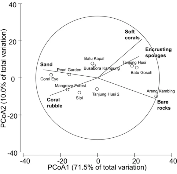
P =0.0001). Differences between sites were significantly related to the percent cover
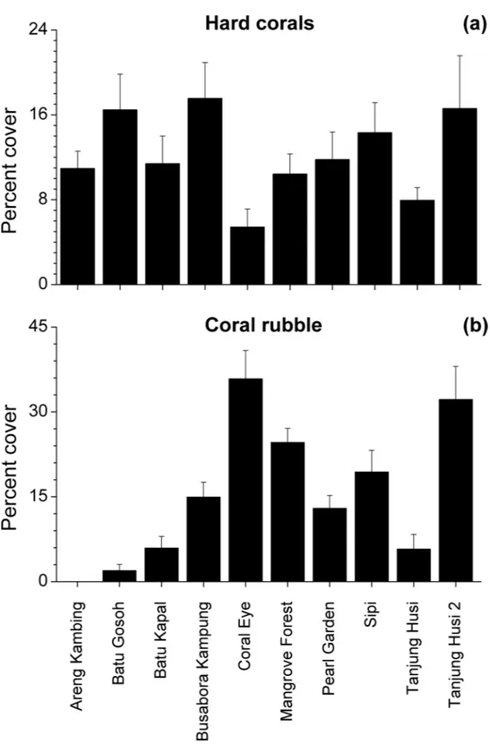
of bare rocks, sand, coral rubble, soft corals and encrusting sponges (Fig. 2). Percent cover of both total hard corals and coral rubble significantly varied among sites but not among depths (Coral rubble, Site:F9,120=16.557,P=0.0001; hard corals, Site:F9,120= 2.0735,P=0.0340). Total hard coral cover varied between 5.4±1.7% at Coral Eye and
17.6±3.4% at Busabora Kampung (Fig. 3A). Coral rubble cover varied from almost zero
at Areng Kambing, a volcanic cliff, to 35.9±5.0% at Coral Eye (Fig. 3B).
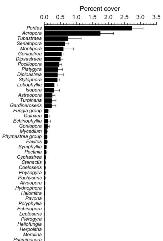
Overall, 42 genera of hard corals were found. The most abundant genera werePorites (mean cover 2.73±0.35%, up to 27.2%) andAcropora(mean cover 1.75±0.42%, up
to 42.0%;Fig. 4). Local high percent cover ofTubastraea(up to 54.4%) andMontipora (up to 43.8%) was also observed. Hard coral assemblages were very heterogeneous and variable among sites and depths (Site×Depth:F18,118 =1.2787,P=0.0044).
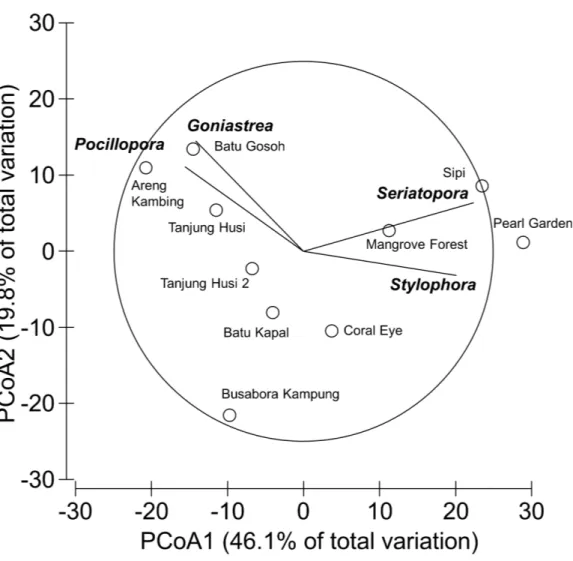
Nevertheless, few hard coral genera significantly contributed to the observable differences among sites. Those that did, included:Seriatopora,Stylophora,Goniastrea, andPocillopora (Fig. 5).
Occurrence of coral diseases and other signs of compromised health
Figure 2 Similarity patterns of benthic assemblages.The PCoA plot shows the multivariate similarity patterns of benthic assemblages, analysed in terms of main groups (i.e., algae, encrusting-, massive-, erected- and boring- sponges, hydroids, anemones, soft- and hard- corals, gorgonians, giant clams, colonial-, solitary- and social- ascidians), among sites. Open circles represent the centroids of the similarities of assemblages found at each site. Superimposed vectors indicate the intensity and direction of the correlations of the benthic variables selected by DistLM procedure (P<0.05 in the marginal test).
signs of compromised health werePoritesfollowed byAcropora, with 14 and 11 categories recorded respectively (Table 3).
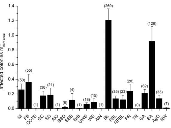
Mean abundances and total number of occurrences of each category of coral diseases and other signs of compromised health are shown inFig. 6. The most abundant type of compromised health recorded during this survey was coral bleaching (BL;Figs. 7Aand7B), followed by skeletal deformations caused by pyrgomatid barnacles (BA;
Fig. 7C), damage caused by fish bites (FB), pigmentation response (PR) and galls caused by cryptochirid crabs (GA;Fig. 7D).
No significant differences were recorded for bleaching across sites or depths, with instances recorded across 69% of the transects (mean 1.21±0.15 affected colonies
m−hard corals2 ) (Fig. 8A,Table 4). Bleaching was shown to affect 26 hard coral genera
Figure 4 Overall scleractinian genera abundances.Mean percent cover (+SE) of each coral genus in the whole study area.
Figure 5 Similarity patterns of hard coral assemblages.The PCoA plot shows the multivariate similar-ity patterns of hard coral assemblages, analysed at the genus level, among study sites. Open circles repre-sent the centroids of the similarities of assemblages found at each site. Superimposed vectors indicate the intensity and direction of the correlations of the genera selected by DistLM procedure (P <0.05 in the
marginal test).
(Table 3), with the most affected genera beingPorites,Dipsastraea,Goniastrea,Platygyra, Seriatopora, AcroporaandPocillopora.
Skeletal deformations caused by pyrgomatid barnacles were found in 51% of transects with a mean abundance of 0.92±0.20 affected colonies m−hard corals2 . Their distribution
was heterogeneous, showing significant differences of abundance among sites (Fig. 8B,
Table 4). Pyrgomatid barnacles were found to occur in 16 genera (Table 3); mostly colonising massive forms of corals belonging to thePoritesgenus.
Fish bites were found in 27% of transects with no significant difference of abundance among sites and depths (0.36±0.11 affected colonies m−hard corals2 ;Fig. 8C,Table 4). Again,
Table 3 Diseases and affected corals.Diseases and other signs of compromised health affecting each scleractinian genus (seeTable 2for the meaning of the acronyms).
Coral
genus
NI FB COTS GC SD AlO BBD SEB BrB UWS WS AtN BL FBL NFBL PR GA BA AgO RW Total
Acropora + + + + + + + + + + + 11
Alveopora + 1
Astreopora + + + + + + + 7
Coeloseris + + + + 4
Ctenactis + + + 3
Cyphastrea + + + + + + 6
Diploastrea + + 2
Dipsastraea + + + + + + 6
Echinophyllia + 1
Echinopora + 1
Favites + + + + + 5
Fungiagroup + + + + + 5
Galaxea + + + 3
Gardineroseris + + + + + 5
Goniastrea + + + + + + + + 8
Goniopora + + 2
Halomitra + 1
Hydnophora + + 2
Isopora + + + + + + 6
Lobophyllia + + 2
Montipora + + + + + 5
Mycedium + + 2
Pachyseris + + 2
Pavona + + + + + 5
Pectinia + + + 3
Phymastreagroup + + + 3
Physogyra + 1
Platygyra + + + + + + 6
Plerogyra + + 2
Pocillopora + + + + + 5
(continued on next page)
P
onti
e
t
al.
(2016),
P
eerJ
,
DOI
10.7717/peerj.2614
Table 3(continued)
Coral
genus
NI FB COTS GC SD AlO BBD SEB BrB UWS WS AtN BL FBL NFBL PR GA BA AgO RW Total
Polyphyllia + + + + 4
Porites + + + + + + + + + + + + + + 14
Seriatopora + + + + 4
Stylophora + + + + + 5
Turbinaria + + + + + + + + 8
Total 20 3 1 5 12 1 3 3 1 8 6 1 26 11 12 3 2 16 12 4
P
onti
e
t
al.
(2016),
P
eerJ
,
DOI
Figure 6 Overall occurrence of coral diseases and other signs of compromised health in the study area.
Mean (+SE) number of affected colonies per seabed surface covered by hard corals for each category, dis-regarding the affected coral genus, in brackets the total number of occurrences (seeTable 2for the mean-ing of the acronyms).
Signs of pigmentation response (PR) i.e., tissue discoloration, often bordering specific lesions or scars, were found in 16% of transects with a mean of 0.24±0.10
affected colonies m−hard corals2 . Their distribution was very heterogeneous, with significant differences found in abundance amongst both sites and depths (Table 4). However PR was only associated with three genera;Acropora,Fungiagroup andPorites(Table 3). It should be noted that PR (as classified in this study) can be caused by a number of factors such as specific coral borers, competitors, algal abrasion, fish bites, breakages, etc. and is therefore a type of ‘‘inflammatory’’ response suggesting a compromised health state but not itself a sign of disease (Beeden et al., 2008). Galls (GA), caused by cryptochirid crabs were encountered only at six of the sites surveyed (out of 10). GA occurred on 15% of transects, with a mean of 0.21±0.05 affected colonies m−2
hard corals. Occasionally
differences of GA abundances occurred between depths (Fig. 8D,Table 4). GA were observed more commonly with branchingSeriatoporaand to a lesser extentStylophora (Table 3).
A number of unidentified signs of tissue loss (named as NI in this manuscript), were found in 20 of the coral genera (Table 3) and at all sites (25% of transects, 0.26±0.08
affected colonies m−hard corals2 ), randomly distributed between site and depth (Table 4).
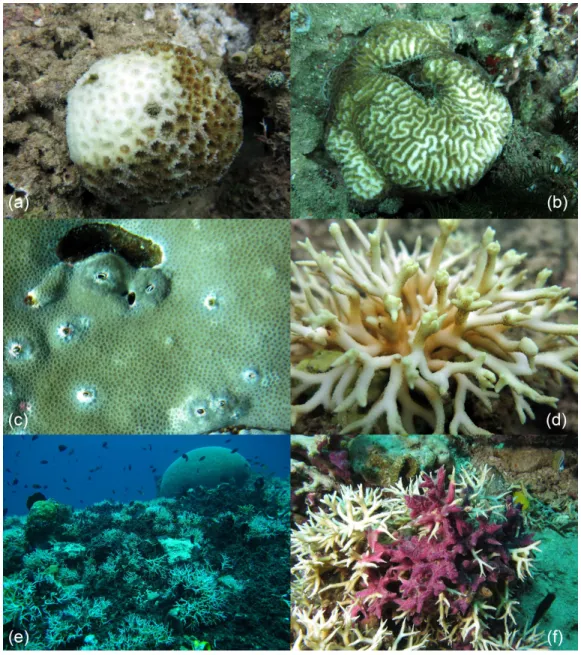
Figure 7 Examples of the most common health impairments found in the study area.Partially bleached colonies ofDipsastraea(A) andPlatygyra(B); skeletal deformations caused by pyrgomatid barnacles in
Porites(C); galls caused by cryptochirid crabs inSeriatopora(D);Terpios hoshinotainfestation, mostly on
Seriatopora, at Kahuko in 2011 (E);Chalinula nematifera, characterised by mauve coloration and white wavy filaments produced by symbiotic fungi, onSeriatopora(F).
Amongst less abundant categories of coral diseases and other signs of compromised health, cases of ulcerative white spot (UWS) were the most prevalent (found at nine out of 10 of the sites surveyed). UWS occurred on 11% of transects, with 0.06±0.02 affected
colonies m−hard corals2 . Eight genera showed signs of UWS (Table 3), withPoritesbeing the most commonly affected and significant differences between sites (Table 4).
Figure 8 Spatial and depth distribution of coral bleaching (A), skeletal deformations caused by pyrgo-matid barnacles (B), fish bites (C), and galls caused by cryptochirid crabs (D).Mean (+SE) number of affected colonies per seabed surface covered by hard corals at each site and investigated depth (−9,−6,−3
m).
12 genera but with relatively low prevalence at each site (Table 3). Focal (FBL) and non-focal bleaching (NFBL, e.g., patches, stripes) together with aggressive overgrowth by sponges and other invertebrates (AgO), e.g., the coral-killing spongesTerpios hoshinota andChalinula nematifera, were found on 11, 12 and 12 coral genera respectively (Table 3), again at relatively low prevalence (Fig. 6).
Interestingly, trematodiasis (TR) was not found on any of the reefs surveyed and black band disease (BBD) was only found on one colony ofGoniastrea, two colonies ofPorites and two ofTurbinaria.Similarly, skeletal eroding band (SEB) was found only on a colony ofCtenactis, a colony ofGalaxeaand two colonies ofPorites. Only one colony ofAcropora and one ofMontiporashowed signs of brown band disease (BrB) and atramentous necrosis (AtN) respectively.
Other occasional findings included oneAcroporaaffected by crown-of-thorns starfish (Acanthaster planci; COTS), oneGardineroserisshowing signs of algal overgrowth (AlO), and a few colonies ofGardineroseris,Platygyra,PoritesandTurbinariainfested by acoelomorph flatworms (RW), more preciselyWaminoasp. (Table 3;Fig. 6).
Spatial distribution patterns of diseases and other signs of compromised health
Patterns of similarities in diseases and other signs of compromised health revealed sig-nificant differences among sites, but not among depths (Table 5;Fig. 9A). The categories
Table 4 Effects of sites and depths on the distribution of coral diseases and other signs of compromised health.Summary of PERMANOVA tests on the abundance (affected colonies m−2
hard corals) of the most common coral diseases and other signs of compromised health, analysed individually,
according to the factors Site, Depth, and their interaction (Site×Depth). SeeTable 2for the meaning of the acronyms.
Site Depth Site×Depth Res
MS F9,118 P MS F2,18 P MS F18,118 P MS
NI 0.6817 0.7194 0.7332 0.3929 0.4257 0.6740 0.9229 0.9739 0.4935 0.9477 FB 2.7547 1.6906 0.0708 0.0266 0.0178 0.9836 1.4972 0.9189 0.5800 1.6294 GC 0.6276 1.1400 0.3154 0.5835 0.9926 0.4127 0.5879 1.0679 0.3522 0.5505 SD 1.0092 0.7667 0.7422 0.5151 0.3747 0.7379 1.3747 1.0443 0.3652 1.3164 UWS 28.8050 1.6294 0.0041** 17.7010 1.0192 0.4588 17.3660 0.9824 0.5456 17.6780
WS 0.2285 0.7742 0.7603 0.3539 1.1990 0.3630 0.2951 0.9999 0.4530 0.2952 BL 0.8230 0.2220 0.9942 1.6280 0.4293 0.6647 3.7922 1.0230 0.4229 3.7069 FBL 0.2643 1.2635 0.2339 0.0813 0.4067 0.6855 0.1997 0.9548 0.5131 0.2092 NFBL 0.4497 0.8634 0.6467 0.4337 0.8365 0.4997 0.5185 0.9955 0.4662 0.5208 PR 1.8391 1.5731 0.0843 4.0477 1.9376 0.1715 2.0911 1.7887 0.0144* 1.1691
GA 1.9667 6.7696 0.0003*** 0.1365 0.2783 0.7676 0.4909 1.6896 0.0449* 0.2905
BA 16.6410 3.0347 0.0018** 8.0038 1.9467 0.1637 4.1084 0.7492 0.8109 5.4836
AgO 0.3706 1.2014 0.2655 0.6377 2.5486 0.0924 0.2501 0.8108 0.7901 0.3084
Notes.
*Indicated significant level ofP<0.05.
**Indicated significant level ofP<0.01. ***Indicated significant level ofP<0.001.
Table 5 Effects of sites and depths on the distribution of coral diseases assemblages.PERMANOVA test on the multivariate similarity patterns obtained by applying the zeroadjusted Bray-Curtis coefficient to square root transformed abundances of diseases and other signs of compromised health (affected colonies m−2
hard corals) according to the factors Site, Depth, and their interaction (Site×Depth).
Source df SS MS F P Perms Denominator
Site 9 48362 5374 2.3929 0.0001 9838 Res
Depth 2 3434 1717 0.6393 0.8225 9932 Site×Depth Site×Depth 18 48361 2687 1.1964 0.0767 9783 Res
Res 118 264990 2246 Total 147 365100
that significantly contribute to the observed similarity pattern were skeletal deformations caused by pyrgomatid barnacles, occurrence of corals showing signs of white syndromes, fish bites, non-focal bleaching and gastropod corallivory. These signs of compromised health increased in abundance closer to the Coral Eye site. In contrast, galls caused by cryptochirid crabs increased towards Sipi and Pearl Garden sites (Fig. 9A).
Figure 9 Similarity patterns of diseases and other signs of compromised health.The PCoA plot shows the multivariate similarity patterns of diseases and other signs of compromised health assemblages among study sites. Open circles represent the centroids of the similarities of assemblages found at each site. Cor-relation vectors superimposed on the PCoA plot represent: (A) diseases and compromised health cat-egories significantly related with the similarity pattern (seeTable 2for the meaning of the acronyms); (B) substrate typologies significantly related with the similarity pattern (selected by DistLM procedure;
P<0.05 in the marginal test).
Correlations between diseases and possible human and natural disturbances
According to information gathered from the local populace and previous surveys (Ponti et al., 2012), the most impacted sites in terms of mechanical disturbance, pollution and fishing, were Busabora Kampung (close to the homonymous village), Pearl Garden (located in the surroundings of a dismissed pearl farm), and Sipi (in front of the newly established metal mine) seeTable 1. At Pearl Garden and Sipi, signs of recent blast fishing were evident. The most relevant recent storm happened a year earlier and hit the southeastern side of the island.
Few significant rank correlations between the abundances of diseases and other signs of compromised health were observed with regard to possible human and natural disturbances. Abundance of colonies affected by skeletal eroding bands increased with the intensity of bio-chemical disturbances (ρ=0.6864,P=0.0284) and decreased with
distance from villages (ρ= −0.7647,P=0.0100). Moreover, instances of damage caused
by sedimentation together with observed instances of non-focal bleaching, decreased with regard to distance from the villages (respectively:ρ= −0.6933,P =0.0262 and ρ=0.7455,P=0.0133), while skeletal deformations caused by pyrgomatid barnacles
decreased with the intensity of bio-chemical disturbances (ρ= −0.6963,P=0.0253).
DISCUSSION
Page & Willis, 2008;Sweet & Séré, 2016), was found to increase towards villages and their bio-chemical disturbances, supporting the hypothesis that its spread is favored by anthropogenic stresses that may compromise corals health (Page & Willis, 2008;Montano et al., 2016).
Coral bleaching was the most common sign of compromised health in the study area, affecting 26 genera. This fits current trends associated with reefs on a global scale (Berkelmans & Oliver, 1999;Carpenter et al., 2008;Burke et al., 2012;Sutthacheep et al., 2013). Although there appears to be no escape for corals from global increases in sea surface temperature (Baker, Glynn & Riegl, 2008), the susceptibility of corals to bleaching appears to vary considerably. For example differences have been reported between species (e.g.,Hoeksema, 1991;Marshall & Baird, 2000;Loya et al., 2001;Montano et al., 2010;Hoeksema & Matthews, 2011) and locations (e.g.,Pineda et al., 2013). The latter variation has been linked to differences in several regional environmental factors (e.g., light intensity and water flow rate) which appear to be influencing the outcome of bleaching intensity (Glynn, 1996;Brown, 1997). Indeed, it has long been ‘known’ that branching corals (e.g., acroporids and pocilloporids) are more sensitive to thermal stress than massive growth forms (Loya et al., 2001;Wooldridge, 2014). However, recent studies have shown quite the opposite can occur (Guest et al., 2012). Observing high levels of bleaching at any given time is often a worrying trend. However, the lack of major diseases in this region is a promising sign, as studies have shown that bleaching events on their own do not necessarily change the coral taxonomic community structure despite often resulting in a reduced amount of total coral cover (Guest et al., 2016).
Next to bleaching, the second most common sign of compromised health around Bangka Island was skeletal deformations caused by pyrgomatid barnacles. Corals with massive growth forms (PoritesandPlatygyrafor example), showed the highest abundances of these skeletal deformations. Interestingly, although such organisms are present on reefs throughout the world, few studies focus on assessing the potential damage they can do to individual colonies (Frank et al., 1995). These coral-inhabiting barnacles are unable to bore directly into the host, but to varying extent, they are able to inhibit or regulate their host’s skeletal growth (Anderson, 1992). Few species are considered able to adversely affect their hosts, nevertheless infestations may damage especially finely branched scleractinian corals (Ross & Newman, 1995). Although this study does not assess the effects high abundances of these organisms have on these reefs, the high abundances found warrants further study to assess if these should be recorded in future baseline surveys of reef health.
Similar to above, the abundances of fish bites, including parrotfish, butterflyfish, filefish, pufferfish, triggerfish, and damselfish families, for example are rarely charac-terised and therefore measured in coral health surveys. This is despite several studies suggesting that corallivorous fishes may be vectors for coral disease (Rogers, 2008). Indeed numerous fish species have been documented to target feeding on coral lesions when present and this has facilitated the hypotheses that they can spread the infectious agents from colony to colony (Chong-Seng et al., 2011). Here, the majority of fish bites observed were associated with massivePoriteswith only few examples associated with colonies of AcroporaandPocillopora. Here instead of highlighting the potential of disease occurrence
due to fish bites, we argue that an intermediate level of predation by fishes on hard coral should actually be considered an index of a balanced fish assemblages in almost all healthy coral reef environments (Cole, Pratchett & Jones, 2008).
In contrast, possible outbreaks of the corallivorous gastropodsDrupellaspp. are raising concerns not only for their direct predatory effects on their preys, but also for their feared involvement as vector in some infectious diseases (e.g.,Turner, 1994;Antonius & Riegl, 1998;Onton et al., 2011;Nicolet et al., 2013). Luckily, cases ofDrupellaaround the island are still relatively few yet we recommend they should still be monitored over time.
A commonly overlooked group of associated fauna affecting the morphology of corals consists of cryptochirid crabs that live in corals ofSeriatoporaand related genera. These tiny crabs are obligate symbionts of living scleractinian corals and have been found to feed on coral tissue and mucus along with inducing gall formation (Kropp, 1990;Terrana et al., 2016). Here, high abundances of cryptochirid crabs were observed at Pearl Garden (the site of an old pearl farm) and Sipi (the location of an active metal mine) (Fig. 9A). Moreover, both sites were also characterised by recent signs of blast fishing. Although the rank correlation between galls caused by cryptochirid crabs and human disturbances was not significant, the presence of many cases in the study area, especially in the most disturbed sites, deserves further investigations. Gall crabs can perhaps also be found in other coral species in the research area, since in nearby Lembeh Strait (North Sulawesi), cryptochirids were observed to occur abundantly in a single, large coral colony ofPavona clavus(Hoeksema & Van der Meij, 2012).
(Fig. 7F).C. nematiferais similarly reportedly to be spreading fast throughout the Indo-Pacific (Ávila & Carballo, 2009) and is thought to have been introduced to this region, possibly by fouling from ships. The presence ofC. nematiferain the study area and along eastern Sulawesi was recently documented byRossi et al. (2015), nevertheless quantitative studies are urgently needed to assess the severity of these sponge outbreaks.
Interestingly, although most studies of this kind focus on the coral genera which show presence of disease signs or other compromised health, few highlight those genera which appear disease-free. In this instance, the majority of genera which showed no signs of compromised health were also those with very low abundances such asHeliofungia, Herpolitha,Leptoseris,Merulina,PsammocoraandSymphyllia.However such findings may simply be down to the low occurrence of these genera. In contrast,Tubastraeawhich was one of the most abundant genera of corals found at our sites was also shown to have a lack of apparent instances of compromised health and further studies should assess if this trend is seen throughout the corals range.
To conclude, although the study area is relatively undisturbed as far as tourism devel-opment, impact on the area by other human activities appear to be having a significant effect on coral health and this warrants further longer term study. Of particular concern is the increase in mining which is occurring in this area along with other off shore islands throughout Sulawesi (Edinger, Siregar & Blackwood, 2007;Caras & Pasternak, 2009; Lasut et al., 2010). However, directly linking such activities to coral health status can be challenging (Rogers, 1990;Bruckner & Bruckner, 1997;Fabricius, 2005;Voss & Richardson, 2006a;Haapkylä et al., 2011;Erftemeijer et al., 2012;Pollock et al., 2014;Heintz, Haapkylae & Gilbert, 2015).
Regardless of asserting the cause of reef decline, the provision of baseline surveys for monitoring coral health status lay the foundations to assess the effects of any such anthropogenic and/or natural effects on reefs over future years. Therefore, such activities should continue to be undertaken with the understanding that return visits are scheduled and conducted in the same manner to allow for direct comparisons between data sets.
ACKNOWLEDGEMENTS
We wish to thank Gianfranco Rossi (Reef Check Italia association) for the training on hard coral identification, and all the Coral Eye staff for the field support. We are very grateful to Simone Montano and another anonymous reviewer for their constructive and generous comments on the manuscript.
ADDITIONAL INFORMATION AND DECLARATIONS
Funding
This research was partially funded by Coral Eye, which hosted FF and covered all field expenses. The non-profit organization Reef Check Italia Onlus sustained the cost for open source publication. The funders had no role in study design, data collection and analysis, decision to publish, or preparation of the manuscript.
Grant Disclosures
The following grant information was disclosed by the authors: Coral Eye.
non-profit organization Reef Check Italia Onlus.
Competing Interests
The authors declare there are no competing interests.
Author Contributions
• Massimo Ponti conceived and designed the experiments, analyzed the data, contributed
reagents/materials/analysis tools, wrote the paper, prepared figures and/or tables, reviewed drafts of the paper.
• Francesca Fratangeli and Nicolò Dondi conceived and designed the experiments,
performed the experiments, analyzed the data, wrote the paper, prepared figures and/or tables, reviewed drafts of the paper.
• Marco Segre Reinach conceived and designed the experiments, performed the
exper-iments, contributed reagents/materials/analysis tools, prepared figures and/or tables, reviewed drafts of the paper.
• Clara Serra performed the experiments, contributed reagents/materials/analysis tools,
reviewed drafts of the paper.
• Michael J. Sweet analyzed the data, contributed reagents/materials/analysis tools, wrote
the paper, reviewed drafts of the paper.
Field Study Permissions
The following information was supplied relating to field study approvals (i.e., approving body and any reference numbers):
A research permit was granted prior to undertaking the survey work by the authority of Lihunu, Likupang eastern districts, Minahasa northern district government (Permit ID: 211/2019/SPP/IX-2013 issued at Lihunu on the 28th September 2013).
Data Availability
The following information was supplied regarding data availability: The raw data has been supplied as aData S1.
Supplemental Information
Supplemental information for this article can be found online athttp://dx.doi.org/10. 7717/peerj.2614#supplemental-information.
REFERENCES
Anderson DT. 1992.Structure, function and phylogeny of coral-inhabiting barnacles (Cirripedia, Balanoidea).Zoological Journal of the Linnean Society106(4):277–339
DOI 10.1111/j.1096-3642.1992.tb01249.x.
Anderson MJ, Robinson J. 2001.Permutation tests for linear models.Australian and New Zealand Journal of Statistic43(1):75–88DOI 10.1111/1467-842X.00156.
Anderson MJ, Ter Braak CJF. 2003.Permutation tests for multi-factorial analysis of variance.Journal of Statistical Computation and Simulation73(2):85–113
DOI 10.1080/0094965021000015558.
Antonius A, Riegl B. 1998.Coral diseases andDrupella cornusinvasion in the Red Sea. Coral Reefs17(1):48–48DOI 10.1007/s003380050093.
Antonius AA, Lipscomb D. 2001.First protozoan coral-killer identified in the Indo-Pacific.Atoll Research Bulletin481:1–21DOI 10.5479/si.00775630.481.
Ávila E, Carballo JL. 2009.A preliminary assessment of the invasiveness of the Indo-Pacific spongeChalinula nematiferaon coral communities from the tropical Eastern Pacific.Biological Invasions11(2):257–264DOI 10.1007/s10530-008-9230-5.
Baker AC, Glynn PW, Riegl B. 2008.Climate change and coral reef bleaching: an ecological assessment of long-term impacts, recovery trends and future outlook. Estuarine Coastal and Shelf Science80(4):435–471DOI 10.1016/j.ecss.2008.09.003. Ban SS, Graham NAJ, Connolly SR. 2014.Evidence for multiple stressor interactions and
effects on coral reefs.Global Change Biology20(3):681–697 DOI 10.1111/gcb.12453. Beeden R, Willis BL, Raymundo LJ, Page CA, Weil E. 2008.Underwater cards for
assessing coral health on Indo-Pacific Reefs. Melbourne: Coral Reef Targeted Research and Capacity Building for Management Program.
Berkelmans R, Oliver JK. 1999.Large-scale bleaching of corals on the Great Barrier Reef. Coral Reefs18(1):55–60DOI 10.1007/s003380050154.
Brown BE. 1997.Coral bleaching: causes and consequences.Coral Reefs16(Suppl.):
S129–S138DOI 10.1007/s003380050249.
Brown BE, Suharsono. 1990.Damage and recovery of coral reefs affected by El Niño related seawater warming in the Thousand Islands, Indonesia.Coral Reefs 8(4):163–170DOI 10.1007/bf00265007.
Bruckner AW, Bruckner RJ. 1997.Outbreak of coral disease in Puerto Rico.Coral Reefs 16(4):260–260DOI 10.1007/s003380050081.
Bruno JF, Petes LE, Harvell CD, Hettinger A. 2003.Nutrient enrichment can increase the severity of coral diseases.Ecology Letters6(12):1056–1061
DOI 10.1046/j.1461-0248.2003.00544.x.
Budd AF, Fukami H, Smith ND, Knowlton N. 2012.Taxonomic classification of the reef coral family Mussidae (Cnidaria: Anthozoa: Scleractinia).Zoological Journal of the Linnean Society166(3):465–529DOI 10.1111/j.1096-3642.2012.00855.x.
Burge CA, Mark Eakin C, Friedman CS, Froelich B, Hershberger PK, Hofmann EE, Petes LE, Prager KC, Weil E, Willis BL, Ford SE, Harvell CD. 2014. Climate change influences on marine infectious diseases: implications for management and society.Annual Review of Marine Science6(1):249–277
DOI 10.1146/annurev-marine-010213-135029.
Burke L, Reytar K, Spalding M, Perry A. 2012.Reefs at risk. Revisited in the coral triangle. Washington, D.C.: World Resources Institute.
Caras T, Pasternak Z. 2009.Long-term environmental impact of coral mining at the Wakatobi marine park, Indonesia.Ocean and Coastal Management 52(10):539–544
DOI 10.1016/j.ocecoaman.2009.08.006.
Carpenter KE, Abrar M, Aeby G, Aronson RB, Banks S, Bruckner A, Chiriboga A, Cortes J, Delbeek JC, Devantier L, Edgar GJ, Edwards AJ, Fenner D, Guzman HM, Hoeksema BW, Hodgson G, Johan O, Licuanan WY, Livingstone SR, Lovell ER, Moore JA, Obura DO, Ochavillo D, Polidoro BA, Precht WF, Quibilan MC, Reboton C, Richards ZT, Rogers AD, Sanciangco J, Sheppard A, Sheppard C, Smith J, Stuart S, Turak E, Veron JEN, Wallace C, Weil E, Wood E. 2008.One-third of reef-building corals face elevated extinction risk from climate change and local impacts.Science321(5888):560–563DOI 10.1126/science.1159196.
Cervino JM, Hauff B, Haslun JA, Winiarski-Cervino K, Cavazos M, Lawther P, Wier AM, Hughen K, Strychar KB. 2012.Ulcerated yellow spot syndrome: implications of aquaculture-related pathogens associated with soft coralSarcophyton ehrenbergi tissue lesions.Diseases of Aquatic Organisms102(2):137–148DOI 10.3354/dao02541. Chong-Seng KM, Cole AJ, Pratchett MS, Willis BL. 2011.Selective feeding by coral reef
fishes on coral lesions associated with brown band and black band disease.Coral Reefs30(2):73–481DOI 10.1007/s00338-010-0707-1.
Clarke KR. 1993.Non-parametric multivariate analyses of changes in community structure.Australian Journal of Ecology18:117–143
DOI 10.1111/j.1442-9993.1993.tb00438.x.
Clarke KR, Somerfield PJ, Chapman MG. 2006.On resemblance measures for ecological studies, including taxonomic dissimilarities and a zero-adjusted Bray-Curtis
coefficient for denuded assemblages.Journal of Experimental Marine Biology and Ecology 330(1):55–80DOI 10.1016/j.jembe.2005.12.017.
Cole AJ, Pratchett MS, Jones GP. 2008.Diversity and functional importance of coral-feeding fishes on tropical coral reefs.Fish and Fisheries9(3):286–307
DOI 10.1111/j.1467-2979.2008.00290.x.
Dalton S, Smith SA. 2006.Coral disease dynamics at a subtropical location, Solitary Islands Marine Park, eastern Australia.Coral Reefs25(1):37–45
DOI 10.1007/s00338-005-0039-8.
De Vantier L, Turak E. 2004.Managing marine tourism in Bunaken National Park and adjacent waters, North Sulawesi, Indonesia. Jakarta: NRM III.
De Voogd NJ, Cleary DFR, Dekker F. 2013.The coral-killing spongeTerpios hoshinota invades Indonesia.Coral Reefs32(3):755–755DOI 10.1007/s00338-013-1030-4. Edinger E. 2012.Gold mining and submarine tailings disposal review and case study.
Oceanography 25(2):184–199.
Edinger EN, Azmy K, Diegor W, Siregar PR. 2008.Heavy metal contamination from gold mining recorded in Porites lobata skeletons, Buyat-Ratototok dis-trict, North Sulawesi, Indonesia.Marine Pollution Bulletin56(9):1553–1569
DOI 10.1016/j.marpolbul.2008.05.028.
Buyat-Ratototok district, North Sulawesi, Indonesia.Environmental Geology 52(4):701–714DOI 10.1007/s00254-006-0506-8.
Elliott J, Patterson M, Summers N, Miternique C, Montocchio E, Vitry E. 2016. How does the proliferation of the coral-killing spongeTerpios hoshinotaaffect benthic community structure on coral reefs?Coral Reefs35(3):1083–1095
DOI 10.1007/s00338-016-1434-z.
Erftemeijer PLA, Riegl B, Hoeksema BW, Todd PA. 2012.Environmental impacts of dredging and other sediment disturbances on corals: a review.Marine Pollution Bulletin64(9):1737–1765DOI 10.1016/j.marpolbul.2012.05.008.
Fabricius KE. 2005.Effects of terrestrial runoff on the ecology of corals and coral reefs: review and synthesis.Marine Pollution Bulletin50(2):125–146
DOI 10.1016/j.marpolbul.2004.11.028.
Fava F, Ponti M, Scinto A, Calcinai B, Cerrano C. 2009.Possible effects of human impacts on epibenthic communities and coral rubble features in the marine Park of Bunaken (Indonesia).Estuarine, Coastal and Shelf Science85(1):151–156
DOI 10.1016/j.ecss.2009.02.028.
Frank U, Brickner I, Rinkevich B, Loya Y, Bak RPM, Achituv Y, Ilan M. 1995. Al-logeneic and xenogeneic interactions in reef-building corals may induce tissue-growth without calcification.Marine Ecology Progress Series124(1–3):181–188
DOI 10.3354/meps124181.
Gittenberger A, Reijnen BT, Hoeksema BW. 2011.A molecularly based phylogeny reconstruction of mushroom corals (Scleractinia: Fungiidae) with taxonomic consequences and evolutionary implications for life history traits.Contributions to Zoology80(2):107–132.
Glynn PW. 1996.Coral reef bleaching: facts, hypotheses and implications.Global Change Biology2(6):495–509DOI 10.1111/j.1365-2486.1996.tb00063.x.
Gower JC. 1966.Some distance properties of latent root and vector methods used in multivariate analysis.Biometrika53:325–338.
Guest JR, Baird AH, Maynard JA, Muttaqin E, Edwards AJ, Campbell SJ, Yewdall K, Affendi YA, Chou LM. 2012.Contrasting patterns of coral bleaching susceptibility in 2010 suggest an adaptive response to thermal stress.PLoS ONE7(3):e33353
DOI 10.1371/journal.pone.0033353.
Guest JR, Low J, Tun K, Wilson B, Ng C, Raingeard D, Ulstrup KE, Tanzil JTI, Todd PA, Toh TC, McDougald D, Chou LM, Steinberg PD. 2016.Coral community response to bleaching on a highly disturbed reef.Scientific Reports6:20717
DOI 10.1038/srep20717.
Haapkylä J, Melbourne-Thomas J, Flavell M, Willis BL. 2013.Disease outbreaks, bleaching and a cyclone drive changes in coral assemblages on an inshore reef of the Great Barrier Reef.Coral Reefs32(3):815–824DOI 10.1007/s00338-013-1029-x. Haapkylä J, Seymour AS, Barneah O, Brickner I, Hennige S, Suggett D, Smith D.
2009a.Association of,Waminoasp. (Acoela) with corals in the Wakatobi Ma-rine Park, South-East Sulawesi, Indonesia.Marine Biology156(5):1021–1027
DOI 10.1007/s00227-009-1145-x.
Haapkylä J, Seymour AS, Trebilco J, Smith D. 2007.Coral disease prevalence and coral health in the Wakatobi Marine Park, south-east Sulawesi, Indonesia. Jour-nal of the Marine Biological Association of the United Kingdom87(2):403–414
DOI 10.1017/s0025315407055828.
Haapkylä J, Unsworth RKF, Flavell M, Bourne DG, Schaffelke B, Willis BL. 2011. Seasonal rainfall and runoff promote coral disease on an inshore reef.PLoS ONE 6(2):e16893DOI 10.1371/journal.pone.0016893.
Haapkylä J, Unsworth RKF, Seymour AS, Melbourne-Thomas J, Flavell M, Willis BL, Smith DJ. 2009b.Spatio-temporal coral disease dynamics in the Wakatobi Marine National Park, South-East Sulawesi, Indonesia.Diseases of Aquatic Organisms87(1– 2):105–115DOI 10.3354/dao02160.
Haapkylae J, Melbourne-Thomas J, Flavell M. 2015.The association between coral communities and disease assemblages in the Wakatobi Marine National Park, south-eastern Sulawesi, Indonesia.Marine and Freshwater Research66(10):948–955
DOI 10.1071/mf14192.
Harvell CD, Mitchell CE, Ward JR, Altizer S, Dobson AP, Ostfeld RS, Samuel MD. 2002.Ecology - Climate warming and disease risks for terrestrial and marine biota. Science296(5576):2158–2162DOI 10.1126/science.1063699.
Harvell D, Jordan-Dahlgren E, Merkel S, Rosenberg E, Raymundo L, Smith G, Weil E, Willis B, Global Envrionm Facility C. 2007.Coral disease, environmental drivers, and the balance between coral and microbial associates.Oceanography 20(1):172–195DOI 10.5670/oceanog.2007.91.
Heintz T, Haapkylae J, Gilbert A. 2015.Coral health on reefs near mining sites in New Caledonia.Diseases of Aquatic Organisms115(2):165–173DOI 10.3354/dao02884. Hill J, Wilkinson C. 2004.Methods for ecological monitoring of coral reefs. A resource for
managers. Townsville: Australian Institute of Marine Science.
Hoeksema BW. 1991.Control of bleaching in mushroom coral populations (Scleractinia: Fungiidae) in the Java Sea: stress tolerance and interference by life history strategy. Marine Ecology Progress Series74:225–237DOI 10.3354/meps074225.
Hoeksema BW. 2012.Distribution patterns of mushroom corals (Scleractinia: Fungi-idae) across the Spermonde Shelf, South Sulawesi.Raffles Bulletin of Zoology 60(1):183–212.
Hoeksema BW, Farenzena ZT. 2012.Tissue loss in corals infested by acoelomorph flat-worms (Waminoasp.).Coral Reefs31(3):869–869DOI 10.1007/s00338-012-0919-7. Hoeksema BW, Matthews JL. 2011.Contrasting bleaching patterns in mushroom
coral assemblages at Koh Tao, Gulf of Thailand.Coral Reefs30(1):95–95
DOI 10.1007/s00338-010-0675-5.
Hoeksema BW, Van der Meij SET. 2012.Gall crab city: an aggregation of endosymbiotic crabs inhabiting a colossal colony ofPavona clavus.Coral Reefs32(1):59–59
Huang D, Benzoni F, Fukami H, Knowlton N, Smith ND, Budd AF. 2014.Taxonomic classification of the reef coral families Merulinidae, Montastraeidae, and Diploas-traeidae (Cnidaria: Anthozoa: Scleractinia).Zoological Journal of the Linnean Society 171(2):277–355DOI 10.1111/zoj.12140.
Johan O, Ginanjar R, Priyadi A. 2015.Coral health levels of wild ornamental coral in East Belitung waters, Indonesia.Nusantara Bioscience7(2):127–132
DOI 10.13057/nusbiosci/n070212.
Katz SM, Pollock FJ, Bourne DG, Willis BL. 2014.Crown-of-thorns starfish pre-dation and physical injuries promote brown band disease on corals.Coral Reefs 33(3):705–716DOI 10.1007/s00338-014-1153-2.
Kropp RK. 1990.Revision of the genera of gall crabs (Crustacea: Cryptochiridae) occurring in the Pacific Ocean.Pacific Science44(4):417–448.
Lafferty KD, Holt RD. 2003.How should environmental stress affect the population dynamics of disease?Ecology Letters6(7):654–664
DOI 10.1046/j.1461-0248.2003.00480.x.
Lasut MT, Yasuda Y, Edinger EN, Pangemanan JM. 2010.Distribution and accumula-tion of mercury derived from gold mining in marine environment and its impact on residents of Buyat Bay, North Sulawesi, Indonesia.Water Air and Soil Pollution 208(1–4):153–164DOI 10.1007/s11270-009-0155-0.
Lindop AMM, Hind EJ, Bythell JC. 2008.The unknowns in coral disease identification: an experiment to assess consensus of opinion amongst experts. In:Proceedings of the 11th International Coral Reef Symposium. Ft Lauderdale, Florida, 7-11 July 2008 Session number 7.
Loya Y, Sakai K, Yamazato K, Nakano Y, Sambali H, Woesik R. Van. 2001.Coral bleaching: the winners and the losers.Ecology Letters4(2):122–131
DOI 10.1046/j.1461-0248.2001.00203.x.
Madduppa H, Schupp PJ, Faisal MR, Sastria MY, Thoms C. 2015.Persistent outbreaks of the black disease spongeTerpios hoshinotain Indonesian coral reefs.Marine BiodiversityEpub ahead of print Dec 16 2015DOI 10.1007/s12526-015-0426-5. Marshall PA, Baird AH. 2000.Bleaching of corals on the Great Barrier Reef: differential
susceptibilities among taxa.Coral Reefs19(2):155–163DOI 10.1007/s003380000086. McArdle BH, Anderson MJ. 2001.Fitting multivariate models to community data:
a comment on distance-based redundancy analysis.Ecology82(1):290–297
DOI 10.1890/0012-9658(2001)082[0290:FMMTCD]2.0.CO2.
Miller J, Sweet MJ, Wood E, Bythell J. 2015.Baseline coral disease surveys within three marine parks in Sabah, Borneo.PeerJ 3:e1391DOI 10.7717/peerj.1391.
Montano S, Giorgi A, Monti M, Seveso D, Galli P. 2016.Spatial variability in distribution and prevalence of skeletal eroding band and brown band dis-ease in Faafu Atoll, Maldives.Biodiversity and Conservation25(9):1625–1636
DOI 10.1007/s10531-016-1145-3.
Montano S, Seveso D, Galli P, Obura DO. 2010.Assessing coral bleaching and recovery with a colour reference card in Watamu Marine Park, Kenya.Hydrobiologia
655(1):99–108DOI 10.1007/s10750-010-0407-4.
Montano S, Strona G, Seveso D, Galli P. 2012.First report of coral diseases in the Republic of Maldives.Diseases of Aquatic Organisms101(2):159–165
DOI 10.3354/dao02515.
Montano S, Strona G, Seveso D, Galli P. 2013.Prevalence, host range, and spatial distribution of black band disease in the Maldivian Archipelago.Diseases of Aquatic Organisms105(1):65–74DOI 10.3354/dao02608.
Montano S, Strona G, Seveso D, Maggioni D, Galli P. 2015.Widespread occur-rence of coral diseases in the central Maldives.Marine and Freshwater Research 67(8):1253–1262DOI 10.1071/mf14373.
Myers RL, Raymundo LJ. 2009.Coral disease in Micronesian reefs: a link between disease prevalence and host abundance.Diseases of Aquatic Organisms87(1–2):97–104
DOI 10.3354/dao02139.
Naumann MS, Mayr C, Struck U, Wild C. 2010.Coral mucus stable isotope composition and labeling: experimental evidence for mucus uptake by epizoic acoelomorph worms.Marine Biology157(11):2521–2531DOI 10.1007/s00227-010-1516-3. Nicolet KJ, Hoogenboom MO, Gardiner NM, Pratchett MS, Willis BL. 2013.The
corallivorous invertebrateDrupellaaids in transmission of brown band disease on the Great Barrier Reef.Coral Reefs32(2):585–595DOI 10.1007/s00338-013-1010-8. Nugues MM, Bak RPM. 2009.Brown-band syndrome on feeding scars of the
crown-of-thorn starfishAcanthaster planci.Coral Reefs28(2):507–510
DOI 10.1007/s00338-009-0468-x.
Nugues MM, Smith GW, Hooidonk RJ, Seabra MI, Bak RPM. 2004.Algal contact as a trigger for coral disease.Ecology Letters7(10):919–923
DOI 10.1111/j.1461-0248.2004.00651.x.
Onton K, Page CA, Wilson SK, Neale S, Armstrong S. 2011.Distribution and drivers of coral disease at Ningaloo reef, Indian Ocean.Marine Ecology Progress Series 433:75–84DOI 10.3354/meps09156.
Page CA, Willis BL. 2008.Epidemiology of skeletal eroding band on the Great Barrier Reef and the role of injury in the initiation of this widespread coral disease.Coral Reefs27(2):257–272DOI 10.1007/s00338-007-0317-8.
Pineda J, Starczak V, Tarrant A, Blythe J, Davis K, Farrar T, Berumen M, Da Silva JCB. 2013.Two spatial scales in a bleaching event: Corals from the mildest and the most extreme thermal environments escape mortality.Limnology and Oceanography 58(5):1531–1545DOI 10.4319/lo.2013.58.5.1531.
Pollock FJ, Lamb JB, Field SN, Heron SF, Schaffelke B, Shedrawi G, Bourne DG, Willis BL. 2014.Sediment and turbidity associated with offshore dredging increase coral disease prevalence on nearby reefs.PLoS ONE9(7):e102498
DOI 10.1371/journal.pone.0102498.
R Core Team. 2016.R: a language and environment for statistical computing. Vienna: R Foundation for Statistical Computing. Available athttp:// www.R-project.org/. Randall CJ, Jordan-Garza AG, Muller EM, Van Woesik R. 2014.Relationships between
the history of thermal stress and the relative risk of diseases of Caribbean corals. Ecology 95(7):1981–1994.
Raymundo LJ, Couch CS, Harvell CD. 2008.Coral disease handbook. Guidelines for assessment, monitoring and management. Melbourne: Coral Reef Targeted Research and Capacity Building for Management Program, 121.
Reichelt-Brushett A. 2012.Risk assessment and ecotoxicology limitations and recom-mendations for ocean disposal of mine waste in the coral triangle.Oceanography 25(4):40–51.
Rogers CS. 1990.Responses of coral reefs and reef organisms to sedimentation.Marine Ecology Progress Series62(1–2):185–202DOI 10.3354/meps062185.
Rogers T. 2008. Corallivorous reef fishes as potential vectors of coral disease based on a study of dietary preferences. In:Independent Study Project (ISP) Collection. Paper 560.Available athttp:// digitalcollections.sit.edu/ isp_collection/ 560.
Ross A, Newman WA. 1995.A coral-eating barnacle, revisited (Cirripedia, Pyrgomati-dae).Contributions to Zoology65(3):129–175.
Rossi G, Montori S, Cerrano C, Calcinai B. 2015.The coral killing spongeChalinula ne-matifera(Porifera: Haplosclerida) along the eastern coast of Sulawesi Island (Indone-sia).Italian Journal of Zoology82(1):143–148DOI 10.1080/11250003.2014.994046. Sabdono A, Radjasa OK, Ambariyanto, Trianto A, Wijayanti DP, Pringgenies D,
Munasik. 2014.An early evaluation of coral disease prevalence on Panjang Island, Java Sea, Indonesia.International Journal of Zoological Research10(2):20–29
DOI 10.3923/ijzr.2014.20.29.
Séré MG, Tortosa P, Chabanet P, Quod J-P, Sweet MJ, Schleyer MH. 2015. Iden-tification of a bacterial pathogen associated with Porites white patch syn-drome in the Western Indian Ocean.Molecular Ecology24(17):4570–4581
DOI 10.1111/mec.13326.
Sutherland KP, Porter JW, Torres C. 2004.Disease and immunity in Caribbean and Indo-Pacific zooxanthellate corals.Marine Ecology Progress Series266:273–302
DOI 10.3354/meps266273.
Sutthacheep M, Yucharoen M, Klinthong W, Pengsakun S, Sangmanee K, Yeemin T. 2013.Impacts of the 1998 and 2010 mass coral bleaching events on the Western Gulf of Thailand.Deep-Sea Research Part II-topical Studies in Oceanography96:25–31
DOI 10.1016/j.dsr2.2013.04.018.
Sweet M, Bythell J. 2012.Ciliate and bacterial communities associated with White Syn-drome and Brown Band Disease in reef-building corals.Environmental Microbiology 14(8):2184–2199DOI 10.1111/j.1462-2920.2012.02746.x.
Sweet MJ, Séré MG. 2016.Ciliate communities consistently associated with coral diseases.Journal of Sea Research113:119–131DOI 10.1016/j.seares.2015.06.008. Terrana L, Caulier G, Todinanahary G, Lepoint G, Eeckhaut I. 2016.Characteristics
of the infestation of Seriatopora corals by the coral gall crab Hapalocarcinus
marsupialis Stimpson, 1859 on the Great Reef of Toliara, Madagascar.Symbiosis 69(2):113–122DOI 10.1007/s13199-016-0391-1.
Thompson A, Schroeder T, Brando VE, Schaffelke B. 2014.Coral community responses to declining water quality: whitsunday Islands, Great Barrier Reef, Australia.Coral Reefs33(4):923–938DOI 10.1007/s00338-014-1201-y.
Tomascik T, Mah AJ, Nontji A, Moosa MK. 1997.The ecology of Indonesian Seas. Part 1. The Ecology of Indonesia Series. Singapore: Periplus Press, p 642.
Trygonis V, Sini M. 2012.photoQuad: a dedicated seabed image processing software, and a comparative error analysis of four photoquadrat methods.Journal of Experi-mental Marine Biology and Ecology424–425:99–108
DOI 10.1016/j.jembe.2012.04.018.
Turner SJ. 1994. The biology and population outbreaks of the corallivorous gastropod Drupellaon Indo-Pacific reefs. In: Ansell AD, Gibson RN, Barnes M, eds. Oceanogra-phy and marine biology: an annual review. Vol. 32. 461–530.
Van der Ent E, Hoeksema BW, De Voogd NJ. 2016.Abundance and genetic variation of the coral-killing cyanobacteriospongeTerpios hoshinotain the Spermonde Archipelago, SW Sulawesi, Indonesia.Journal of the Marine Bi-ological Association of the United Kingdom96(Special Issue 02):453–463
DOI 10.1017/S002531541500034X.
Veron JEN. 2000.Corals of the World. Townsville: Australian Institute of Marine Science. Voss JD, Richardson LL. 2006a.Coral diseases near Lee Stocking Island, Bahamas:
patterns and potential drivers.Diseases of Aquatic Organisms69(1):33–40
DOI 10.3354/dao069033.
Voss JD, Richardson LL. 2006b.Nutrient enrichment enhances black band disease progression in corals.Coral Reefs25(4):569–576DOI 10.1007/s00338-006-0131-8. Wallace CC, Done BJ, Muir PR. 2012.Revision and catalogue of worldwide staghorn
corals Acropora and Isopora (Scleractina: Acroporidae) in the Museum of Tropical
Queensland. South Brisbane: Queensland Museum.
Weil E, Irikawa A, Casareto B, Suzuki Y. 2012.Extended geographic distribution of several Indo-Pacific coral reef diseases.Diseases of Aquatic Organisms98(2):163–170
DOI 10.3354/dao02433.
Weil E, Rogers CS. 2011. Coral reef diseases in the Atlantic-Caribbean. In: Dubinsky Z, Stambler N, eds.Coral reefs: an Ecosystem in transition. Dordrecht: Springer, 465–491.
Willis BL, Page CA, Dinsdale EA. 2004. Coral disease on the Great Barrier Reef. In: Rosenberg ELY, ed.Coral health and disease. Berlin: Springer-Verlag, 69–104. Winkler R, Antonius A, Renegar D. Abigail. 2004.The skeleton eroding band
disease on coral reefs of Aqaba, Red Sea.Marine Ecology 25(2):129–144
DOI 10.1111/j.1439-0485.2004.00020.x.
Wooldridge SA. 2014.Differential thermal bleaching susceptibilities amongst coral taxa: re-posing the role of the host.Coral Reefs33(1):15–27