Submitted22 July 2016
Accepted 1 September 2016
Published28 September 2016
Corresponding author
Yutaka Kodama,
Academic editor
Tokuko Haraguchi
Additional Information and Declarations can be found on page 13
DOI10.7717/peerj.2513
Copyright
2016 Kimura and Kodama
Distributed under
Creative Commons CC-BY 4.0
OPEN ACCESS
Actin-dependence of the chloroplast cold
positioning response in the liverwort
Marchantia polymorpha
L.
Shun Kimura and Yutaka Kodama
Center for Bioscience Research and Education, Utsunomiya University, Utsunomiya, Tochigi, Japan
ABSTRACT
The subcellular positioning of chloroplasts can be changed by alterations in the envi-ronment such as light and temperature. For example, in leaf mesophyll cells, chloro-plasts localize along anticlinal cell walls under high-intensity light, and along pericli-nal cell walls under low-intensity light. These types of positioning responses are in-volved in photosynthetic optimization. In light-mediated chloroplast positioning re-sponses, chloroplasts move to the appropriate positions in an actin-dependent man-ner, although some exceptions also depend on microtubule. Even under low-intensity light, at low temperature (e.g., 5◦C), chloroplasts localize along anticlinal cell walls; this phenomenon is termed chloroplast cold positioning. In this study, we analyzed whether chloroplast cold positioning is dependent on actin filaments and/or micro-tubules in the liverwortMarchantia polymorphaL. When liverwort cells were treated with drugs for the de-polymerization of actin filaments, chloroplast cold position-ing was completely inhibited. In contrast, chloroplast cold positionposition-ing was not af-fected by treatment with a drug for the de-polymerization of microtubules. These ob-servations indicate the actin-dependence of chloroplast cold positioning inM. poly-morpha. Actin filaments during the chloroplast cold positioning response were visu-alized by using fluorescent probes based on fluorescent proteins in living liverwort cells, and thus, their behavior during the chloroplast cold positioning response was documented.
SubjectsCell Biology, Plant Science
Keywords chloroplast movement, bryophyte, chloroplast cold positioning, actin, cytoskeleton, bioimaging, nucleus, peroxisome, organelle, plant cell
INTRODUCTION
positions also change with temperature alterations. When the temperature was shifted from room temperature (e.g., 20◦C) to low temperature (e.g., 5◦C), chloroplasts re-localize from periclinal cell walls to anticlinal cell walls, even if under weak light conditions. Over one century ago, cold-induced chloroplast relocation movements (cold positioning) were analyzed in the moss Funaria hygrometrica(Senn, 1908). Recently, we also found that cold positioning occurs in the ferns A. capillus-venerisandPteris cretica as well as the liverwort M. polymorpha(Kodama et al., 2008;Ogasawara et al., 2013). Although a blue-light photoreceptor, phototropin2, was reported to mediate chloroplast cold positioning inA. capillus-veneris(Kodama et al., 2008), other factors involved in cold positioning have not been found.
It is well known that actin cytoskeletal filaments are involved in light-induced chloroplast positioning (Gabryś, 2004;Wada & Suetsugu, 2004;Suetsugu & Wada, 2007; Kong & Wada, 2011). The anti-actin drugs (actin polymerization inhibitors) such as cytochalasin and latrunculin, which depolymerize actin filaments, prevent light-induced chloroplast positioning in green alga and vascular plants (Wagner, Haupt & Laux, 1972;Malec, Rinaldi & Gabryś, 1996;Kandasamy & Meagher, 1999). Recently, in a study of light-induced chloroplast positioning, short actin filaments were found on the chloroplast periphery in transgenic A. thaliana expressing a genetically encoded fluorescent probe (GFP-mTalin) that binds to actin filaments (Kadota et al., 2009). The short actin filaments are now called chloroplast actin (cp-actin) filaments. Cp-actin filaments evenly distribute at the periphery of unmoving chloroplasts; they reorganize on the chloroplast in response to light before and during relocation with a biased distribution of cp-actin filaments at the front region of the chloroplast (Kadota et al., 2009). Cp-actin filaments exhibit rapid dynamic changes during light-induced chloroplast positioning (Kadota et al., 2009;Kong et al., 2013), therefore, they seem to be an important machinery in the light-induced chloroplast positioning observed in A. thaliana(Kadota et al., 2009),A. capillus-veneris(Tsuboi & Wada, 2012) and the mossPhyscomitrella patens (Yamashita et al., 2011). InP. patens, an involvement of microtubules in addition to actin filaments has been reported in light-induced chloroplast positioning (Sato, Wada & Kadota, 2001). WhenP. patensis exposed to the anti-microtubule drug (microtubule polymerization inhibitor) Cremart, the red-light-induced chloroplast positioning response was completely inhibited (Sato, Wada & Kadota, 2001). Blue-light-induced chloroplast positioning in P. patens was partially inhibited by Cremart, and completely inhibited by simultaneous treatment of Cremart and cytochalasin B (Sato, Wada & Kadota, 2001). However, the molecular mechanism of temperature-dependent chloroplast positioning is not completely understood, and it is unknown whether the chloroplast cold positioning response is mediated via cytoskeletal filaments such as actin and/or microtubules.
MATERIALS AND METHODS
Plant materials and growth conditions
The male strain (Tak-1) ofM. polymorphawas used in this study. As previously described (Ogasawara et al., 2013), the thalli were cultured on M51C medium with 1% agar (M51C agar), and asexually maintained under 75µmol photons m−2s−1continuous white light (FL40SW, NEC Corporation, Tokyo, Japan). One-day-old gemmalings (immature thalli grown from gemmae) obtained from approximately 1-month-old wild type or transgenic thalli (G1 generation) were used for all experiments.
Treatments of temperature, light, and inhibitors
One-day-old gemmalings were incubated at 22◦C or 5◦C in the temperature-controlled incubators (IJ100 or IJ101, Yamato Scientific Co., Ltd., Tokyo, Japan). In the incubators, illuminators with white- or blue-colored light-emitting diodes (LEDs) (OptoSupply Limited, Hong Kong, China) were set-up to irradiate the gemmalings with weak light (Ogasawara et al., 2013; Y Fujii et al., 2016, unpublished data). The gemmalings were incubated at 22◦C for 2 h followed by transfer at 5◦C for 24 h to induce chloroplast cold positioning.
Before induction of chloroplast cold positioning, an inhibitor (1-µM latrunculin A, 50-µM cytochalasin B, or 10-µM oryzalin) was applied at 22◦C for 2 h. To prepare the drugs with appropriate concentrations for use, stock solutions stored at −20◦C were diluted with sterile water. The stock solutions were 0.1-mM latrunculin A in ethanol, 20-mM cytochalasin B in dimethyl sulfoxide (DMSO) protected from light, and 10-mM oryzalin in DMSO protected from light.
For analysis of chloroplast accumulation response, dark positioning response of chloroplast was induced under dark at 22◦C for 4 days, and then the cells were treated with an inhibitor, 1-µM latrunculin A or 10-µM oryzalin, under the dark at 22◦C for 2 h. The treated cells were transferred to weak blue-light (25µmol photons m−2s−1) condition and incubated for 12 h to induce chloroplast accumulation response. For analysis of chloroplast avoidance response, chloroplast accumulation response was induced under weak blue-light (25 µmol photons m−2s−1) condition at 22◦C for 24 h, and the cells were treated with an inhibitor, 1-µM latrunculin A or 10-µM oryzalin, under the weak light condition at 22◦C for 2 h. The treated cells were transferred to strong blue-light (50µmol photons m−2s−1) condition and incubated for 3 h to induce chloroplast avoidance response.
Plasmid constructions
To visualize actin filaments and microtubules inM. polymorpha, binary vectors adapted to the gateway cloning system (Invitrogen, CA, USA) (Ishizaki et al., 2015a.) were used to performAgrobacterium-mediated transformations.
For Lifeact-Citrine, the cDNA fragment for the Lifeact peptide (MGVADLIKKFE-SISKEE) (Riedl et al., 2008) was produced by PCR with the following oligo primers:
[5′-GGGGACAAGTTTGTACAAAAAAGCAGGCTTCATGGGCGTGGCCGACCTGATC
CTCTCGAACTT-3′]. The cDNA fragment for Citrine was amplified by PCR with pDONR207-Citrine (Tsuboyama & Kodama, 2014) as a template with the following oligo primers: [5′-GGTGGCTCTGGAGGTATGGTGAGCAAGGGC-3′ and 5′
-GGGGACCACTTTGTACAAGAAAGCTGGGTCTCACTTGTACAGCTCGTCC-3′]. The two fragments for Lifeact and Citrine were mixed, and this solution was then used as a template for the second PCR to fuse the two fragments with the following oligo primers: [5′-GGGGACAAGTTTGTACAAAAAAGCAGGCTTCATGGGCGTGGCCGAC CTGATCAAGAAGTTCGAGAGCATC-3′ and 5′-GGGGACCACTTTGTACAAGAAAG CTGGGTCTCACTTGTACAGCTCGTCC-3′]. The resulting fusion fragment for Lifeact-Citrine was cloned into the pDONR207 plasmid using the BP reaction, and the sequence was checked. There is no linker sequence between Lifeact and Citrine. The fusion gene for Lifeact-Citrine was transferred into pMpGWB403 (Ishizaki et al., 2015a.).
To construct the fusion gene for Citrine-mTalin, the cDNA fragment for mTalin was amplified by PCR with a template DNA containing the cDNA of mTalin with the following oligo primers: [5′-GGGGACAAGTTTGTACAAAAAAGCAGGCTTCAGCGGA GCAGGAGCAGGA-3′and 5′-GGGGACCACTTTGTACAAGAAAGCTGGGTCTTAGTG CTCGTCTCGAAGC-3′]. The amplified fragment was cloned into the pDONR207 plasmid using the BP reaction, and the sequence was confirmed. The cloned gene for mTalin was transferred into the pMpGWB105 vector (Ishizaki et al., 2015a.), which harbors theCitrine gene,generating aCitrine-mTalinfusion gene. The amino acid sequence of a linker between Citrine and mTalin is (AVITSLYKKAGF).
For visualization of microtubules, the cDNA fragment for MpTubulin beta 3 (accession number: KJ948117) (Buschmann et al., 2016) was amplified by PCR with the cDNA library as a template using the following oligo primers: [5′-GGGGACAAGTTTGTACAA AAAAGCAGGCTTCATGAGAGAAATTCTCCAC-3′and 5′-GGGGACCACTTTGTACAA GAAAGCTGGGTCTTAGTTGGCTTCAAGCTCT-3′]. The cDNA library prepared from theM. polymorphaTak-1 strain was used. The amplified cDNA fragment for MpTubulin was cloned into the pDONR207 plasmid using the BP reaction. After the sequence was checked, the fragment was transferred into the pMpGWB105 vector (Ishizaki et al., 2015a.), which harbors theCitrinegene, generating aCitrine-MpTubulinfusion gene. The amino acid sequence of a linker between Citrine and MpTubulin is (AVITSLYKKAGF).
Genetic transformation ofMarchantia polymorpha
Agrobacterium-mediated genetic transformation ofM. polymorphawas performed by G-and T-AgarTrap procedures (Tsuboyama-Tanaka & Kodama, 2015;Tsuboyama-Tanaka, Nonaka & Kodama, 2015). In this study, Tak-1 was used as the material to produce these transformants. For all experiments, transgenic G2 gemmae were used. Several transformants were independently produced for each construct (Lifeact-Citrine, Citrine-mTalinorCitrine-MpTubulin), and it was confirmed that the chloroplast cold positioning response was normally induced in these transformants.
Microscopic observation and analysis
previously (Ogasawara et al., 2013). Excitation filter 480/40 nm and barrier filter LP 510 nm were used for observation, and the P/A ratio method (Kodama et al., 2008;Ogasawara et al., 2013) was utilized for evaluation of chloroplast positioning inM. polymorpha. Because procedure of the P/A ratio method forM. polymorphawas previously reported (Ogasawara et al., 2013), we briefly describe in this paper. Chloroplast position was quantified by the brightness ratio of chlorophyll fluorescence from chloroplasts along the anticlinal and periclinal cell walls. Fluorescent intensities from 30 points (0.625 mm each) and 30 areas (39.1 mm2each) were measured along the anticlinal and periclinal cell walls, respectively. After subtraction of background fluorescence from each of the averaged fluorescent intensities, the P/A ratio with a standard deviation was obtained as an average of experiments repeated five times (the raw data inData S1).
To visualize the fluorescence of Lifeact-Citrine and Citrine-mTalin for actin filaments and Citrine-MpTubulin for microtubules, a confocal laser scanning microscope SP8X system (Leica Microsystems, Wetzlar, Germany) was used with a 514-nm laser obtained from a highly flexible pulsed white-light laser. Citrine and chlorophyll fluorescence were detected by the hybrid detector and the conventional photomultiplier tube, respectively (Kodama, 2016). To reject chlorophyll autofluorescence when Citrine fluorescence was observed, the time-gated imaging method (Kodama, 2016) was employed with a gating time set at 0.5–12.0 ns. For capturing images, the scan speed was set at 100 Hz (100 lines/s) and line averages were four times. To visualize the fluorescence of Citrine in the nucleus and peroxisomes (Ogasawara et al., 2013), another confocal laser scanning microscope SP2 system (Leica Microsystems, Wetzlar, Germany) was used with a 514-nm laser (Argon laser).
RESULTS AND DISCUSSION
Effects of inhibitors for cytoskeletal filaments in M. polymorpha
To determine whether chloroplast cold positioning is dependent on actin filaments and/or microtubules, we treated gemmalings (thallus grown from gemma) with anti-actin and anti-microtubule drugs, and observed the chloroplast cold positioning response in the cells. The degree of the response was evaluated by the P/A ratio method, which is a quantitative method for chloroplast positioning (Kodama et al., 2008; Ogasawara et al., 2013). Chloroplast position was quantified by the brightness ratio of chlorophyll fluorescence from chloroplasts along the anticlinal and periclinal cell walls, and chloroplast position was stated as the numeric value, P/A ratio. A procedure of the P/A ratio method forM. polymorphawas previously reported (Ogasawara et al., 2013).
Figure 1 Effect of inhibitors for actin filaments in organelle cold positioning.(A) Procedure of inhibitor treatments for actin filaments. (B) Representative images of chloroplast position with the inhibitors, 1-µM latrunculin A (Lat) and 50-µM cytochalasin B (Cyto). As controls, 1% ethanol (Et) and
Figure 1 (. . . continued)
(C) Quantification of chloroplast position (B) by measurement of the P/A ratio. Bars indicate standard de-viations. (D) Representative images of cold-mediated intracellular positions of nuclei at 5◦C, with 1-µM
latrunculin A (Lat) as the inhibitor and 1% ethanol (Et) as a control. Scale bar represents 25µm. Lines are
drawn along the cell shape. Fluorescence of Citrine-labeled nuclei (Ogasawara et al., 2013) and chlorophyll were colored in green and red, respectively. (E) Representative images of cold-mediated intracellular po-sitions of peroxisomes at 5◦C. Scale bar represents 25µm. Lines are drawn along the cell shape.
Fluores-cence of Citrine-labeled peroxisomes (Ogasawara et al., 2013) and chlorophyll were colored in green and red, respectively.
was not induced (Fig. 1B), and the average P/A ratio remained unchanged at 22◦C and 5◦C (Fig. 1C). To avoid unforeseen side effects of latrunculin A, another anti-actin drug (cytochalasin B) was also tested. Disruption of the actin filaments with 50-µM cytochalasin B was also confirmed inM. polymorpha(see below). When 1-day-old gemmalings were pre-treated with 50-µM cytochalasin B at 22◦C for 2 h (Fig. 1A), cold positioning was completely inhibited (Figs. 1Band1C). In the control experiment with 0.25% dimethyl sulfoxide (DMSO), we successfully observed the induction of cold positioning (Figs. 1B
and1C). These results indicate that the induction of the chloroplast cold positioning response is dependent on actin filaments inM. polymorpha. Our previous study reported that, in addition to chloroplasts, the nucleus and peroxisomes also change their subcellular localization in response to cold temperatures (Kodama et al., 2008;Ogasawara et al., 2013). Transformants ofM. polymorpha, wherein the nuclei or peroxisomes are visualized by fluorescent proteins, have been previously produced (Ogasawara et al., 2013). In this study, the transformants were treated with 1-µM of the anti-actin drug latrunculin A. Similar to chloroplasts, latrunculin A inhibited cold-induced relocations of the nucleus and peroxisomes in M. polymorpha (Figs. 1D and1E); thus, cold-induced organelle relocation appears to be mediated via actin filaments inM. polymorpha. Because light-induced positioning responses of nuclei and peroxisomes are dependent on light-light-induced positioning responses of the attached chloroplasts (Higa et al., 2014;Oikawa et al., 2015), the cold-induced relocations of the nuclei and peroxisomes might be dependent on the relocation of the chloroplasts inM. polymorpha.
We next examined the effect of the anti-microtubule drug oryzalin on cold positioning (Fig. 2A). When transgenic gemmalings expressing Citrine-MpTubulin (Fig. 2B) were pre-treated with 10-µM oryzalin, disruption of microtubules was observed (Fig. 2C). MpTubulin-fused fluorescent protein has been previously reported to visualize
microtubules inM. polymorpha(Buschmann et al., 2016). Note that the previous study used
β-tubulin 1 fromM. polymorpha, while we employedβ-tubulin 3 fromM. polymorpha (Buschmann et al., 2016). Treatments with oryzalin did not affect the cold positioning responses of chloroplasts (Figs. 2Dand2E), the nucleus or peroxisomes (Fig. 2F), indicating no involvement of microtubules in the cold positioning response.
Figure 2 Effect of inhibitors for microtubules in organelle cold positioning.(A) Procedure of inhibitor treatments for microtubules. (B) Schematic illustration of Citrine-MpTubulin. N and C indicate the amino and carboxyl termini, respectively. Bar represents 50 amino acids. (C) Disruption of microtubules visualized by Citrine-MpTubulin with 10-µM oryzalin (Ory) treatment. Fluorescence of
Citrine-MpTubulin and chlorophyll were colored in yellow and red, respectively. Scale bars represent 25
µm. (D) Representative images of chloroplast position with the inhibitor, 10-µM oryzalin (Ory). Scale
bar represents 50µm. (E) Quantification of chloroplast position (D) by measurement of the P/A ratio.
Bars indicate standard deviations. (F) Representative images of cold-mediated intracellular positions of nuclei and peroxisomes at 5◦C, with 10-µM oryzalin (Ory) as the inhibitor. Scale bars represent
25µm. Lines are drawn along the cell shape. Fluorescence of Citrine-labeled organelles (nuclei and
peroxisomes) (Ogasawara et al., 2013) and chlorophyll were colored in green and red, respectively. (G) Disruption of microtubules visualized by Citrine-MpTubulin at 5◦C for 2 h. Scale bars represent 10µm.
(H) Chloroplast positioning after treatment of 10-µM oryzalin (Ory) at 22◦C for 0 h, 2 h, or 24 h. Scale
bar represents 50µm.
chloroplast cold positioning. In other words, disruption of microtubules may trigger to induce the relocation of chloroplast from periclinal to anticlinal cell walls. However, oryzalin-induced disruption did not change chloroplast positioning, although we observed for 24 h at 22◦C (Fig. 2H). Taken together, we concluded that microtubule is not involved in the chloroplast cold positioning response inM. polymorpha.
Visualization of actin filaments during chloroplast cold positioning
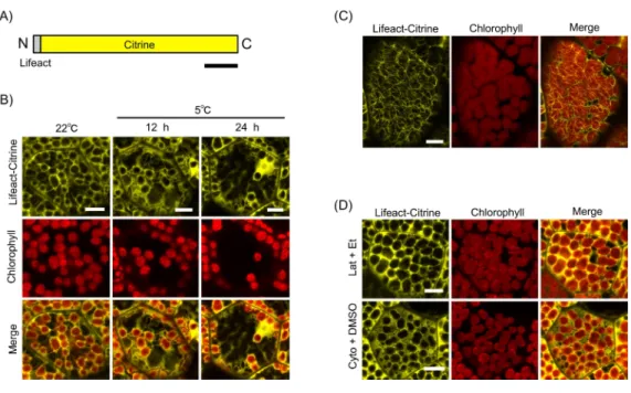
Figure 3 Lifeact-based visualization of actin filaments during chloroplast cold positioning.(A) Schematic illustration of Lifeact-Citrine. N and C indicate the amino and carboxyl termini, respectively. Bar represents 50 amino acids. (B) Visualization of actin filaments mediated by Lifeact-Citrine probe during chloroplast cold positioning in 1-day-old gemmaling. Fluorescence of Lifeact-Citrine and chlorophyll were colored in yellow and red, respectively. Scale bars represent 10µm. (C) Visualization
of actin filaments by Lifeact-Citrine probe in 4-day-old gemmaling. Scale bar represents 10µm. (D)
Disruption of actin filaments visualized by Lifeact-Citrine probe with treatment of 1-µM latrunculin A
(Lat) and 50-µM cytochalasin B (Cyto). Scale bars represent 10µm.
cell walls and the long F-actin structures at periclinal cell walls were reduced together with the chloroplasts (Fig. 3B). Eventually, we could not observe cp-actin-like structures around the chloroplast during the cold positioning response (Fig. 3B).
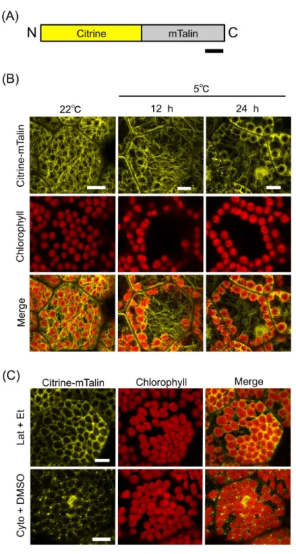
A previous study of A. thaliana demonstrated that mouse talin (mTalin) (Kost, Spielhofer & Chua, 1998), which is another actin binding protein, is suitable for the visualization of cp-actin as compared with the Lifeact peptide (Kong et al., 2013). In the mossP. patens, cp-actin filaments have been successfully visualized by the mTalin-fused fluorescent protein (Yamashita et al., 2011). Therefore, mTalin was considered to be suitable for observing cp-actin filaments. We fused mTalin with Citrine (Citrine-mTalin) (Fig. 4A) and transformed Citrine-mTalin into M. polymorpha(Fig. 4B). Surprisingly, the actin filaments visualized by Citrine-mTalin were totally different from that by Lifeact-Citrine in the 1-day-old cells (Figs. 3Band 4B), and numerous long F-actin structures were clearly observed in the transgenic cells expressing Citrine-mTalin (Fig. 4B). In this context, a type of F-actin bound by Citrine-mTalin may differ from that bound by Lifeact-Citrine. In addition, the structure was disrupted by anti-actin drugs (1-µM latrunculin A and 50-µM cytochalasin B), visualizing F-actin with Citrine-mTalin (Fig. 4C). In the 1-day-old transgenic gemmalings expressing the Citrine-mTalin, the chloroplast accumulation response was induced at 22◦C, and the long F-actin structures were visualized as likely covering the chloroplasts (Fig. 4B). However, as with the case of Lifeact-Citrine (Fig. 3B), cp-actin-like short F-actin structures were not detected by Citrine-mTalin (Fig. 4B). When cold positioning was induced, any biased distribution of the actin filaments between the front and rear of chloroplasts in its directional movement was also undetectable (Fig. 4B). However, the reduction of the long F-actin structures at periclinal cell walls was clearly observed during chloroplast relocation in the transgenic liverworts expressing Citrine-mTalin, confirming the results with Lifeact-Citrine (Fig. 4B).
In the present study, by using Lifeact-Citrine and Citrine-mTalin as fluorescent probes, we detected long F-actin structures, but not short F-actin structures, such as cp-actin filaments, around chloroplast in M. polymorpha. It remains to be known why these particular structures of cp-actin were not detected inM. polymorpha. It is possible that the actin-based machinery involved in the chloroplast positioning observed inM. polymorpha may differ from the already reported cp-actin filaments as cp-actin-independent but actin-mediated mechanisms for light-induced positioning have recently been suggested (Suetsugu et al., 2016). As a possibility, cp-actin filaments may not form in chloroplast cold positioning. Based on our observations (Fig. 3Band4B), long F-actin structures at periclinal cell walls may be involved in chloroplast cold positioning ofM. polymorphabut further experiments are necessary to make this conclusion.
Figure 4 mTalin-based visualization of actin filaments during chloroplast cold positioning.(A) Schematic illustration of Citrine-mTalin. N and C indicate the amino and carboxyl termini, respectively. Bar represents 50 amino acids. (B) Representative images of actin filaments visualized by Citrine-mTalin during chloroplast cold positioning. Fluorescence of Citrine-mTalin and chlorophyll were colored in yellow and red, respectively. Scale bars represent 10µm. (C) Disruption of actin filaments visualized by
Citrine-mTalin with treatment of 1-µM latrunculin A (Lat) and 50-µM cytochalasin B (Cyto). Scale bars
Figure 5 Actin-dependency in the light-induced chloroplast positioning responses.(A) Procedure of inhibitor treatments to analyze chloroplast accumulation response. wBL, weak blue-light. (B) Representa-tive images of chloroplast position after induction of the accumulation response with the inhibitor, 1-µM
latrunculin A (Lat) or 10-µM oryzalin (Ory). As controls, 1% ethanol (Et) and 0.1 % DMSO were used.
Scale bar represents 50µm. (C) Quantification of chloroplast position (B) by measurement of the P/A
ra-tio. Bars indicate standard deviations. (D) Procedure of inhibitor treatments to analyze chloroplast avoid-ance response. wBL, weak blue-light; sBL, strong blue-light. (E) Representative images of chloroplast posi-tion after inducposi-tion of the avoidance response with the inhibitor, Lat or Ory. As controls, 1% ethanol (Et) and 0.1% DMSO were used. Scale bar represents 50µm. (F) Quantification of chloroplast position (E) by
measurement of the P/A ratio. Bars indicate standard deviations.
analyze temperature-induced responses, we took only photographs because technically it was not possible to record movies with fluorescence images under cold conditions. In our experiments, cells were observed by confocal laser scanning microscopy after temperature treatment in a conventional incubator, suggestive of a time-lag (within minutes) between the treatment and the observation. The reorganization of cp-actin filaments has been reported to occur on a minute time-scale (Kadota et al., 2009), thus cp-actin filaments may have already disappeared in the cells when they were scanned in our study. To find the expected cp-actin filaments under temperature alteration, the use of temperature-controlled fluorescence microscopy or confocal microscope equipped with a time-lapse video recording system may be necessary. Fluorescence observation by confocal laser scanning microscopy under temperature controlled conditions has been reported previously (Holzinger et al., 2007), thus these technical limitations should be resolved in the future by adding a time-lapse recording system.
In summary, our findings indicate that chloroplast cold positioning is actin-dependent; however, the actin-based machinery (i.e., cp-actin-like machinery associated with chloroplasts or long F-actin structures along periclinal cell walls) necessary for the cold positioning response ofM. polymorpha could not be conclusively identified in the present study. Future work will include temperature-controlled fluorescence microscopy with a time-lapse video recording system to determine the machinery. Currently, M. polymorpha is being developed as a model liverwort, and various molecular biological techniques such as transformation and genome editing have been developed (Ishizaki et al., 2015b). We believe that future research using these molecular techniques, in addition to temperature-controlled fluorescence microscopy, will allow the identification of the actin-based machinery critical for the cold positioning response.
ACKNOWLEDGEMENTS
We thank Dr. Akeo Kadota (Tokyo Metropolitan University, Japan) for critical reading of the manuscript and valuable comments. We also thank Drs. Sam-Geun Kong (Kongju National University, Korea) and Masamitsu Wada (Tokyo Metropolitan University, Japan) for providing the cDNA fragment of mouse Talin, Dr. Takayuki Kohchi (Kyoto University, Japan) for providing the Tak-1 strain ofM. polymorpha, Dr. Kimitsune Ishizaki (Kobe University, Japan) for providing information of MpTubulin sequences, and Ms. Rieko Saijo (Utsunomiya University, Japan) for maintenance of transgenic liverworts.
ADDITIONAL INFORMATION AND DECLARATIONS
Funding
Grant Disclosures
The following grant information was disclosed by the authors: Hayashi Rheology Memorial Foundation.
Japan Society for the Promotion of Science (JSPS) KAKENHI: 23870002, 26840088. Sasagawa Scientific Research Grant of the Japan Science Society (Y.K).
CDI Research Project of Utsunomiya University (Y.K).
Competing Interests
The authors declare there are no competing interests.
Author Contributions
• Shun Kimura performed the experiments, analyzed the data, contributed reagents/ma-terials/analysis tools, prepared figures and/or tables, reviewed drafts of the paper. • Yutaka Kodama conceived and designed the experiments, analyzed the data, contributed
reagents/materials/analysis tools, wrote the paper, prepared figures and/or tables, reviewed drafts of the paper.
Data Availability
The following information was supplied regarding data availability: The raw data has been supplied as aData S1.
Supplemental Information
Supplemental information for this article can be found online athttp://dx.doi.org/10.7717/ peerj.2513#supplemental-information.
REFERENCES
Buschmann H, Holtmannspötter M, Borchers A, O’Donoghue MT, Zachgo S. 2016.Microtubule dynamics of the centrosome-like polar organizers from the basal land plantMarchantia polymorpha.New Phytologist 209:999–1013
DOI 10.1111/nph.13691.
Era A, Tominaga M, Ebine K, Awai C, Saito C, Ishizaki K, Yamato KT, Kohchi T, Nakano A, Ueda T. 2009.Application of lifeact reveals F-actin dynamics in Ara-bidopsis thalianaand the liverwort,Marchantia polymorpha.Plant and Cell Physiology
50:1041–1048DOI 10.1093/pcp/pcp055.
Gabryś H. 2004.Blue light-induced orientation movements of chloroplasts in higher plants: recent progress in the study of their mechanisms.Acta Physiologiae Plantarum
26:473–478DOI 10.1007/s11738-004-0038-3.
Hardham AR, Gunning BE. 1978.Structure of cortical microtubule arrays in plant cells. The Journal of Cell Biology77:14–34DOI 10.1083/jcb.77.1.14.
Higa T, Suetsugu N, Kong S-G, Wada M. 2014.Actin-dependent plastid movement is required for motive force generation in directional nuclear movement in plants. Proceedings of the National Academy of Sciences of the United States of America
Holzinger A, Buchner O, Lütz C, Hanson MR. 2007.Temperature-sensitive formation of chloroplast protrusions and stromules in mesophyll cells ofArabidopsis thaliana. Protoplasma230:23–30DOI 10.1007/s00709-006-0222-y.
Ishizaki K, Nishihama R, Ueda M, Inoue K, Ishida S, Nishimura Y, Shikanai T, Kohchi T. 2015a.Development of gateway binary vector series with four different selection markers for the liverwortMarchantia polymorpha.PLoS ONE10:e0138876
DOI 10.1371/journal.pone.0138876.
Ishizaki K, Nishihama R, Yamato KT, Kohchi T. 2015b.Molecular genetic tools and techniques forMarchantia polymorpharesearch.Plant and Cell Physiology
57:262–270DOI 10.1093/pcp/pcv097.
Kadota A, Yamada N, Suetsugu N, Hirose M, Saito C, Shoda K, Ichikawa S, Kagawa T, Nakano A, Wada M. 2009.Short actin-based mechanism for light-directed chloroplast movement inArabidopsis.Proceedings of the National Academy of Sciences of the United States of America106:13106–11DOI 10.1073/pnas.0906250106.
Kagawa T, Wada M. 1996.Phytochrome- and blue-light-absorbing pigment-mediated directional movement of chloroplasts in dark-adapted prothallial cells of fernAdiantumas analyzed by microbeam irradiation.Planta198:488–493
DOI 10.1007/BF00620067.
Kandasamy MK, Meagher RB. 1999.Actin-organelle interaction: association with chloroplast inArabidopsisleaf mesophyll cells.Cell Motility and the Cytoskeleton
44:110–118
DOI 10.1002/(SICI)1097-0169(199910)44:2<110::AID-CM3>3.0.CO;2-O.
Kodama Y. 2016.Time gating of chloroplast autofluorescence allows clearer fluorescence imagingin planta.PLoS ONE 11:e0152484DOI 10.1371/journal.pone.0152484.
Kodama Y, Tsuboi H, Kagawa T, Wada M. 2008.Low temperature-induced chloroplast relocation mediated by a blue light receptor, phototropin 2, in fern gametophytes. Journal of Plant Research121:441–448DOI 10.1007/s10265-008-0165-9.
Komatsu A, Terai M, Ishizaki K, Suetsugu N, Tsuboi H, Nishihama R, Yamato KT, Wada M, Kohchi T. 2014.Phototropin encoded by a single-copy gene mediates chloroplast photorelocation movements in the liverwortMarchantia polymorpha. Plant Physiology166:411–427DOI 10.1104/pp.114.245100.
Kong S-G, Arai Y, Suetsugu N, Yanagida T, Wada M. 2013.Rapid severing and motility of chloroplast-actin filaments are required for the chloroplast avoidance response in Arabidopsis.The Plant Cell25:572–590DOI 10.1105/tpc.113.109694.
Kong SG, Wada M. 2011.New insights into dynamic actin-based chloroplast photorelo-cation movement.Molecular Plant 4:771–781DOI 10.1093/mp/ssr061.
Kost B, Spielhofer P, Chua NH. 1998.A GFP-mouse talin fusion protein labels plant actin filamentsin vivoand visualizes the actin cytoskeleton in growing pollen tubes. The Plant Journal16:393–401DOI 10.1046/j.1365-313x.1998.00304.x.
Malec P, Rinaldi RA, Gabryś H. 1996.Light-induced chloroplast movements in Lemna trisulca. Idntification of the motile system.Plant Science120:127–137
Ogasawara Y, Ishizaki K, Kohchi T, Kodama Y. 2013.Cold-induced organelle re-location in the liverwortMarchantia polymorphaL.Plant, Cell and Environment
36:1520–1528DOI 10.1111/pce.12085.
Oikawa K, Matsunaga S, Mano S, Kondo M, Yamada K, Hayashi M, Kagawa T, Kadota A, Sakamoto W, Higashi S, Watanabe M, Mitsui T, Shigemasa A, Iino T, Hosokawa Y, Nishimura M. 2015.Physical interaction between peroxisomes and chloroplasts elucidated byin situlaser analysis.Nature Plants30:15035
DOI 10.1038/nplants.2015.35.
Riedl J, Crevenna AH, Kessenbrock K, Yu JH, Neukirchen D, Bista M, Bradke F, Jenne D, Holak TA, Werb Z, Sixt M, Wedlich-Soldner R. 2008.Lifeact: a versatile marker to visualize F-actin.Nature Methods5:605–607DOI 10.1038/nmeth.1220.
Sato Y, Wada M, Kadota A. 2001.Choice of tracks, microtubules and/or actin filaments for chloroplast photo-movement is differentially controlled by phytochrome and a blue light receptor.Journal of Cell Science114:269–279.
Senn G. 1908.Die gestalts-und lageveränderung der pflanzen-chromatophoren. Leipzig: Wilhelm-Engelmann.
Suetsugu N, Higa T, Gotoh E, Wada M. 2016.Light-induced movements of chloroplasts and nuclei are regulated in both cp-actin-filament-dependent and -independent manners inArabidopsis thaliana.PLoS ONE11:e0157429
DOI 10.1371/journal.pone.0157429.
Suetsugu N, Kagawa T, Wada M. 2005.An auxilin-like J-domain protein, JAC1, regu-lates phototropin-mediated chloroplast movement.Plant Physiology139:151–162
DOI 10.1104/pp.105.067371.
Suetsugu N, Wada M. 2016.Evolution of the cp-actin-based motility system of chloro-plasts in green plants.Frontiers in Plant Science7:561 DOI 10.3389/fpls.2016.00561.
Suetsugu N, Wada M. 2007.Chloroplast photorelocation movement mediated by phototropin family proteins in green plants.Biological Chemistry 388:927–935
DOI 10.1515/BC.2007.118.
Tsuboi H, Wada M. 2012.Distribution pattern changes of actin filaments during chloroplast movement inAdiantum capillus-veneris.Journal of Plant Research
125:417–428DOI 10.1007/s10265-011-0444-8.
Tsuboyama S, Kodama Y. 2014.AgarTrap: a simplifiedAgrobacterium-mediated transformation method for sporelings of the liverwortMarchantia polymorphaL. Plant and Cell Physiology 55:229–236DOI 10.1093/pcp/pct168.
Tsuboyama-Tanaka S, Kodama Y. 2015.AgarTrap-mediated genetic transformation using intact gemmae/gemmalings of the liverwortMarchantia polymorphaL.Journal of Plant Research128:337–344DOI 10.1007/s10265-014-0695-2.
Tsuboyama-Tanaka S, Nonaka S, Kodama Y. 2015.A highly efficient AgarTrap method for genetic transformation of mature thalli of the liverwortMarchantia polymorpha L.Plant Biotechnology31:333–336 DOI 10.5511/plantbiotechnology.15.0813a.
Wada M, Suetsugu N. 2004.Plant organelle positioning.Current Opinion in Plant Biology
Wagner G, Haupt W, Laux A. 1972.Reversible inhibition of chloroplast move-ment by cytochalasin B in the green alga mougeofia.Science176:808–809
DOI 10.1126/science.176.4036.808.