Submitted23 April 2016
Accepted 15 July 2016
Published30 August 2016
Corresponding author
Michael A. Savka, [email protected]
Academic editor
Qi Liu
Additional Information and Declarations can be found on page 15
DOI10.7717/peerj.2332
Copyright
2016 Gan et al.
Distributed under
Creative Commons CC-BY 4.0
OPEN ACCESS
Genome sequencing-assisted identification
and the first functional validation of
N
-acyl-homoserine-lactone synthases
from the Sphingomonadaceae family
Han Ming Gan1,2, Lucas K. Dailey3, Nigel Halliday4, Paul Williams4,
André O. Hudson3and Michael A. Savka3
1School of Science, Monash University Malaysia, Bandar Sunway, Selangor, Malaysia
2Genomics Facility, Tropical Medicine Biology Platform, Monash University Malaysia, Bandar Sunway, Selangor, Malaysia
3Thomas H. Gosnell School of School of Life Sciences, Rochester Institute of Technology, Rochester, NY, USA
4School of Life Sciences, Centre for Biomolecular Sciences, University of Nottingham, Nottingham, UK
ABSTRACT
Background. Members of the genusNovosphingobiumhave been isolated from a variety of environmental niches. Although genomics analyses have suggested the presence of genes associated with quorum sensing signal production e.g., theN-acyl-homoserine lactone (AHL) synthase (luxI) homologs in variousNovosphingobiumspecies, to date, noluxIhomologs have been experimentally validated.
Methods. In this study, we report the draft genome of the N-(AHL)-producing bacteriumNovosphingobium subterraneumDSM 12447 and validate the functions of predictedluxIhomologs from the bacterium through inducible heterologous expres-sion inAgrobacterium tumefaciensstrain NTL4. We developed a two-dimensional thin layer chromatography bioassay and used LC-ESI MS/MS analyses to separate, detect and identify the AHL signals produced by theN. subterraneumDSM 12447 strain. Results. Three predicted luxI homologs were annotated to the locus tags NJ75_2841 (NovINsub1), NJ75_2498 (NovINsub2), and NJ75_4146 (NovINsub3). Inducible
heterologous expression of eachluxI homologs followed by LC-ESI MS/MS and two-dimensional reverse phase thin layer chromatography bioassays followed by biolumi-nescent ccd camera imaging indicate that the three LuxI homologs are able to produce a variety of medium-length AHL compounds. New insights into the LuxI phylogeny was also gleemed as inferred by Bayesian inference.
Discussion. This study significantly adds to our current understanding of quorum sensing in the genusNovosphingobiumand provide the framework for future charac-terization of the phylogenetically interesting LuxI homologs from members of the genus
Novosphingobiumand more generally the family Sphingomonadaceae.
SubjectsBioinformatics, Genomics, Microbiology
INTRODUCTION
Quorum sensing (QS) is commonly employed by bacteria to monitor population cell density to synchronize gene expression (Fuqua, Winans & Greenberg, 1994; Miller & Bassler, 2001;Schuster et al., 2013;Waters & Bassler, 2005). In one type of QS system from Gram-negative bacteria, the bacteria produce and detect chemical signals calledN -acyl-homoserine lactones (AHLs). These signals are produced by an AHL synthase, a member of the LuxI-type protein family, and are usually detected by a transcriptional regulator belong-ing to the LuxR-type family. A typical AHL-QS system contains an AHL synthase (LuxI) and the transcriptional activator/repressor protein (LuxR) that are usually in a genomic con-text regarding proximity (Fast & Tipton, 2012). Upon reaching a concentration threshold that corresponds to a given the cell density, the AHL signal(s) is detected by the cognate LuxR and activates population-wide-responses leading to the coordination of gene activation or repression. In Gram-negative bacteria, AHL-dependent QS regulation is used to regulate diverse responses such as; the production of bioluminescence, the activation of virulence factors, conjugation, the production of antimicrobial metabolites and the production of polysaccharides (Fuqua & Greenberg, 2002;Miller & Bassler, 2001;Waters & Bassler, 2005).
Besides the presence of the canonicalluxI/luxRpairs, many bacteria contain additional
luxRtranscriptional regulators that do not haveluxIgenes in their vicinity on the chromo-some. These unpairedluxRgenes have been coined solos or orphans and are orthologous to QS LuxR-type transcriptional regulators in that LuxR solos contain the AHL-binding domain at the N terminus and a DNA-binding helix-turn-helix (HTH) domain at the C terminus (Case, Labbate & Kjelleberg, 2008;Cude & Buchan, 2013;Fuqua, 2006;Gonzalez & Venturi, 2013;Subramoni & Venturi, 2009;Tsai & Winans, 2010). Generally speaking, the solo LuxR-type transcriptional activators increase the regulatory range by responding to endogenously produced AHLs, by listening-in or eavesdropping on exogenous signals produced by other bacteria or to alternative signals produced by the bacterial community or colonised host (Chatnaparat et al., 2012)
Members of theNovosphingobiumgenus have been isolated from a variety of terrestrial and aquatic environments, including grapevine crown gall tumor surface,Populus deltoides
rhizosphere, marine subsurface and muddy sediments, pulp and paper wastewater, polluted waters in addition to other sites (Gan et al., 2009;White, Sutton & Ringelberg, 1996) Despite numerous successfulin-silicoidentifications of sphingomonadluxIhomolog(s) in publicly available databases, no sphingomonad luxI homologs have ever been experimentally validated to date (Gan et al., 2012;Gan et al., 2015;Gan et al., 2013). During our on-going work of AHL signal detection in members of the genusNovosphingobium, N. subterraneum
DSM 12447 strain was distinguished by its ability to accumulate substantially higher amounts of AHL signals as compared to other sphingomonad strains tested in our lab.
Hence, to fill this existing gap in the quorum-sensing field, we aim to useN. subterraneum
DSM 12447 to (1) develop a two-dimensional reverse-phase thin layer chromatography
(TLC) bioassay to separate and detect the multiple AHLs produced byN. subterraneum
by heterologously expressed LuxI homologs using mass spectrometry analysis, (3) sequence the whole genome of strain DSM 12447 to identify its AHL biosynthetic genes and elucidate its evolutionary relationship with other LuxI homologs through Bayesian inference, and (4) functionally characterize the identified AHL synthase(s) through regulated heterologous expression in theAgrobacteriumstrain NTL4.
MATERIALS AND METHODS
Bacterial strains, growth media and biosensor strains
Novosphingobium subterraneum DSM 12447 (previouslySphingomonas subterraneum
DSM12447) was provided by Andreas Stolz (Institut fur Mikrobiologie at the Universitat Stuttgart, Stuttgart, Germany). This strain was isolated from terrestrial subsurface and was shown to catabolize a variety of natural recalcitrant and anthropogenic compounds including naphthalene, toluene, biphenyl, dibenzothiophene and fluorine (Balkwill et al., 1997;Takeuchi, Hamana & Hiraishi, 2001). The bacterial strains and plasmids used in this work are listed inTable 1.
Novosphingobium subterraneumDSM 12447 strain was grown in tryptone soy broth
(TSB), potato dextrose (PD) or R2A medium (Difco Laboratories, Detroit, MI) at 28◦
C. Agrobacteria AB minimal media (Chilton et al., 1974) at 28◦
C was used to grow AHL-dependent biosensor strains AgrobacteriumNTL4 and A136. For AHL signal induction bioassays,E. coliJM109 andAgrobacteriumNTL4 strains harboring empty vector pSRKKm or pSRKKm with clonednovI genes were grown on Luria-Bertani broth (LB) medium at 37◦
C and 28◦
C and supplemented with kanamycin at 25 and 50µg/ml, respectively.
For AHL signal detection bioassays,Agrobacterium tumefaciensNTL4 (pZLR4) and A136 (pCF218, pMV26) were grown in AB medium supplemented with 0.2% (w/v) dextrose and 0.01% (w/v) yeast extract and gentamycin (10µg/ml) for NTL4 (pZLR4) (Cha et al., 1998)
and kanamycin (25µg/ml) and tetracycline (5µg/ml) for A136 (pCF218, pMV26) (Sokol et
al., 2003)E. coli-based biosensors JM109 (pSB401), JM109 (pSB1075) and JM109 (pSB536) were grown in LB media with appropriate antibiotic for plasmid maintenance (Winson et al., 1998).Chromobacterium violaceumCV026 biosensor was growth in tryptone yeast extract/potato dextrose (1:1) agar media for T-streak bioassays (McClean et al., 1997).
Each AHL-dependent bacterial biosensor strain used in this work along with its AHL receptor protein and cognate AHL signal is listed in Table 1. All media and growth conditions for AHL detection bioassays are as previously described by our laboratory (Gan et al., 2009;Lowe et al., 2009).
Biosensor detection
Reverse-phase (RP) one-dimensional (1-D) TLC plates were used to determine AHL signal profiles. Concentrated acidified ethyl acetate (aEtOAc) extracts were spotted on to the C18 RP-TLC plate origin in 2-µL volumes and representing from 0.5 to 2-mL supernatant
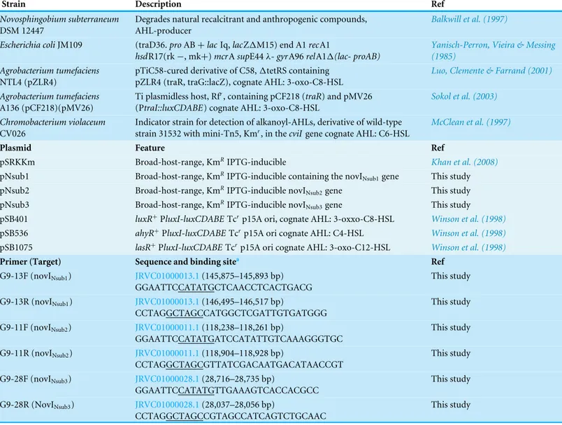
Table 1 Bacterial strains plasmids and primers used in this study.
Strain Description Ref
Novosphingobium subterraneum DSM 12447
Degrades natural recalcitrant and anthropogenic compounds, AHL-producer
Balkwill et al. (1997)
Escherichia coliJM109 (traD36.proAB+lacIq,lacZ1M15) end A1recA1
hsdR17(rk−, mk+)mcrAsupE44λ-gyrA96relA11(lac- proAB)
Yanisch-Perron, Vieira & Messing (1985)
Agrobacterium tumefaciens NTL4 (pZLR4)
pTiC58-cured derivative of C58,1tetRS containing pZLR4 (traR, traG::lacZ), cognate AHL: 3-oxo-C8-HSL
Luo, Clemente & Farrand (2001)
Agrobacterium tumefaciens A136 (pCF218)(pMV26)
Ti plasmidless host, Rfr, containing pCF218 (traR) and pMV26 (PtraI::luxCDABE) cognate AHL: 3-oxo-C8-HSL
Sokol et al. (2003)
Chromobacterium violaceum CV026
Indicator strain for detection of alkanoyl-AHLs, derivative of wild-type strain 31532 with mini-Tn5, Kmr, in thecviIgene cognate AHL: C6-HSL
McClean et al. (1997)
Plasmid Feature Ref
pSRKKm Broad-host-range, KmRIPTG-inducible Khan et al. (2008)
pNsub1 Broad-host-range, KmRIPTG-inducible containing the novI
Nsub1gene This study
pNsub2 Broad-host-range, KmRIPTG-inducible novI
Nsub2gene This study
pNsub3 Broad-host-range, KmRIPTG-inducible novI
Nsub3gene This study
pSB401 luxR+PluxI-luxCDABETcrp15A ori, cognate AHL: 3-oxxo-C8-HSL Winson et al. (1998)
pSB536 ahyR+
PluxI-luxCDABETcrp15A ori cognate AHL: C4-HSL Winson et al. (1998)
pSB1075 lasR+
PluxI-luxCDABETcrp15A ori cognate AHL: 3-oxo-C12-HSL Winson et al. (1998)
Primer (Target) Sequence and binding sitea Ref
G9-13F (novINsub1) JRVC01000013.1(145,875–145,893 bp)
GGAATTCCATATGCTCAACCTCACTGACG
This study
G9-13R (novINsub1) JRVC01000013.1(146,495–146,517 bp)
CCTAGGCTAGCCATGGCTCGATTGTGATGGG
This study
G9-11F (novINsub2) JRVC01000011.1(118,238–118,261 bp)
GGAATTCCATATGATCCATATTGTCAAAGGGTGC
This study
G9-11R (novINsub2) JRVC01000011.1(118,904–118,928 bp)
CCTAGGCTAGCGTTATCGACAATGACATAACCGT
This study
G9-28F (novINsub3) JRVC01000028.1(28,716–28,735 bp)
GGAATTCCATATGTTGAAAGTCACCACGCC
This study
G9-28R (NovINsub3) JRVC01000028.1(28,037–28,056 bp)
CCTAGGCTAGCCGTAGCCATCAGTCTGCAAC
This study
Notes.
aUnderlined nucleotide bases indicate restriction enzyme sites for cloning.
were identified with appropriate reference compounds. This involves determining and comparing retardation factors (Rf) of unknown samples to AHL reference compounds (Shaw et al., 1997).
Development of two-dimensional (2-D) thin layer chromatography for AHLs
The AHL extract was initially spotted onto the bottom left corner of the C18 RP-TLC plate The amount needed was estimated based on the AHL signal strength obtained from multiple independent 1-D RP-TLC runs. The spotted TLC plate was eluted with 70:30 (v/v) methanol: water as the first mobile-phase in a glass tank. The mobile-phase was allowed to rise until the top of the TLC plate before removing the plate to dry overnight. Then, the TLC plate was rotated 90◦
water as the second mobile-phase until it reached the top of the TLC plate. After drying, the TLC plate was overlaid with TraR-dependentAgrobacteriumbiosensor strain A136 using the same procedure as used for 1-D TLCs.
AHL identification and quantification by LC-MS/MS
Equipment
Chromatography was achieved using a Shimadzu series 10AD VP LC system. The column oven was maintained at 50◦
C. The HPLC Column used was a Phenomenex Gemini C18 column (3.0µm , 100×3.0 mm) with an appropriate guard column. Mobile phase A was
0.1% (v/v) formic acid in water, and mobile phase B 0.1% (v/v) formic acid in methanol. The flow rate throughout the chromatographic separation was 450 µL/min. The binary
gradient began initially at 10% B and increased linearly to 99% B over 12 min and remained at 99% B for 1 min. A rapid decrease to 10% B occurred over 0.1 min, and stayed at this composition for 1.9 min. Total run time per sample was 15 min.
The MS system used was an Applied Biosystems Qtrap 4,000 hybrid triple-quadrupole linear ion trap mass spectrometer equipped with an electrospray ionisation (ESI) interface. Instrument control, data collection and analysis were conducted using Analyst software. Source parameters were set as: curtain gas: 20.0, ion source potential: 5,000 V, temperature: 450◦
C, nebulizer gas: 20.0, and auxiliary gas: 15.0.
AHL standards
Synthetic standards of C4, C6, C8, C10, C12, C14, C4, C6, C8, 3-oxo-C10, 3-oxo-C12, 3-oxo-C14, 3-OH-C4, 3-OH-C6, 3-OH-C8, 3-OH-3-oxo-C10, 3-OH-C12 and 3-OH-C14 AHLs were synthesised according to established procedures (Chhabra et al., 2003;Chhabra et al., 1993).
Sample preparation
Dried extracts were stored at−20◦C. Prior to analysis, each sample extract was reconstituted
in 100 µl of methanol +0.1% (v/v) formic acid. The injection volume was 5 µl.
Analysis method
Initial analysis was conducted with the MS operating in precursor ion scan mode screening for precursor ions that give rise to a product ion of m/z=102 (a fragment ion that is
common to all AHLs), upon collision induced fragmentation (Table 2). Comparison of detected peak areas with an AHL mix sample of known concentration was used to gauge a useful calibration range for the subsequent quantification of detected AHLs. Samples were rerun with the MS in MRM (multiple reaction monitoring) mode, analysing the LC eluent for specific AHLs detected in the previous analysis. The quantification was conducted by comparing peak areas of detected peaks with a six point calibration line constructed by analysing (in triplicate) mixed AHL calibration samples containing C8, 3-OH-C8 and 3-OH-C10 AHLs at 0.5, 1.0, 2.0, 5.0, 10 and 20µM.
Whole genome sequencing of N. subterraneumDSM 12447
Genomic DNA of strain DSM12447 was extracted using the GenEluteTM(Sigma-Aldrich,
Table 2 Mass transitions used for the MRM detection of common AHLs.
Acyl chain length
Carbon 3 substitution
MRMs Retention
time/min
Unsubstituted 172–102 3.18
Oxo 186–102 2.02
C4
OH 188–102 1.58
Unsubstituted 200–102 4.53
Oxo 214–102 4.08
C6
OH 216–102 3.93
Unsubstituted 228–102 5.20
Oxo 242–102 4.66
C8
OH 244–102 4.49
Unsubstituted 256–102 5.87
Oxo 270–102 5.31
C10
OH 272–102 5.05
Unsubstituted 284–102 6.65
Oxo 298–102 5.98
C12
OH 300–102 5.72
Unsubstituted 312–102 7.44
Oxo 326–102 6.77
C14
OH 328–102 6.50
XT (Illumina, San Diego, CA, USA) according to the manufacturer’s instructions. Whole genome sequencing was performed using the MiSeq (Illumina, San Diego, CA, USA) at the Monash University Malaysia Genomics Facility. The raw data for each bacterium were error-corrected and assembled using Spades v2.5 (default setting) (Bankevich et al., 2012). The generated contigs were scaffolded and gap-closed using SSPACE and GAPFiller, respectively (Boetzer et al., 2011;Boetzer & Pirovano, 2012). Genome annotation was performed using Prokka and InterProScan5 (Jones et al., 2014;Seemann, 2014).
Identification and phylogenetic analyses of LuxI homologs
Amplification and cloning of 3 putative AHL synthase genes
Primers and plasmids used in this study are listed inTable 1. Amplification of theluxI
homologs was performed using Q5 polymerase mastermix (New England Biolabs, Ipswich, MA, USA) according to the manufacturer’s instructions. Approximately 150 ng of the purified PCR amplicons were mixed with 50 ng of pSRKKm vector (Khan et al., 2008) and double digested with NheI and NdeI (New England Biolabs, Ipswich, MA, USA) for 1 h. After heat inactivation, the digested products were purified using magnetic beads (Omega Biotek, Norcross, GA, USA) and ligated with Electroligase (New England Biolabs, Ipswich, MA, USA) for 30 min. The ligated products were transformed intoA. tumefaciensNTL4 andEscherichia coliJM109 using electroporation.
Inducible expression ofluxI homologs and detection of AHLs from solid media
Bacterial culture (A. tumefaciensNTL4 andEscherichia coliJM109 grown for 96- and 48 h, respectively) supernatants were resuspended from LB plates supplemented with antibiotic kanamycin and filter-sterilized IPTG at 0, 10, 100 and 1000µM containing pSRKKm
with and without the clonedluxIhomologs (Table 1) were extracted with acidified ethyl acetate (aEtOAc) (1 mL of glacial acetic acid per 200 mL of ethyl acetate) for 60 min with shaking (150 r.p.m.). The extracts were then centrifuged to separate the aqueous and ethyl acetate phases. The ethyl acetate phase was recovered and dried in a Savant Speed Vac. Twenty-, thirty- or fifty-fold concentrated extracts were prepared and used in AHL detection bioassays.
Acyl-homoserine lactone extractions for induction assays
NTL4 (pNsub1,2 or 3) strains were grown in 20 mL of LB (50 µg/ml kanamycin)
supplemented with different amount of inducer isopropyl β-D-1-thiogalactopyranoside (IPTG) inducer to final concentration of 0,10,100, or 1,000µM and incubated overnight
in a shaking incubator at 28 ◦
C. The next day, 20 ml of ethyl acetate was added to each of the twelve tubes and shaken at room temperature for 2 h. The tubes were then centrifuged at 5,000 rpm for 10 min to separate the liquid layers and then the top layer of ethyl acetate was aspirated off and stored. This layer was separated into several 1.5 ml micro centrifuge tubes and the ethyl acetate evaporated off using a speed-vac. The residue in the tube was then resuspended in 75µl of fresh ethyl acetate to bring all samples to a 20×-volume
equivalent extract concentration.
RESULTS
N. subterraneum DSM 12447 produces multiple distinct AHL signals
Table 3 Detection ofN-acyl-homoserine lactones by five different AHL-dependent biosensor strains.
AHL receptora
AhyRb LuxRb TraRb LasRb CviRc
Novosphingobium subterraneumDSM 12447 − ++ + + + − +
Notes.
aAhyR, AHL receptor fromAeromonas hydrophilia; LuxR,Vibrio fisheri; TraR,Agrobacterium tumefaciens; LasR,Pseudomonas
aeruginosa; CviR,Chromobacterium violaceum.
bScores for bioluminescence-based biosensor detection of AHL in strain extracts:−,<2-fold higher than background levels of relative light units (RLU) bioluminescence;+>2-fold higher than background RLUs;++>50 to 75-fold higher than back-ground RLUs;+ + +>75-fold higher than background RLUs.
cViolacein pigment (purple) production in T-streak bioassays on PDA/TYE (1:1) agar media:+, visible pigment production;−, no pigment production.
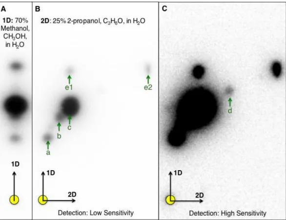
detection. The additional separation of AHL using 25% 2-propanol as the second mobile phase coupled with detection using a luminescence-based reporter rendered significant improvement in AHL signal detection and identification. Based on 2-D RP-TLC of extracts prepared from N. subterraneumDSM 12447 strain followed by AHL detection usingA. tumefaciensA136 overlay, six distinct putative AHL signals were identified (Figs. 1B–1C).
Whole genome sequencing of N. subterraneumDSM 12447 identified 3 putativeluxI homologs (novI) that share a commonnovRnovIphyH
gene synteny
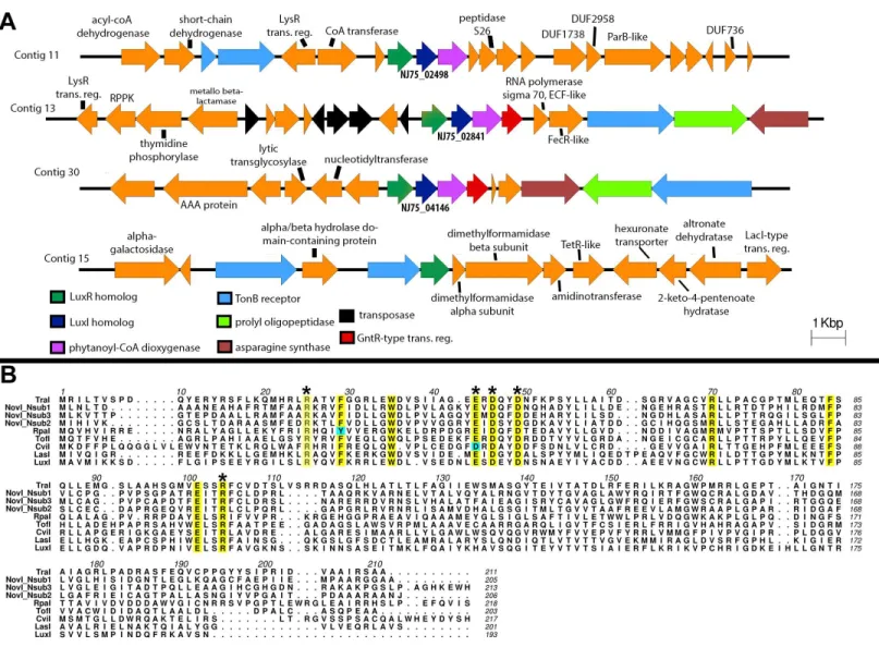
The draft genome of strain DSM 12447 has a GC content of 63.2% and consists of 54 contigs with a total genome length of 4,885,942 bp (N50 of 181,386 bp). Anti-SMASH analysis
(Weber et al., 2015) revealed three LuxI-type AHL synthase genes that are separately located in three different contigs (Fig. 2A). Protein alignment of the putative LuxI homologs with known LuxI homologs shows that these homologs contain the highly conserved amino acid signatures which are crucial for the function of AHL synthesis (Asterisk signs inFig. 2B). We propose the names, NovINsub1, NovINsub2 and NovINsub3 for locus tags NJ75_2841,
NJ75_2498 and NJ75_4146, respectively. Among autoinducer proteins within the genus
Novosphingobium, NovINsub1, NovINsub2and NovINsub3show 62.4%, 51.1% and 62.2%
pro-tein identity to LuxI homologs of N. sp AP12 (PMI02_00996), N. sp. Leaf2 (ASE49_1606) and N. sp. AAP1 (IP65_14795), respectively. Beyong the genus Novosphingobium, NovINsub1, NovINsub2 and NovINsub3 show 61.5%, 100% and 60.8% protein identity to
the LuxI homologs of Sphingobium sp AP49 (PMI04_04262), Sphingopyxis sp. H050 (ATE67_10720, and Sphingobium japonicum UT26S (SJA_C1-29990)), Analysis of the gene neighbourhood of all three novI genes reveals a conserved novR-novI-phyH
arrangement (Fig. 2A). The gene organization of novINsub1 and novINsub3 differ slightly
in that novINsub3contains an additional convergently oriented gene coding for GntR-like
transcriptional regulator directly downstream of phyH along with the same other genes of similar composition as in novINsub1 but in the opposite orientation. It is also worth
noting that several transposase-coding genes were tightly clustered upstream of novINsub3
suggesting that novINsub3maybe a result of replicative transposition (Fig. 2B). In addition,
Figure 1 One-dimensional (1-D)- and two-dimensional (2-D)-reverse phase thin layer chromatogra-phy (RP-TLC) separation and TraR-LuxCDABE-based detection ofNovosphingobium subterraneum
AHL signals.Ten microliters of 20×extract prepared fromN. subterraneumNBRC 16086 strain grown on solid media was spotted to the 1-D and 2-D chromatographs (circle in lower left of each image). (A) Conventional 1-D TLC showing the detection of three AHL signals byAgrobacterium tumefaciensA136 (pCF218, pMV28). The CCD camera setting: low at 0, Gamma at 1.0 and high at 42,500 unless noted. (B) Resolution of additional AHL signals by A.tumefaciensA136 (pCF218, pMV28) as a result of the devel-opment of 2-D RP-TLC separation conditions for AHLs. For (B), the CCD camera high setting was at 47,771. (C). Improved coupled charge detection (CCD) camera detection of AHL signals of the same 2-D RP-TLC overlaid as in (B), the high setting was decreased to 3,000. Arrows denote detected signal and identical alphabetical letters denote AHL signals with similar retardation factor.
IPR016032 (Signal transduction response regulator, C-terminal effector), IPR011991 (Winged helix-turn-helix DNA-binding domain), and IPR000792 (Transcription regulator LuxR, C-terminal) in the translated protein (Fig. 2B).
Phylogenetic analysis of all functionally validated acyl-homoserine lactone synthases reveals new insight into their evolutionary
relatedness
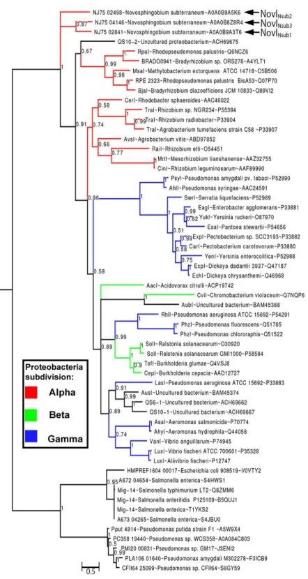
By rooting MAG-14 homologs as the outgroup, Bayesian inference of the newly identified NovI proteins do not exhibit close evolutionary relatedness to any of the selected LuxI homologs and instead occupy a very basal position in the phylogenetic tree. NovINsub1
andNovINsub3 formed a monophyletic clade among themselves with moderate strong
posterior probability support (pp =0.87) (Fig. 3). The relatedness of NovINsub1 and
Figure 2 Identification and gene organization of three functional LuxI homologs and one newly identified luxR solo.(A) Alignment of three identifiedNovosphingobium subterraneumLuxI homologs with other previously reported functional LuxI homologs. Conserved sites are highlighted in yellow and cyan-colored residues indicate deviation from consensus sequence. (B) Gene organization of the identifiedluxIhomologs and also a newly identifiedluxRsolo.
in their gene organization (Figs. 3and2A). A majority of the functionally validated LuxI homologs isolated from metagenomic libraries did not demonstrate novel phylogenetic position and instead formed monophyletic clustering with known LuxI homologs with strong posterior probability support. CviI from Chromobacterium violaceumshared the most common ancestor with metagenome-derived AubI with maximal posterior probability support while LasI from Pseudomonas aeruginosais sister taxa to the clades containing AusI, QS6-1 and QS10-1 (pp=0.91). One notable exception is QS10-S (accession number: ACH69675) that formed a weakly supported (pp=0.67) monophyletic cluster with the
clade containing LuxI homologs from the generaRhodopseudomonas, Bradyrhizobiumand
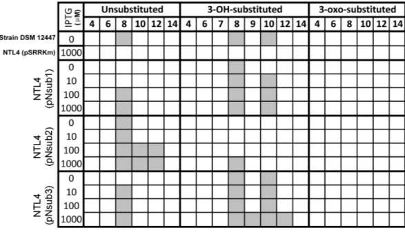
Figure 4 Screening of AHL signals production by individual heterologously expressed NovI homolog against a wide range of authentic standards.Colored columns indicate presence of detectable signal (>0 signal peak area unit). Numerical values in the second row indicate the acyl-chain length of AHL.
First functional validation of three LuxI homologs from the family sphingomonadaceae
All three identified NovI homologs led to the accumulation of AHL signals in culture medium when they were heterologously expressed in Agrobacterium tumefaciensstrain NTL4 (Fig. 4). Using an inducible expression, the effect of low-, medium- and high-expression on AHL accumulation pattern i.e., detection of additional AHL signals previously not observed in the wild type, can be better studied. In this system, the addition of inducer, IPTG, to the culture medium results in the de-repression of the cloned genes within the cells of the population (Khan et al., 2008). Out of the 20 screened AHL signals, a total of 7 AHL unsubstituted and OH-substituted signals can be identified at the highest IPTG induction of three clonednovI. The signals detected include C8, C8-OH and C10-OH for NovInsub1; C8, C10, C12 for NovInsub2; and C8, C8-OH, C9-OH, C10-OH and C12-OH
for Novnsub3. It is worth noting that, in the absence of IPTG inducer, basal levels of AHL
signals were detected in the growth medium, suggesting suboptimal gene repression by the lacR repressor inAgrobacterium tumefacienshost. In comparison to the 2-D RP-TLC analysis (Fig. 1), LC-MS/MS analysis corroborated the presence of six AHLs and extended it to seven AHLs through inducible heterologous expression of the three individual NovI homologs ofN. subterraneumDSM 12447 (Fig. 4).
Dissimilar amount and ratio of major AHLs accumulation in liquid media during the heterologous expression of NovINsub1and NovINsub3
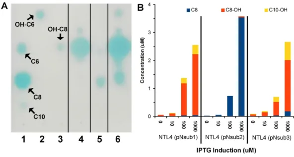
Figure 5 Identification and quantification of AHL signals produced through the individual heterolo-gous expression ofNovosphingobium subterraneumLuxI homologs. (A) One-dimensional TLC-based detection. Lane 1, Unsubstituted medium length AHL signals; Lane 2, hydroxy-C6 AHL; Lane 3, 3-hydroxy-C8 AHL; Lane 4, NTL4 (pNsub1) 2µl of 20×EtOAc extract; Lane 5, NTL4(pNsub2) 6µl of 20
×EtOAc extract; Lane 6, NTL4(pNsub3) 1µl of 20×EtOAc extract. (B) Concentration of three major AHL molecules, C8-OH, C8 and C10-OH, were calculated based on standard curves calibrated with differ-ent concdiffer-entrations of authdiffer-entic AHL standards.
prepared samples for a six-point calibration curves ranging from 0.5µM–20µM for C8,
C8-OH, and C10-OH. AHL signals. C8, C8-OH, and C10-OH, are the signals present in the highest concentrations in culture extracts. This approach identified and quantified the three main signals produced by the three NovI homologs showing that NovINsub2mainly
produces C8 while C8-OH and C10-OH are the major AHLs synthesized by NovINsub1and
NovINsub3. The ratio of C8-OH to C10-OH consistently differ by at least 2-fold in NovINsub1
and NovINsub3(6.6 vs 2.8) at various IPTG induction concentrations (Fig. 5B). The strong
overlap of AHL accumulation profile between NovINsub1and NovINsub3provides additional
evidence supporting their close evolutionary relatedness.
DISCUSSION
identification. In a futurein vivostudy, it would be interesting to explore if the change in AHL profiles are correlated to the alterations of a variety of growth media.
The conservednovI-novR-phyH synteny adds to the growing association of such a synteny with the luxI homologs of Sphingomonadaceae family as reported previously (Gan et al., 2013). This arrangement has also been discovered in metagenomic sampling as described by Hao and colleagues (Hao et al., 2010). In eukaryotes, PhyH is localized in the peroxisome and catalyzes the alpha-oxidation of phytanic acid to pristanic acid through the elimination of one carbon (Van den Brink & Wanders, 2006). The frequent association ofphyH with variousnovI/Rleads Hao and coworkers (Hao et al., 2010) to speculate that its transcription maybe regulated by quorum sensing. The increasing observation ofphyH
linkage with thenovI/Rwarrants future work investigating its transcriptional regulation by quorum sensing and more importantly the yet-to-be described enzymatic reaction that PhyH catalyzes in bacteria.
Similarly utilizing Mig14 family protein (PF07395) from the acetyltransferase-like clan (CL0257) as the outgroup, our phylogenetic inference does not support the the basal position of clade containing the YenI, EagI, EsaI proteins (Christensen et al., 2014). Such striking differences may stem from the lack of LuxI homologs sampling from the Sphingomonadaceae family, a potential source of new phylogenetic signal, and possibly the usage of different phylogenetic inference method e.g., distance-based vs model-based. The similar neighborhood joining methods employed byChristensen et al. (2014) was also implemented in two major LuxI phylogeny studies (Gan et al., 2013;Gray & Garey, 2001;Lerat & Moran, 2004). The newly constructed Bayesian tree incorporating complex model for across-site heterogeneities in addition to improved taxon sampling represents a significant improvement over previously reported phylogenetic (Lartillot, Brinkmann & Philippe, 2007;Lartillot & Philippe, 2004;Zwickl & Hillis, 2002). That being said, the basal position of LuxI homologs from the genusNovosphingobiumwas similarly observed in a previously reported neighborhood-joining tree for LuxI homologs (Gan et al., 2013). It will be interesting to see if the tree topology will remain consistent as more LuxI homologs are being functionally validated and included into the phylogenetic analysis in the future.
The close evolutionary relationship of NovINsub1 and NovINsub3corroborates with their
overlapping AHL profile i.e., when heterologously expressed, both produce mainly C8-OH and C10-OH but at a different ratio. The presence of various genes coding for transposases upstream of novINsub1along with its high relatedness to novISub3suggests recent replicative
transposition. The slight dissimilarity in AHL production efficiency between NovISub1
and NovISub3could be explained by an on-going ‘‘neofunctionalization’’ process given
CONCLUSIONS
Novosphingobium subterraneumDSM 12447 accumulated different medium-length AHL
compounds with a majority of them being C8-OH. Whole genome sequencing and annotation identified threeluxIhomologs in strain DSM 12447 with two of them exhibiting higher relatedness as evidenced by their monophyletic clustering, shared gene synteny and overlapping AHL signal profile based on heterologous expression inAgrobacterium
tumefaciensNTL4. This work provides the first functional validation of LuxI homologs in
the family Sphingomonadacea.
ADDITIONAL INFORMATION AND DECLARATIONS
Funding
The American Society for Microbiology (ASM) provided a summer 2015 stipend to LKD and MAS under the ASM Undergraduate Research Fellowship program to LKD. MAS, AOH and HMG received support from Thomas H. Gosnell School of Life Sciences (GSOLS) and the College of Science (COS) at the Rochester Institute of Technology (RIT). HMG received support from the Monash University Malaysia Tropical Medicine and Biology Multidisciplinary Platform. The funders had no role in study design, data collection and analysis, decision to publish, or preparation of the manuscript.
Grant Disclosures
The following grant information was disclosed by the authors: ASM Undergraduate Research Fellowship program.
Thomas H. Gosnell School of Life Sciences (GSOLS).
College of Science (COS) at the Rochester Institute of Technology (RIT).
Monash University Malaysia Tropical Medicine and Biology Multidisciplinary Platform.
Competing Interests
The authors declare there are no competing interests.
Author Contributions
• Han Ming Gan conceived and designed the experiments, performed the experiments,
analyzed the data, contributed reagents/materials/analysis tools, wrote the paper, prepared figures and/or tables, reviewed drafts of the paper.
• Lucas K. Dailey performed the experiments, contributed reagents/materials/analysis
tools, wrote the paper, prepared figures and/or tables.
• Nigel Halliday conceived and designed the experiments, performed the experiments,
analyzed the data, wrote the paper, reviewed drafts of the paper.
• Paul Williams conceived and designed the experiments, analyzed the data, contributed
reagents/materials/analysis tools, wrote the paper, reviewed drafts of the paper.
• André O. Hudson conceived and designed the experiments, analyzed the data, wrote the
• Michael A. Savka conceived and designed the experiments, analyzed the data, contributed
reagents/materials/analysis tools, wrote the paper, prepared figures and/or tables, reviewed drafts of the paper.
DNA Deposition
The following information was supplied regarding the deposition of DNA sequences: This Whole Genome Shotgun project for N. subterraneum DSM 12447 has been deposited at DDBJ/EMBL/GenBank under the accession JRVC00000000. The version described in this paper is versionJRVC01000000.
REFERENCES
Balkwill DL, Drake GR, Reeves RH, Fredrickson JK, White DC, Ringelberg DB, Chandler DP, Romine MF, Kennedy DW, Spadoni CM. 1997.Taxonomic study of aromatic-degrading bacteria from deep-terrestrial-subsurface sediments and description of Sphingomonas aromaticivorans sp. nov., Sphingomonas subterranea sp. nov., and Sphingomonas stygia sp. nov.International Journal of Systematic
Bacteriology47:191–201 DOI 10.1099/00207713-47-1-191.
Bankevich A, Nurk S, Antipov D, Gurevich AA, Dvorkin M, Kulikov AS, Lesin VM, Nikolenko SI, Pham S, Prjibelski AD, Pyshkin AV, Sirotkin AV, Vyahhi N, Tesler G, Alekseyev MA, Pevzner PA. 2012.SPAdes: a new genome assembly algorithm and its applications to single-cell sequencing.Journal of Computational Biology
19:455–477DOI 10.1089/cmb.2012.0021.
Boetzer M, Henkel CV, Jansen HJ, Butler D, Pirovano W. 2011.Scaffolding pre-assembled contigs using SSPACE.Bioinformatics27:578–579
DOI 10.1093/bioinformatics/btq683.
Boetzer M, Pirovano W. 2012.Toward almost closed genomes with GapFiller.Genome
Biology13:R56DOI 10.1186/gb-2012-13-6-r56.
Capella-Gutierrez S, Silla-Martinez JM, Gabaldon T. 2009.trimAl: a tool for automated alignment trimming in large-scale phylogenetic analyses.Bioinformatics
25:1972–1973DOI 10.1093/bioinformatics/btp348.
Case RJ, Labbate M, Kjelleberg S. 2008.AHL-driven quorum-sensing circuits: their frequency and function among the Proteobacteria.The ISME Journal2:345–349
DOI 10.1038/ismej.2008.13.
Cha C, Gao P, Chen YC, Shaw PD, Farr SK. 1998.Production of acyl-homoserine lac-tone quorum-sensing signals by gram-negative plant-associated bacteria.Molecular
Plant-Microbe Interactions11:1119–1129DOI 10.1094/MPMI.1998.11.11.1119.
Chatnaparat T, Prathuangwong S, Ionescu M, Lindow SE. 2012.XagR, a LuxR ho-molog, contributes to the virulence of Xanthomonas axonopodis pv. glycines to soy-bean.Molecular Plant-Microbe Interactions25:1104–1117
DOI 10.1094/MPMI-01-12-0008-R.
molecule N-(3-oxododecanoyl)-L-homoserine lactone as immune modulators.
Journal of Medicinal Chemistry 46:97–104DOI 10.1021/jm020909n.
Chhabra SR, Stead P, Bainton NJ, Salmond GP, Stewart GS, Williams P, Bycroft BW. 1993.Autoregulation of carbapenem biosynthesis in Erwinia carotovora by analogues of N-(3-oxohexanoyl)-L-homoserine lactone.Journal of Antibiotics
46:441–454DOI 10.7164/antibiotics.46.441.
Chilton MD, Currier TC, Farrand SK, Bendich AJ, Gordon MP, Nester EW. 1974.
Agrobacterium tumefaciensDNA and PS8 bacteriophage DNA not detected in crown
gall tumors.Proceedings of the National Academy of Sciences of the United States of
America71:3672–3676DOI 10.1073/pnas.71.9.3672.
Christensen QH, Brecht RM, Dudekula D, Greenberg EP, Nagarajan R. 2014.Evolution of acyl-substrate recognition by a family of acyl-homoserine lactone synthases.PLoS ONE9:e112464DOI 10.1371/journal.pone.0112464.
Cude WN, Buchan A. 2013.Acyl-homoserine lactone-based quorum sensing in the roseobacter clade: complex cell-to-cell communication controls multiple physiolo-gies.Frontiers in Microbiology4:336 DOI 10.3389/fmicb.2013.00336.
Fast W, Tipton PA. 2012.The enzymes of bacterial census and censorship.Trends in Biochemical Sciences37:7–14DOI 10.1016/j.tibs.2011.10.001.
Fuqua C. 2006.The QscR quorum-sensing regulon of pseudomonas aeruginosa: an or-phan claims its identity.Journal of Bacteriology188:3169–3171
DOI 10.1128/JB.188.9.3169-3171.2006.
Fuqua C, Greenberg EP. 2002.Listening in on bacteria: acyl-homoserine lactone signalling.Nature Reviews Molecular Cell Biology3:685–695 DOI 10.1038/nrm907. Fuqua WC, Winans SC, Greenberg EP. 1994.Quorum sensing in bacteria: the
LuxR.-LuxI family of cell density-responsive transcriptional regulators.Journal of Bacteriol-ogy176:269–275.
Gan HM, Buckley L, Szegedi E, Hudson AO, Savka MA. 2009.Identification of an rsh gene from aNovosphingobiumsp. necessary for quorum-sensing signal accumula-tion.Journal of Bacteriology191:2551–2560DOI 10.1128/JB.01692-08.
Gan HM, Chew TH, Hudson AO, Savka MA. 2012.Genome sequence of Novosph-ingobiumsp. strain Rr 2-17, a nopaline crown gall-associated bacterium isolated from Vitis vinifera L. grapevine.Journal of Bacteriology194:5137–5138
DOI 10.1128/JB.01159-12.
Gan HM, Gan HY, Ahmad NH, Aziz NA, Hudson AO, Savka MA. 2015.Whole genome sequencing and analysis reveal insights into the genetic structure, diversity and evolutionary relatedness of luxI and luxR homologs in bacteria belonging to the Sphingomonadaceae family.Frontiers in Cellular and Infection Microbiology4:188
DOI 10.3389/fcimb.2014.00188.
Gan HM, Hudson AO, Rahman AY, Chan KG, Savka MA. 2013.Comparative genomic analysis of six bacteria belonging to the genusNovosphingobium: insights into marine adaptation, cell–cell signaling and bioremediation.BMC Genomics14:431
Gonzalez JF, Venturi V. 2013.A novel widespread interkingdom signaling circuit.Trends in Plant Science18:167–174DOI 10.1016/j.tplants.2012.09.007.
Gray KM, Garey JR. 2001.The evolution of bacterial LuxI and LuxR quorum sensing regulators.Microbiology147:2379–2387DOI 10.1099/00221287-147-8-2379. Hao Y, Winans SC, Glick BR, Charles TC. 2010.Identification and characterization
of new LuxR/LuxI-type quorum sensing systems from metagenomic libraries.
Environmental Microbiology12:105–117DOI 10.1111/j.1462-2920.2009.02049.x.
Jones P, Binns D, Chang HY, Fraser M, Li W, McAnulla C, McWilliam H, Maslen J, Mitchell A, Nuka G, Pesseat S, Quinn AF, Sangrador-Vegas A, Scheremetjew M, Yong SY, Lopez R, Hunter S. 2014.InterProScan 5: genome-scale protein function classification.Bioinformatics30:1236–1240DOI 10.1093/bioinformatics/btu031. Katoh K, Standley DM. 2014.MAFFT: iterative refinement and additional methods.
Methods in Molecular Biology 1079:131–146DOI 10.1007/978-1-62703-646-7_8.
Khan SR, Gaines J, Roop II RM, Farr SK. 2008.Broad-host-range expression vectors with tightly regulated promoters and their use to examine the influence of TraR and TraM expression on Ti plasmid quorum sensing.Applied and Environmental
Microbiology74:5053–5062DOI 10.1128/AEM.01098-08.
Lartillot N, Brinkmann H, Philippe H. 2007.Suppression of long-branch attraction arte-facts in the animal phylogeny using a site-heterogeneous model.BMC Evolutionary
Biology7(Suppl 1):S4DOI 10.1186/1471-2148-7-S1-S4.
Lartillot N, Philippe H. 2004.A Bayesian mixture model for across-site heterogeneities in the amino-acid replacement process.Molecular Biology and Evolution21:1095–1109
DOI 10.1093/molbev/msh112.
Lartillot N, Rodrigue N, Stubbs D, Richer J. 2013.PhyloBayes MPI: phylogenetic
reconstruction with infinite mixtures of profiles in a parallel environment.Systematic
Biology62:611–615DOI 10.1093/sysbio/syt022.
Lerat E, Moran NA. 2004.The evolutionary history of quorum-sensing systems in bacteria.Molecular Biology and Evolution21:903–913DOI 10.1093/molbev/msh097. Lowe N, Gan HM, Chakravartty V, Scott R, Szegedi E, Burr TJ, Savka MA. 2009.
Quorum-sensing signal production byAgrobacteriumvitis strains and their tumor-inducing and tartrate-catabolic plasmids.FEMS Microbiology Letters296:102–109
DOI 10.1111/j.1574-6968.2009.01627.x.
Luo ZQ, Clemente TE, Farrand SK. 2001.Construction of a derivative ofAgrobacterium tumefaciensC58 that does not mutate to tetracycline resistance.Molecular
Plant-Microbe Interactions14:98–103.
Maddison WP, Maddison DR. 2015.Mesquite: a modular system for evolutionary analysis. 3.0.Available athttp:// mesquiteproject.org.
McClean KH, Winson MK, Fish L, Taylor A, Chhabra SR, Camara M, Daykin M, Lamb JH, Swift S, Bycroft BW, Stewart GS, Williams P. 1997.Quorum sensing and
Chromobacterium violaceum: exploitation of violacein production and inhibition
for the detection ofN-acylhomoserine lactones.Microbiology143:3703–3711
Miller MB, Bassler BL. 2001.Quorum sensing in bacteria.Annual Review of Microbiology
55:165–199DOI 10.1146/annurev.micro.55.1.165.
Schuster M, Sexton DJ, Diggle SP, Greenberg EP. 2013.Acyl-homoserine lactone quorum sensing: from evolution to application.Annual Review of Microbiology
67:43–63DOI 10.1146/annurev-micro-092412-155635.
Scott RA, Weil J, Le PT, Williams P, Fray RG, Von Bodman SB, Savka MA. 2006. Long- and short-chain plant-produced bacterialN-acyl-homoserine lactones become components of phyllosphere, rhizosphere, and soil.Molecular Plant-Microbe
Interactions19:227–239DOI 10.1094/MPMI-19-0227.
Seemann T. 2014.Prokka: rapid prokaryotic genome annotation.Bioinformatics
30:2068–2069DOI 10.1093/bioinformatics/btu153.
Shaw PD, Ping G, Daly SL, Cha C, Cronan Jr JE, Rinehart KL, Farr SK. 1997.Detecting and characterizingN-acyl-homoserine lactone signal molecules by thin-layer chromatography.Proceedings of the National Academy of Sciences of the United States
of America94:6036–6041DOI 10.1073/pnas.94.12.6036.
Sokol PA, Sajjan U, Visser MB, Gingues S, Forstner J, Kooi C. 2003.The CepIR quorum-sensing system contributes to the virulence of Burkholderia cenocepacia respiratory infections.Microbiology149:3649–3658DOI 10.1099/mic.0.26540-0. Subramoni S, Venturi V. 2009.LuxR-family ‘solos’: bachelor sensors/regulators of
signalling molecules.Microbiology155:1377–1385DOI 10.1099/mic.0.026849-0. Sullivan MJ, Petty NK, Beatson SA. 2011.EasyFig: a genome comparison visualizer.
Bioinformatics27:1009–1010DOI 10.1093/bioinformatics/btr039.
Takeuchi M, Hamana K, Hiraishi A. 2001.Proposal of the genus Sphingomonas sensu stricto and three new genera, Sphingobium,Novosphingobiumand Sphingopyxis, on the basis of phylogenetic and chemotaxonomic analyses.International Journal of
Systematic and Evolutionary Microbiology51:1405–1417
DOI 10.1099/00207713-51-4-1405.
Tsai CS, Winans SC. 2010.LuxR-type quorum-sensing regulators that are detached from common scents.Molecular Microbiology77:1072–1082
DOI 10.1111/j.1365-2958.2010.07279.x.
Van den Brink DM, Wanders RJ. 2006.Phytanic acid: production from phytol, its break-down and role in human disease.Cellular and Molecular Life Science63:1752–1765
DOI 10.1007/s00018-005-5463-y.
Waters CM, Bassler BL. 2005.Quorum sensing: cell-to-cell communication in bacteria.
Annual Review of Cell and Developmental Biology21:319–346
DOI 10.1146/annurev.cellbio.21.012704.131001.
Weber T, Blin K, Duddela S, Krug D, Kim HU, Bruccoleri R, Lee SY, Fischbach MA, Muller R, Wohlleben W, Breitling R, Takano E, Medema MH. 2015.antiSMASH 3.0-a comprehensive resource for the genome mining of biosynthetic gene clusters.
Nucleic Acids Research43:W237–W243DOI 10.1093/nar/gkv437.
White DC, Sutton SD, Ringelberg DB. 1996.The genus Sphingomonas: physiology and ecology.Current Opinion in Biotechnology7:301–306
Winson MK, Swift S, Fish L, Throup JP, Jorgensen F, Chhabra SR, Bycroft BW, Williams P, Stewart GS. 1998.Construction and analysis of luxCDABE-based plas-mid sensors for investigatingN-acyl homoserine lactone-mediated quorum sensing.
FEMS Microbiology Letters163:185–192DOI 10.1111/j.1574-6968.1998.tb13044.x.
Yanisch-Perron C, Vieira J, Messing J. 1985.Improved M13 phage cloning vectors and host strains: nucleotide sequences of the M13mp18 and pUC19 vectors.Gene
33:103–119.
Zwickl DJ, Hillis DM. 2002.Increased taxon sampling greatly reduces phylogenetic error.