BRUNA PENA SOLLERO
TRANSCRIPTIONAL PROFILING DURING PIG SKELETAL MUSCLE
DEVELOPMENT OF A BRAZILIAN LOCAL BREED AND TWO
COMMERCIAL GENETIC GROUPS
Thesis
presented
to
the
Animal Science Graduate Program of
the Universidade Federal de Viçosa, in
partial fulfillment of the requirements
for degree of Doctor Scientiae.
VIÇOSA
BRUNA PENA SOLLERO
TRANSCRIPTIONAL PROFILING DURING PIG SKELETAL MUSCLE
DEVELOPMENT OF A BRAZILIAN LOCAL BREED AND TWO
COMMERCIAL GENETIC GROUPS
Thesis presented to the
Animal Science Graduate Program of
the Universidade Federal de Viçosa,
in partial fulfillment of the
requirements for degree of Doctor
Scientiae.
APROVADA: October 19, 2010.
Prof. Fabyano Fonseca e Silva
(Co-adviser)
Prof. Flávia Maria Lopes Passos
Prof. Paulo Sávio Lopes
(Co-adviser)
Prof. Catherine W. Ernst
ii
“
Do not go where the path may lead.
Go instead where there is no path
and leave a trail
”
iii
ACKNOWLEDGEMENTS
To Universidade Federal de Viçosa and the Animal Science Department, for
providing me the opportunity to conclude an important step in my life;
To CNPq, for the scholarship here and in United States (fellowship program);
To my parents and sisters, for all their affection and unconditional support;
To my adviser, Professor Simone Eliza Facioni Guimarães, for her support, for
enriching my knowledge, increasing my confidence and being my confirmation that “a good
example is the best sermon”;
To my co-adviser Fabyano Fonseca, for all the time he spent giving me fundamental
advice that enabled me to develop and complete this work;
To Professor Paulo Sávio for his guidance while co-advising me;
To Professor José Domingos Guimarães, for his essential support;
Many thanks to the Department of Animal Science at Michigan State University, the
USDA CSREES National Research Initiative, and Michigan Agricultural Experiment Station;
To Dr. Ernst for the opportunity to work with her research group and develop part of
this project at the Meat Animal Muscle Biology and Molecular Genetics Laboratory (MSU);
To Dr. Steibel and Dr. Tempelman;
To Valencia, Nancy, IgSeo, Michelle and all my friends in US (Julia, Kristi, Oscar,
Julie, Rita, Erik, Koushik) for their company, help, friendship and patience;
To Professor Glória Regina Franco and graduate student Priscila Grinberg at the
Departamento de Genética Bioquímica of the Universidade Federal de Minas Gerais, for the
opportunity to create the third step of this project and for their collaboration;
To all the others Professors, employees and students from the Animal Science
Department, for making workdays a pleasant living environment, sharing ideas and enjoying
great moments;
To my friends from LABTEC, for all these years of friendship and complicity:
Débora, Carlos, Lincoln, Kleibe, Katiene, Ana Paula, Renata, Priscila, Lucas, Erica, Mayara and
Carol; also Nicola and Miguel. Specially, many thanks to dear Marcos, for his willingness to
participate- his collaboration was essential!
To all my other friends and relatives, for sharing with me happiness, fears, victories
and overruns!
iv
BIOGRAPHY
Bruna Pena Sollero, daughter of Paulo Antônio Sollero and Dolores Maria Pena
Sollero, born on September 1, 1981 in Brasilia, Distrito Federal, Brazil. She began her studies
in Animal Science at the Universidade Federal de Viçosa in 2000, studying many different
areas and participating in many different research groups and projects, especially with the
Program for Breeding Swine.
In January of 2005, she chose to major in Animal Science, began pursuing a Master of
Agricultural Science at the Universidade de Brasília, Brazil, in March of that same year. This
degree was completed in partnership with Embrapa Recursos Genéticos e Biotecnologia
(Embrapa Genetic Resources and Biotechnology) in the area of Animal Genetic Resources
Conservation. Ms. Sollero completed her final examination on November 10, 2006 to obtain
the title of Magister Scientiae in Animal Production.
In March of 2007 she began the doctoral graduation program in Animal Breeding at the
Animal Science Department of the Universidade Federal de Viçosa. During her doctoral
program she spent one year, from September of 2008 to September of 2009, at the Meat Animal
Muscle Biology and Molecular Genetics Laboratory at Michigan State University in the United
States. It was there that she developed a large part of her doctoral research project. Ms. Sollero
completed her dissertation defense on October 19, 2010, to obtain the title of Doctor Scientiae
v
SUMMARY
ABSTRACT
RESUMO
vi
viii
GENERAL INTRODUCTION
REFERENCES
01
06
CHAPTER I (ARTICLE I)
ABSTRACT
RESUMO
INREODUCTION
MATERIAL AND METHODS
REUSLTS
DISCUSSION
ACKNOWLEDGEMENTS
REFERENCES
08
09
10
12
15
20
24
32
CHAPTER II (ARTICLE II)
ABSTRACT
RESUMO
INTRODUCTION
MATERIAL AND METHODS
RESULTS
DISCUSSION
REFERENCES
38
39
40
41
45
54
63
CHAPTER III
ABSTRACT
RESUMO
INTRODUCTION
69
70
71
REVIEW
Background correction
Loess normalization method
Robust Spline normalization method
Quantile normalization method
False Discovery Rate
72
73
74
76
77
MATERIAL AND METHODS
Experimental design
Data processing
Clustering analysis and Gene Ontology analysis
79
80
81
RESULTS
Identifying genes differentially expressed
Clustering analysis
Gene Ontology analysis
82
87
93
DISCUSSION
105
REFERENCES
111
GENERAL CONCLUSIONS
116
APPENDIX 1
118
APPENDIX 2
119
vi
ABSTRACT
SOLLERO, Bruna Pena, D.Sc., Universidade Federal de Viçosa, October, 2010.
Transcriptional profiling during pig skeletal muscle development of a Brazilian
local breed and two commercial genetic groups. Adviser: Simone Eliza Facioni
Guimarães. Co-Advisers: Paulo Sávio Lopes e Fabyano Fonseca e Silva.
Skeletal muscle development is a complex process involving the coordinated expression of
thousands of genes. The aim of this study was to identify differentially expressed genes in
longissimus dorsi (LD) muscle of pigs and their expression pattern through five time-points of
the development. Firstly, a microarray analysis using the Pigoligoarray containing 20,400
oligonucleotides was done to compare two genetic groups of pigs that differ in muscularity
(North American Yorkshire x Landrace (YL) crossbred pigs and Piau pigs -a naturalized
Brazilian breed) at 40 and 70 days (d) of gestation (developmental stages encompassing primary
and secondary fiber formation). A total of 486 oligonucleotides were differentially expressed (FC ≥ 1.5; FDR ≤ 0.05) between 40 and 70 d gestation in either YL or Piau pigs, and a total of 1,300 oligonucleotides were differentially expressed (FC ≥ 1.5; FDR ≤ 0.05) between YL and Piau pigs at either age. Gene ontology annotation and pathway analyses determined functional
classifications for differentially expressed genes and revealed breed-specific developmental
expression patterns. Thirteen genes were selected for confirmation by qRT-PCR analyses and
expression patterns for most of these genes were confirmed, providing further insight into the
roles of these genes in pig muscle development. The relative abundance of transcripts tended to
be greater for the Piau pigs at 70 d of gestation suggesting that gene expression in LD muscle of
YL pigs may be more delayed than in Piau pigs. The second experiment focused to evaluate the
expression profile during prenatal (21, 40, 70 and 90 days pos conception) and early postnatal
(10 weeks) stages of the same muscle between the local Brazilian pig breed (Piau) and a
Brazilian white composite line. Based on qRT-PCR analyses, others fourteen genes related to
intrinsic biological processes during myogenesis were investigated, and nine genes out of them
were differentially expressed between both genetic groups (P<0.05). Significant different levels
of expression were also observed through the five time points and patterns of genes related to
cell proliferation, cell differentiation, energy metabolism, and maintenance were distinct in a
breed-specific way. A new reference gene for expression profile investigation of pig muscle not
used before for this purpose is proposed: DDIT3 (DNA-damage-inducible transcript 3).
Furthermore, this research evaluated the application of three different protocols for data
normalization and assessment of genes differentially expressed in microarray analyses. A
trade-off among the results was observed when comparing the three protocol analyses tested by
vii
microarray analysis applied in the first chapter, detected genes as DE only in two (C40vsP40
and P40vsP70) out of the four contrasts under evaluation (C40vsP40, P40vsP70, C70vsP70 and
C40vsC70). The results strengthened the idea that the Robust Spline normalization method was
able to identify more genes differentially expressed (FDR-False Discovery Rate ≤0.05) related
with important GO terms involved with the muscle development process (especially in
breed-contrasts) in comparison with the Loess method. In addition, normexp background correction
method was advisable, working clearly better than the traditional subtraction method. It was
suggested that the q-value FDR method can be too flexible to detect genes differentially
expressed in small microarray experiments, while the BH FDR method can be too restrictive for
that – although pointing out genes DE with higher fold change values and with similar
functionalities related to the myogenesis process in context. In general, this study which was the
first whole-genome expression evaluation of the Brazilian native Piau pigs, revealed both
developmental and breed-specific patterns of gene expression in fetal pig skeletal muscle
including genes not previously associated with myogenesis, and these information can
contribute to future pig genetic improvement efforts. Also, comparisons among protocol
analyses for microarray data are suggested for cleaning and improving the quality of the
viii
RESUMO
SOLLERO, Bruna Pena, D.Sc., Universidade Federal de Viçosa, outubro de 2010.
Perfil transcricional durante o desenvolvimento muscular esquelético de suínos
de uma raça local Brasileira e dois grupos genéticos comerciais. Orientadora:
Simone Eliza Facioni Guimarães. Co-orientadores: Paulo Sávio Lopes e Fabyano
Fonseca e Silva.
O desenvolvimento esquelético muscular é um processo complexo que envolve a expressão
coordenada de milhares de genes. O objetivo deste estudo foi identificar genes diferencialmente
expressos no músculo longissimus dorsi (LD) de suínos, bem como seus padrões de expressão
entre cinco estádios do desenvolvimento. Primeiramente, uma análise de microarranjo
utilizando o Pigoligoarray, contendo 20.400 oligonucleotídeos, foi realizada para comparar dois
grupos genéticos de suínos que diferem em capacidade de crescimento muscular e conteúdo de
carne magra (grupo cruzado comercial Yorkshire-Landrace-YL, proveniente dos Estados
Unidos e suínos Piau- raça Brasileira naturalizada) aos 40 e 70 dias (d) de gestação (estádios do
desenvolvimento decorrentes da formação de fibras musculares primárias e secundárias). Um total de 486 oligonucleotídeos foram diferencialmente expressos (FC ≥ 1.5; FDR ≤ 0.05) entre 40 e 70 dias de gestação no grupo YL ou Piau, e um total de 1.300 oligonucleotíedos foram diferencialmente expressos (FC ≥ 1.5; FDR ≤ 0.05) entre suínos YL e Piau em ambas as idades pré-natais. Anotações baseadas no Gene Ontology (GO) e análises de vias metabólicas
determinaram classificações funcionais para os genes diferencialmente expressos e revelaram
padrões de expressão raça-específicos. Treze genes foram selecionados para confirmação por
análises de qRT-PCR e os padrões de expressão para a maioria destes genes foram confirmados,
gerando informações relevantes sobre seus papéis desempenhados durante o processo do
desenvolvimento muscular de suínos. Na raça Piau a abundância relativa de transcritos aos 70 d
de gestação foi tendencialmente maior, sugerindo que a expressão gênica no músculo LD de
suínos YL pode ser atrasada em relação à raça local. O segundo experimento objetivou avaliar
perfis de expressão durante cinco períodos pré-natais (21, 40, 70 e 90 dias pós-concepção) e um
período pós-natal (10 semanas pós-nascimento) no mesmo músculo esquelético (LD) entre a
raça Brasileira local Piau e uma linhagem comercial também brasileira. Baseado em análises de
qRT-PCR, outros quatorze genes relacionados a processos biológicos intrínsecos ao processo de
miogênese foram investigados, e nove destes genes foram significativamente (P<0.05)
diferencialmente expressos entre ambos os grupos genéticos de suínos. Níveis significativos de
expressão gênica também foram observados entre os cinco estádios avaliados e padrões de
genes relacionados à proliferação celular, diferenciação celular, metabolismo de energia e
manutenção da formação muscular foram distintos de forma raça-específica. Um novo gene
ix
proposto: DDIT3 (DNA-damage-inducible transcript 3). A última etapa desta pesquisa
objetivou avaliar a aplicação de três protocolos para normalização e detecção de genes
diferencialmente expressos em análises de microarranjos. Resultados conflitantes foram
observados quando comparado os três protocolos analisados por distintos métodos estatísticos.
Os dois protocolos propostos, em relação à primeira análise de microarranjo aplicada no
primeiro capítulo, detectaram genes como diferencialmente expressos em apenas dois
(C40vsP40 e P40vsP70) dos quarto contrastes sob avaliação (C40vsP40, P40vsP70, C70vsP70 e
C40vsC70). Os resultados demonstraram que o método de normalização Robust Spline foi capaz
de identificar um maior número de genes diferencialmente expressos (FDR-Taxa de Falsas
Descobertas ≤0.05) relacionados a importantes GO terms envolvidos no processo de
desenvolvimento muscular (especialmente em contrastes entre idades) em comparação ao
método Loess. Além do mais, o método de correção background normexp foi recomendado,
uma vez que se mostrou mais acurado do que o método tradicional de Subtração. Foi sugerido
que o método FDR q-value pode ser muito flexível na detecção de genes diferencialmente
expressos em pequenos experimentos de microarranjo, enquanto o método BH pode ser muito
restritivo – ainda que capaz de apontar genes DE com maiores valores de expressão relativa e
funcionalmente relacionados ao processo de miogênese em contexto. Em geral, este estudo, que
pela primeira vez avaliou em nível genômico a expressão de genes da raça Piau, revelou
padrões de expressão gênica raça e idade-específicos durante o desenvolvimento muscular fetal,
incluindo genes não descritos previamente como associados a tal processo. Tais informações
podem contribuir para futuros avanços do melhoramento genético de suínos. Também,
comparações entre diferentes protocolos de análises de microarranjos foram sugeridas, a fim de
1
General IntroductionAccording to Abipecs (Associação Brasileira da Indústria Produtora e Exportadora de Carne
Suína, 2010), the growing demand for protein food sources is a worldwide reality and the pork is
continuously increasing its importance on the meat market.
For the meat-producing industries, emphasis has been given on increasing the understanding of
the physiological process and molecular pathways associated with skeletal muscle growth and
development (Reecy et al., 2006). In addition to the knowledge that factors operating prenatally
theoretically determine the maximum size to which this tissue can growth (Wigmore & Stickland, 1983),
skeletal muscle is essential for a wide range of functions in livestock animals, including breathing,
locomotion, maintenance of posture and thermogenesis (Dauncey & Gilmour, 1996).
Pig breeding companies are focused on the organization and genetic structures of “artificial pig
populations”; defining specific breeding goals, their weighting in an overall breeding objective and the
choice of optimal selection criteria. Challenges imposed to these companies resulted from the evolution of
genetic improvement combined with constant changing in the production systems. As discussed by van
der Steen et al., (2005), in particular, highly heritable traits, such as milk production in dairy cows and
lean percentage in pigs and broilers, are predominantly improved through the genetic route. Nevertheless,
behind each choice of selection criteria strong researches focusing on the molecular genetics aspects of a
certain population is constantly under evaluation.
The process of domestication, as described for instance by Jonsson (1991), includes selective
breeding for specific characteristics, and may be considered as a first step in genetic improvement. An
idea of the distance covered from this primary process can be gained by comparing the modern domestic
pig to its wild counterpart. In this scenario, genetic groups, which were not submitted to the same
selection process or still maintain the original genetic bases from the beginning, are attractive genetic
2
The Piau Brazilian naturalized pig breed, originated from other breeds introduced by Portuguese
settlers in the 16th century, is also influenced by Dutch and African pig breeds (Vianna, 1985). These
animals are considered to be fat-type, used to be reared in small farms, supplying farmers with meat and a
large amount of fat. Piau was the first Brazilian breed to be registered at Associação Brasileira de
Criadores de Suínos in 1986, and is one of the most recognized local breeds in Brazil (Mariante et al.,
2003; Sollero et al., 2008). Since 1998 the Pig Breeding Farm at Universidade Federal de Viçosa
allocates Piau pigs for research purposes and many trials have been carried out using this breed, such as
QTL mapping using divergent crosses (Guimarães & Lopes, 2000; Pires et al., 2008; Paixão et al., 2008;
Silva et al., 2009) and association studies between expression of candidate genes and intramuscular
content (Serrão et al., 2009).
The pig genome has been analyzed to address biomedical, agricultural and fundamental
biological questions, using more and more sensitive and comprehensive tools. The first draft of the
domesticated pig genome announced in November of the last year increased even more the perspectives
(
http://www.thaindian.com/newsportal/health/first-draft-of-the-swine-genome-sequence-revealed_100269189.html#ixzz0zKVWsYZj). We know that there is a finite number of genes for pigs (approximately 30,000), a number though that is still very large, and require more detailed studies in the
transcriptome of this specie. Great advances in our understanding of the porcine transcriptome have
occurred over the past decade, and especially in the past few years (Tuggle et al., 2007). One of the most
common tool used to carry out the measurement of RNA transcript in a particular tissue, for instance,
include complementary oligonucleotide microarrays (Barrett & Kawasaki, 2003).
Although the increased number of publications, level of execution and interpretation of
microarray experiments, researchers are still in the very early stages of understanding RNA expression
profiles considering complex biological processes such as myogenesis. In general, there are many
protocols and types of systems available, but the basic technique of microarray involves extraction of
3
In the case of two-color microarrays, the RNA (or, in some protocols, isolated messenger RNA)
under evaluation is stained with different fluorescences in order to distinguish them as control and tested
group. The labeled cDNA/cRNA groups are then hybridized to a microarray slide which contains probes
designed to represent the transcriptional variety of the specific tissue or biological system under
investigation (Hoen et al., 2003). When the hybridization step finish, the excess is washed off and the
microarray is scanned under laser light detecting specific absorbance waves which designate each
fluorescence. In oligonucleotide microarrays, all probes have been designed to be theoretically similar
regarding to hybridization temperature and binding affinity. Each microarray measures each contrasting
samples and provides an absolute measurement level for each RNA molecule hybridized to each probe.
The end of the process results in thousand measurements of gene expression per biological samples
simultaneously.
Theoretically, the oligonucleotides synthesized to compose microarray platforms have very
similar melting temperatures or G-C (guanosine – cytosine) content, very little homology with other
oligonucleotides, are entirely contained within an exon, and have no repetitive – or harping sequences and
other characteristics (Stears et al., 2003; Woo et al., 2004; Zhao et al., 2005; Sethi et al., 2008) which
justify the adoption of oligonucleotide microarrays.
In accordance with White & Salamonsen, (2005) the main disadvantage of using oligonucleotide
microarrays is their high cost. However, initiatives like The Swine Protein-Annotated Oligonucleoide
Microarray, has been developed as open source collaboration between investigators and institutions with
an interest in pig physiology. The sequences of the oligonucleotides and the annotation of the consensus
sequences are provided at no cost to the entire research community. In this manner, this type of array
become economically attractive, and thus, constantly used in research. Based on a novel 70-mer
oligonucleotide microarray for profiling expression of the pig (Sus scrofa) genome, this microarray
platform is enable to proceed rapid and simultaneous comparison of mRNA levels for thousands of genes
4
The main issue in microarray experiments is data analysis and the consequent extraction of
biological knowledge. Transcriptome analysis itself is complicated by multiple factors such as the limited
number of possible experiment replications - which is always lower than the number of variables, i.e.
genes under investigation (Draghici et al., 2001) - and the limited knowledge of gene regulation and gene
product function in different conditions or systems. Moreover, the multiplicity of problems in which
thousands of hypotheses are tested simultaneously within one experiment (Dudoit et al., 2002) is another
tricky characteristic in microarray analyses. Because of that, it is also advisable to compare different
protocol analyses of microarray experiments with the objective of cleaning and improving the quality of
the measures of gene expression.
Whatever information embedded in a microarray experiment appears to be entangled in a
complex mix of various types of noise. This has caused some researchers to call for establishing industrial
manufacturing standards through validation of the technology (Verducci et al., 2006). Others welcome the
diversity of platforms and analytic methods as complementary forms of discovery, relying on alternative
PCR-based technologies for validation of expression levels. Quantitative real time PCR analysis is being
a feasible way to address and confirm the differential expression of genes with more sensitivity and
specificity. The qRT-PCR is a complex assay which all physical and chemical components of the reaction
are interdependent (Bustin, 2000). Based on the reverse transcription of the RNA template into cDNA,
followed by its exponential amplification in a traditional PCR reaction, there are a wide range of choices
of methodologies differing in processivity, fidelity, thermal stability and ability to read the template
strand. The SYBR® Green is one of the methods of choice due to inexpensive (particular to each Lab) and
sensitivity. It is a fluorogenic dye that exhibits little fluorescence when in solution, but emits a strong
fluorescent signal upon binding to double-stranded DNA after a primer pair annealing. The Taqman®
method (Perkin-Elmer-Applied Biosystem) utilizes the 5‟-nuclease activity of the DNA polymerase to
hydrolyse a hybridization probe bound to its target amplicon (together with the primer, after denaturation)
5
Introducing the biological process to be considered in this research, basically, the development of
skeletal muscle fibers during fetal growth (myogenesis) in mammals takes place in two waves known as
primary and secondary fiber formation (Wigmore & Evans, 2002). During each wave, myoblasts
proliferate and fuse to form new muscle fibers. Primary fibers are formed de novo, whereas secondary
fibers form around a primary fiber. In pigs, this process takes place at approximately 30-60 days of
gestation and 54-90 days of gestation for primary and secondary fibers, respectively (Wigmore &
Stickland, 1983).
The goal of this study was to use whole-genome microarray and quantitative real time PCR
molecular technologies to identify both developmental and breed-specific patterns of gene expression in
the longissimus dorsi (LD) muscle of pig breeds (or genetic groups) with distinctly different muscularity.
Skeletal muscle development involves the synchronized expression and interaction of many genes, and
this study aims to provide additional insight into the process of muscle development, covering the early
stages of myogenesis until the beginning of postnatal stage development of pigs. We also discussed the
application of different protocol analyses in microarray data, in order to investigate specific steps of the
6
ReferencesBarrett, JC and ES Kawasaki (2003) Microarrays: the use of oligonucleotides and cDNA for the analysis of gene expression. Drug Discov Today 8:134-41.
Bustin, SA (2002) Quantification of mRNA using real-time reverse transcription PCR (RT-PCR): trends and problems. Journal of Molecular Endocrinology 29:23-39.
Butte, A (2002) The use and analysis of microarray data. Nature Reviews 1: 951-960.
Dauncey, MJ and Gilmour RS (1996) Regulatory factors in the control of muscle development.
Proceedings of the Nutrition Society 55: 543-559.
Draghici S., AK, B Ho and S Shams (2001) Experimental design, analysis of variance and slide quality assessment in gene expression arrays. Current Opinion in Drug Discovery and Development, 4 (3):332-337.
Dudoit, S, YH Yang, MJ Callow and TP Speed (2002) Statistical method for identifying with differential expression in replicated cDNA microarray experiments. Statistica Sinica, 12: 111-139.
Guimarães, S. E. F., Lopes, P. S. (2000) Use of native genetic resources as a tool for the genomic mapping in swine. In: Rare Breeds International Symposium, Brasília, DF, Brazil.
Hoen, PAC, F de Kort, GJB van Ommen and JT den Dunnen (2003) Fluorescent labelling of cRNA for microarray applications. Nucleic Acids Research 31: e20. doi: 10.1093/nar/gng020.
Jonsson P (1991) Evolution and domestication, an introduction. In: K Maijala (ed) Genetic Resources of
Pig, Sheep and Goat. World Animal Science, 12, Elsevier, Amsterdam, pp. 1-10.
Mariante, A. S., Castro, S. T. R., Albuquerque, M. S. M., Paiva, S. R., Germano, J. L. (2003). Pig Biodiversity in Brazil. Archivos de Zootecnia da Universidade de Córdoba 52: 245-248.
Paixão D.M., Guimarães S.E.F., Silva Filho M. I., Lopes P.S, Pereira M.S., Sollero B.P. (2008) Detecção de locos de características quantitativas nos cromossomos 16, 17 e 18 de suínos. Brazilian Journal of Animal Science 37, 1781-87.
Pires, A. V., Lopes, P. S., Guimarães, S. E. F., Guimarães, C. T., Peixoto, J. O. (2008). Mapeamento de locos de características quantitativas, no cromossomo seis suíno, associados às características de cortes de carcaça. Arquivo Brasileiro de Medicina Veterinária e Zootecnia, 60, 725-732.
Reecy, JM, DM Spurlock and CH Stahl (2006) Gene expression profiling: Insights into skeletal muscle growth and development. Journal of Animal Science 84: 150-154.
Serão, NVL, (2009) Association of candidate gene expression with intramuscular fat content in the porcine longissimus dorsi muscle. M.Sc. dissertation, Universidade Federal de Viçosa: Viçosa.
7
Silva, K. M. , Guimarães, S. E. F. , Lopes, P. S. , Nascimento, C. S. , Lopes, M. S. , Weller, M. M. d C. A. (2009). Mapeamento de Locos de Características Quantitativas para Desempenho no Cromossomo 4 de suínos. Revista Brasileira de Zootecnia / Brazilian Journal of Animal Science 38, 474-479.
Sollero B.P., Paiva S.R., Faria D.A., Guimarães S.E.F., Castro S.T.R., Egito A.A., Albuquerque M.S.M., Piovezan U. Bertani G.R. & Mariante A. da S. (2008) Genetic diversity of Brazilian pig breeds evidenced by microsatellite markers. Livestock Science 123: 8-15.
Stears, RL, T Martinsky and M Schena (2003) Trends in microarray analysis. Nature Medicine 9: 140-145.
Steibel J.P., Wysocki M., Lunney L.K., Ramos A.M., Hu Z.-L., Rothschild M.F. & Ernst C.W. (2009) Assessment of the swine protein-annotated oligonucleotide microarray. Animal Genetics 40: 883-93.
Tuggle, CK, Y Wang and O Couture (2007) Advances in Swine Transcriptomics. International Journal of
Biological Science 3:132-152.
van der Steen, HAM, GFW Prall and GS Plastow (2005) Application of genomics to the pork industry.
Journal of Animal Science 83: 1-8.
Verducci, JS, VF Melfi, S Lin, Z Wang, S Roy and C K Sen (2006) Microarray analysis of gene
expression: considerations in data mining and statistical treatment. Physiology Genomics 25: 355–
363.
Vianna, A. T. (1985). Os suínos. (14th ed.). São Paulo, Brasil: Nobel.
White, CA and LA Salamonsen (2005) A guide to issues in microarray analysis: application to
endometrial biology. Reproduction 130::1–13. doi: 10.1530/rep.1.00685.
Wigmore, PMC and NC Stickland (1983) Muscle development in large and small pig fetuses. Journal of
Anatomy 137: 235–245.
Wigmore P.M. & Evans D.J. (2002) Molecular and cellular mechanisms involved in the generation of fiber diversity during myogenesis. International Review of Cytology 216: 175-232.
Woo, Y, J Affourth, S Daigle, A Viale, K Johnson, J Naggert and G Churchill (2004) A comparison of cDNA, Oligonucleotides and Affymetrix GeneChip Expression Microarray Platforms. Journal of
Biomoleclar Techniques 15: 276-284.
8
CHAPTER I
(
ARTICLE I
)
Transcriptional profiling during fetal skeletal muscle development of Piau and
Yorkshire-Landrace crossbred pigs
B.P. Sollero
*,†, S.E.F. Guimarães
†, V.D. Rilington
*, R.J. Tempelman
*, N.E. Raney
*, J.P. Steibel
*,
J.D. Guimarães
‡, P.S. Lopes
†, M.S. Lopes
†and C.W. Ernst
**
Department of Animal Science, Michigan State University, East Lansing, 48824, USA; †Animal Science
Department, Universidade Federal de Viçosa, Viçosa, 36571-000, Brazil; ‡Veterinary Department,
Universidade Federal de Viçosa, Viçosa, 36571-000, Brazil
Abstract
Skeletal muscle development is a complex process involving the coordinated expression of thousands of
genes. The aim of this study was to identify differentially expressed genes in longissimus dorsi (LD)
muscle of pigs at 40 and 70 days (d) of gestation (developmental stages encompassing primary and
secondary fiber formation) in Yorkshire-Landrace (YL) crossbred pigs and Piau pigs (a naturalized
Brazilian breed), two breed types that differ in muscularity. Fetuses were obtained from gilts at each
gestational age (n = 3 YL; n = 4 Piau), and transcriptional profiling was performed using the
Pigoligoarray microarray containing 20,400 oligonucleotides. A total of 486 oligonucleotides were differentially expressed (FC ≥ 1.5; FDR ≤ 0.05) between 40 and 70 d gestation in either YL or Piau pigs, and a total of 1,300 oligonucleotides were differentially expressed (FC ≥ 1.5; FDR ≤ 0.05) between YL and Piau pigs at either age. Gene ontology annotation and pathway analyses determined functional
classifications for differentially expressed genes and revealed breed-specific developmental expression
patterns. Thirteen genes were selected for confirmation by qRT-PCR analyses and expression patterns for
most of these genes were confirmed, providing further insight into the roles of these genes in pig muscle
development. This study revealed both developmental and breed-specific patterns of gene expression in
fetal pig skeletal muscle including genes not previously associated with myogenesis, and this information
can contribute to future pig genetic improvement efforts.
9
ResumoO desenvolvimento do músculo esquelético é um processo complexo que envolve a expressão coordenada
de milhares de genes. O objetivo deste estudo foi identificar genes diferencialmente expressos no
músculo Longissimus dorsi de suínos aos 40 e 70 dias de gestação (estádios do desenvolvimento
decorrente da formação de fibras primárias e secundárias) em uma raça comercial resultante do
cruzamento entre Yorkshire-Landrace (YL) e suínos Piau (raça naturalizada Brasileira), dois grupos
genéticos divergentes em musculosidade. Fetos foram obtidos de fêmeas em cada idade gestacional (n =
3 YL; n = 4 Piau), e o perfil transcricional foi avaliado utilizando o microarranjo Pigoligoarray contendo
20.400 oligonucleotídeos. Um total de 486 oligonucleotídeos foram diferencialmente expressos (FC ≥
1,5; FDR ≤ 0,05) entre 40 e 70 dias de gestação nos grupos YL ou Piau, e um total de 1.300
oligonucleotídeos foram diferencialmente expressos (FC ≥ 1,5; FDR ≤ 0,05) entre os suínos YL e Piau em
ambas as idades gestacionais. De acordo com anotações propostas pelo Gene Ontology e por meio de
análises de vias metabólicas, pôde-se determinar a classificação funcional dos genes diferencialmente
expressos, e padrões de expressão raça-específico foram revelados. Treze genes foram selecionados para
confirmar os resultados do microarranjo por meio de análises de qRT-PCR e os padrões de expressão da
maioria dos genes foram confirmados, provendo informações relevantes sobre seus papéis
desempenhados durante o processo do desenvolvimento muscular de suínos. Este estudo revelou padrões
de expressão gênica tanto raça quanto idade-específicos durante o desenvolvimento fetal do músculo
esquelético, incluindo genes até então não associados com o processo de miogênese. Tais informações
podem contribuir para futuros avanços do melhoramento genético de suínos.
10
Introduction
The most abundant tissue in animals is skeletal muscle, which typically accounts for 40 to 65% of
the carcass weight in meat animal species. Development, growth and function of skeletal muscle are
dynamic processes critical to animal survival, and involve the coordinated expression of thousands of
genes. Development of skeletal muscle fibers during fetal growth in mammals takes place in two waves
known as primary and secondary fiber formation (Wigmore & Evans, 2002). During each wave,
myoblasts proliferate and fuse to form new muscle fibers. Primary fibers are formed de novo, whereas
secondary fibers form around a primary fiber. In pigs, this process takes place at approximately 30-60
days (d) of gestation and 54-90 d of gestation for primary and secondary fibers, respectively (Wigmore &
Stickland, 1983).
Several previous studies have evaluated gene expression patterns during pig fetal or embryonic
development (Yelich et al. 1997; Wilson et al. 2000; Wesolowski et al. 2004). In addition, several
studies have examined transcriptional profiles during various stages of pig skeletal muscle development
in different breeds (Zhao et al. 2003; Lin & Hsu 2005; te Pas et al. 2005; Cagnazzo et al. 2006; Muráni et
al. 2007; Tang et al. 2007; Li et al. 2008; Lobjois et al. 2008). It appears from these studies that there are
breed-specific patterns of gene expression in developing pig skeletal muscle. In addition, as
transcriptional profiling resources improve for exploring global gene expression patterns in the pig, it will
be possible to gain further insight into the genes that are expressed during primary and secondary
myofiber formation in pigs.
Piau is a naturalized Brazilian breed and genetic diversity exists within its current population
(Sollero et al. 2009). Peixoto et al. (2006) have described this breed. The high fat content of Piau
carcasses can be desirable in crossbreeding programs for improving meat quality traits such as marbling.
Historically, this breed was used in the 1940s and 1950s in some genetic improvement programs due to
its fat and meat characteristics (Vianna, 1956). Piau pigs have also recently been used for development of
11
reproductive traits (Peixoto et al. 2006; Paixão et al. 2008; Silva et al. 2008). However, investigation of
Piau pigs at the transcriptome level has not been explored.
In this study, we use a whole-genome microarray to identify differentially expressed genes in
longissimus dorsi (LD) muscle of Piau and Yorkshire-Landrace (YL) crossbred pigs at 40 and 70 d of
gestation. The two developmental ages examined encompass the two waves of primary and secondary
myofiber formation in pigs, and the two genetic groups allow comparison of breeds with distinctly
different muscularity. Differentially expressed genes were functionally characterized, and selected genes
are confirmed by quantitative real-time PCR analyses to increase understanding of the myogenesis
12
Material and methods
Animals and tissue sampling
Animal handling was done in accordance with regulations approved by the institutional animal
welfare and ethics/protection commission of the Universidade Federal de Viçosa (UFV; DVT-UFV
02/2008) or by the Michigan State University (MSU) Institutional Animal Care and Use Committee
(11/04-141-00). At the UFV Pig Breeding Farm, pregnant Piau gilts at either 40 d of gestation (n = 4) or
70 d of gestation (n = 4) were aborted using the following protocol: intramuscular injections of 1 mL
Prelobam® (PGAF-α)-plus 1 mL Estrogen, followed 12 h later by 2 mL Orastina® (Ocitocine).
Longissims dorsi (LD) muscle samples were isolated from fetuses and placed in sterile tubes containing
RNAlater® (Qiagen). Samples were stored at 4ºC overnight and at -80ºC prior to RNA isolation. Pig
fetuses were also collected from Yorkshire x Landrace (YL) crossbred gilts at 40 (n=3) and 70 (n=3) d of
gestation by slaughtering gilts in the federally inspected MSU Meats Laboratory. Samples of LD muscle
were obtained from fetuses, flash frozen in liquid nitrogen and stored at -80ºC.
RNA isolation and preparation of fluorescently labeled aRNA
Total RNA from approximately 30 mg RNAlater®-stabilized LD tissue from Piau fetuses was
isolated using the RNeasy Mini Kit (Qiagen) according to the manufacturer‟s protocol. Total RNA from
three Piau fetuses per litter was pooled for use in subsequent assays. Total RNA from approximately 1.0
g of LD tissue pooled from three fetuses from each of the YL gilts was extracted using TRIzol® reagent
(Invitrogen Corp.) according to the manufacturer‟s instructions. Total RNA concentrations were
measured using a NanoDrop spectrophotometer (NanoDrop Technologies, Inc.), and quality and integrity
were determined with an RNA 6000 Labchip® kit using an Agilent 2100 Bioanalyzer (Agilent
Technologies, Inc.). Total RNA (1 µg) was reverse transcribed with an oligo(dT) primer and
13
purification, 20 μg of each allyl-modified aRNA were dye coupled using NHS ester CyTM
3 and CyTM5
dyes (GE Healthcare).
Oligonucleotide microarray hybridization and image processing
Transcriptional profiling was performed using the Swine Protein-Annotated Oligonucleotide
Microarray (Pigoligoarray; www.pigoligoarray.org), which includes 20,400 70-mer oligonucleotides. We
have previously evaluated this microarray for use in pig functional genomics studies and reported on the
high quality of the array annotation (Steibel et al. 2009b). Hybridization and washing procedures were
performed as previously described by our group (Steibel et al. 2009b). Images were processed at two
different wavelengths (Cy3 and Cy5 fluorescence) to detect relative transcript abundance using an Axon
GenePix® 4000B scanner (Molecular Devices) and the Gene Pix Pro 6.0 software (Molecular Devices).
After spot alignment, non-normalized and non-background corrected median intensity values of each dye
channel as well as normalized Cy5/Cy3 log ratios were stored as comma-separated values for analysis,
and data were submitted to the National Center for Biotechnology Information‟s Gene Expression
Omnibus database [GEO:
GSE21412
].Experimental design and statistical analysis
In order to study the joint effects of age and breed, a factorial design was used to allow
investigation of interactions between both factors. Gene expression patterns across ages and breeds were
evaluated using a connected loop design (Tempelman 2005) with 13 microarray slides. Dyes were
balanced so that each RNA sample was measured once with each dye.
Microarray data was normalized in the LIMMA software (Dudoit & Yang 2003) using a
within-array global loess normalization (Yang et al. 2002). The Cy3 and Cy5 log normalized intensities were
derived from the loess-normalized log Cy5/Cy3 intensities and analyzed using a linear mixed model (SAS
14
Yijklm = + Dyei + Breedj + Agek + BreedxAgejk + Sample(BreedxAge)l:jk + Arraym + eijklm,where Yijklm denotes the normalized log intensity for the l
th
sample within the jth breed and kth age and
labeled with the ith dye, is the overall mean, Dyei is the fixed effect of the i
th
dye (i = Cy3, Cy5), Breedj
is the fixed effect of the jth breed (j = YL, Piau), Agek is the fixed effect of the k
th
age (k = 40, 70),
BreedxAgejk pertains to the fixed interaction effects of the j th
breed with the kth age, Sample(BreedxAge)l:jk
is the random effect of the lth sample (l = 1, 2, 3 for both ages of YL; l = 1, 2, 3, 4 for both ages of Piau)
within the jth breed and kth age, Arraym is the effect of the m
th
array (m = 1, 2 …, 13), and eijklm is the
residual. The software SAS® PROC MIXED was used for this analysis (Littell et al. 2006). In order to
limit the occurrence of false positives, P-values were adjusted using the false discovery rate (FDR)
procedure outlined by Storey and Tibshirani (2003).
Genes identified to be differentially expressed (FDR ≤ 0.05) between developmental ages or
between breed types were evaluated using the DAVID software (Database for Annotation, Visualization
and Integrated Discovery; http://david.abcc.ncifcrf.gov/; Dennis et al. 2003; Huang et al. 2009) in order
to determine functional classifications based on gene ontology categories. Differentially expressed genes
were further evaluated using the Ingenuity Pathways Analysis software (Ingenuity Systems) to identify
biological pathways.
Real-time quantitative PCR (qRT-PCR) analysis
Quantitative real-time PCR (qRT-PCR) analyses using TaqMan® technology (Applied
Biosystems) were used to confirm microarray results. Thirteen genes observed to be differentially
expressed either between ages or between breeds were chosen for confirmation: carbonic anhydrase III
muscle specific (CA3), catenin (cadherin-associated protein) beta 1 (CTNNB1), cathepsin L2 (CTSL2),
delta-like 1 homolog (DLK1), F-box protein 32 (FBXO32), myozenin 1 (MYOZ1), nebulin-related
anchoring protein (NRAP), ornithine decarboxylase 1 (ODC1), sarcopilin (SLN), signal transducer and
15
ubiquitin specific peptidase 13 (isopeptidase T-3) (USP13). Each of the thirteen assays consisted of twounlabeled PCR primers and a FAM™ dye-labeled TaqMan®
MGB (minor groove binder) probe. Ten of
the primer-probe sets were commercially available from Applied Biosystems, whereas the remaining
three (USP13, ODC1 and TIMP3) were custom designed by Applied Biosystems for this project (Custom
TaqMan® Gene Expression Assay). Hypoxanthine phosphoribosyltransferase 1 (HPRT1; Von der Hardt
et al., 2009; Lobjois et al., 2008) was used as a control gene for these analyses, and transcript abundance
of HPRT1 was consistent across all samples. TaqMan® primer-probe information is provided in Table S1.
Total RNA (2 µg) from LD muscle samples (n=14) was reverse transcribed using random primers with
the High Capacity cDNA Kit (Applied Biosystems) according to the manufacturer‟s instructions. Assays
were performed in triplicate using 50 or 90 ng cDNA and the TaqMan 2X Universal PCR Master Mix (20
µL final volume per reaction) in an ABI Prism 7500 sequence detection system (Applied Biosystems).
Cycling conditions were 95°C for 10 min, followed by 40 cycles of 95°C for 15 sec and 60°C for 1 min.
Transcription levels for each gene were normalized using the HPRT1 gene. The cycles to threshold (Ct)
data were analyzed using the %QPCR_MIXED macro developed in SAS (v. 9.1.3) based on a linear
mixed model (Steibel at al., 2009a) and applied in this case for a completely randomized design with two
factors.
Results
Identification of differentially expressed genes
A total of 1,043 oligonucleotides with HUGO Gene Nomenclature Committee (HGNC)
annotation were found to be differentially expressed (FDR ≤ 0.05) in LD muscle of fetuses between 40
and 70 d gestation in either YL or Piau pigs. Only 268 of these oligos were differentially expressed
between 40 and 70 d gestation fetuses in both breed types, whereas 261 were significant only in the YL
pigs and 514 were significant only in the Piau pigs. Limiting the number of significantly differentially
16
(Shi et al. 2008) resulted in 197 oligos differentially expressed between 40 and 70 d of gestation in both
breed types, 85 oligos differentially expressed in YL pigs only, and 204 differentially expressed in Piau
pigs only (data not shown).
A total of 4,112 oligos were differentially expressed (FDR ≤ 0.05) between YL and Piau breed
types at either 40 or 70 d of gestation. Of these, 1,728 oligos were differentially expressed between the
two breed types at both 40 and 70 d of gestation, 1,508 oligos were significant only at 40 d of gestation,
and 876 were significant only at 70 d of gestation. Limiting the number of significantly differentially
expressed oligos (FDR ≤ 0.05) to only those exhibiting FC differences ≥ 1.5 resulted in 840 oligos
differentially expressed between YL and Piau at both ages, 282 oligos differentially expressed only at 40
d of gestation, and 178 differentially expressed only at 70 d of gestation (data not shown).
Gene ontology annotation
In order to gain insight into the biological functions of differentially expressed transcripts in LD
muscle between 40 and 70 d of gestation in the YL and Piau pigs, gene ontology (GO) annotations were
evaluated using the DAVID software (Dennis et al. 2003; Huang et al. 2009). Category enrichment
information based on biological process (BP), cellular component (CC) and molecular function (MF) GO
terms are included in Table S3. Reduced lists are provided in Tables 1, 2 and 3 for BP, CC and MF GO
terms, respectively.
The GO annotation was performed by considering four sets of differentially expressed genes;
genes differentially expressed between developmental ages for each breed type and genes differentially
expressed between the two breed types at each developmental age. The most highly enriched BP
categories for the age comparisons in both breeds were muscle system related (Table 1) including muscle
system process, muscle contraction, cytoskeletal organization and muscle organ development.
Interestingly, the BP categories of phosphate metabolic process, and phosphorus metabolic process
included 22 genes for the age comparison in the YL pigs, but these categories had no differentially
17
comparison at 40 d of gestation were regulation of transcription, and transcription. However, these
categories included no differentially expressed genes for the age comparisons in either breed or for the
breed comparison at 70 d of gestation.
The most enriched GO CC categories for both the age and breed comparisons were
non-membrane bound organelle and cytoskeleton (Table 2). For MF, the most enriched terms for the age
comparisons were cytoskeletal protein binding and structural molecule activity (Table 3). The MF
category of calcium ion binding included 31 differentially expressed genes in the age comparison for the
Piau pigs; however there were no differentially expressed genes in this category for the age comparison of
the YL pigs. The most enriched MF categories for the breed comparisons included nucleotide binding
and DNA binding. Thus, the GO annotation indicates that the differentially expressed genes identified in
this study are categorized into functional categories that would be expected for fetal LD muscle. In
addition, several GO categories were identified to be enriched with differentially expressed genes for one
breed type but not for the other, suggesting breed-specific developmental gene expression patterns.
Pathway analysis
The 467 genes observed to be differentially expressed in LD muscle between 40 and 70 d of
gestation in either breed type (FDR ≤ 0.05; FC ≥ 1.5) were further functionally evaluated using the
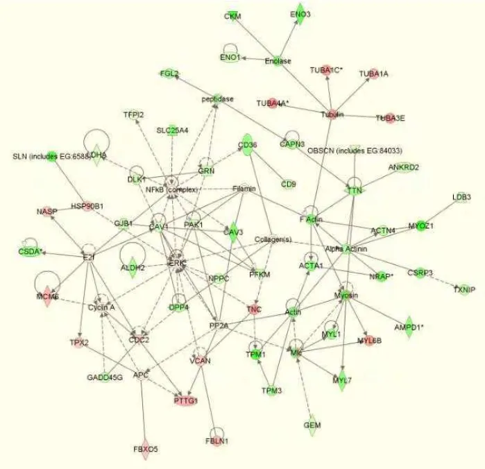
Ingenuity Pathways Analysis (IPA) software. This gene set was categorized into a total of 136 networks
by the IPA software. A network containing 54 genes related to skeletal and muscular system
development was observed for genes that were differentially expressed between 40 and 70 d gestation in
both the YL and Piau breed types (Fig. 1). In this network, 39 genes were more highly expressed at 70 d
of gestation (green color), whereas 15 genes were more highly expressed at 40 d of gestation (red color).
While the same genes were observed to exhibit increased or decreased mRNA abundance in both the Piau
and YL breed types in this network, the FC differences varied for some genes as depicted by the intensity
of the green and red colors between panels (a) and (b) of Fig. 1. Interestingly, several genes exhibit
18
expression at 40 d in the YL pigs (higher red intensity) suggesting the potential for a breed-specific
expression pattern.
An additional network that included differentially expressed genes between 40 and 70 d gestation
in both Piau and YL pigs was related to cellular function and maintenance (data not shown). For this
network, 10 genes were more highly expressed at 70 d of gestation and 4 genes were more highly
expressed at 40 d of gestation. A third informative network, cellular morphology and cellular assembly,
involved 53 genes that were differentially expressed between the two ages only in the Piau breed (data not
shown). Thirty-five of these genes exhibited increased expression at 70 d of gestation, whereas 18
exhibited higher expression at 40 d of gestation.
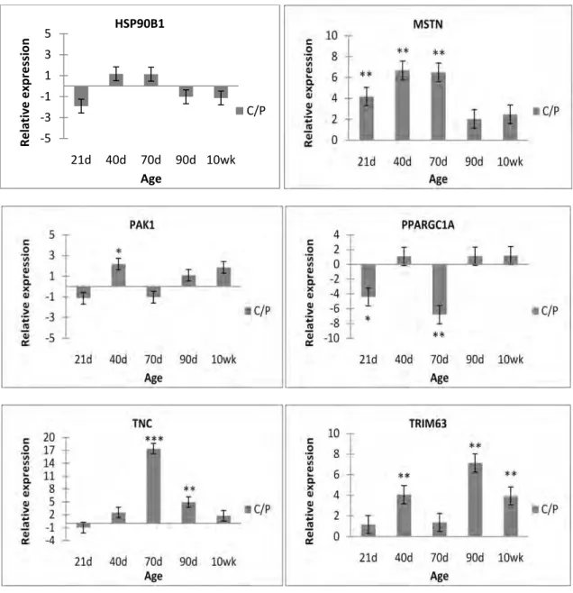
Quantitative RT-PCR confirmation of differentially expressed genes
A total of 13 differentially expressed genes were selected for confirmation by qRT-PCR.
Selected genes represented various functional groups including myofibrillar genes (MYOZ1 and NRAP),
proliferation or differentiation (DLK1 and ODC1), metabolic processes (SLN and CA3), extracellular
matrix (TIMP3 and TNC), signal transduction or transcription activation (STAT1 and CTNNB1), and
phosphorylation-dependent ubiquitination process or proteolysis mechanisms (CTSL2, FBXO32 and
USP13). Also, five of these genes (DLK1, NRAP, MYOZ1, SLN and TNC) were included in the skeletal
and muscular system development network shown in Fig. 1.
Nine of the selected genes exhibited differential expression in LD between 40 and 70 d of
gestation in at least one breed type using the Pigoligoarray, and expression patterns for all of these genes
were confirmed by qRT-PCR (P ≤ 0.01; Fig. 2). CA3, DLK1, MYOZ1, NRAP and SLN all exhibited
higher mRNA abundance at 70 d of gestation in both the YL and Piau breed types on the microarray, and
these expression patterns were confirmed by qRT-PCR. Expression of TNC was observed to be higher in
LD at 40 d of gestation in both YL and Piau pigs on the microarray and this was also confirmed by
qRT-PCR. Microarray results for CTSL2 indicated higher expression at 40 d of gestation for Piau pigs, but no
19
and USP13 exhibited higher expression at 70 d of gestation in Piau pigs on the microarray, but no
significant difference was observed for the YL pigs using FDR ≤ 0.05. Results of the qRT-PCR analyses
for these genes confirmed that expression was higher in LD at 70 d for the Piau pigs, and also indicated
expression to be higher at 70 d for the YL pigs.
Results for qRT-PCR analyses of six genes significantly differentially expressed by microarray
analysis in LD at either 40 or 70 d of gestation or at both ages when comparing YL and Piau pigs are
shown in Fig. 3. This gene set includes two of the genes that were also differentially expressed between
ages (FBXO32 and NRAP). Differential expression of most of these genes was confirmed. Expression of
NRAP was observed to be higher in LD of Piau pigs at both 40 and 70 d of gestation on the microarray
and this result was confirmed by qRT-PCR. FBXO32 exhibited significantly higher expression in LD
from Piau pigs than in LD from YL pigs at 70 d of gestation, but no difference in expression between
breed types was observed at 40 d of gestation, and this result was also confirmed by qRT-PCR. Results
for ODC1 on the microarray indicated that expression was higher in Piau pigs at 70 d of gestation, but no
breed type differences were observed at 40 d of gestation with an FDR ≤ 0.05. Results for ODC1 using
qRT-PCR confirmed higher expression in Piau pigs at 70 d of gestation and also indicated that expression
was significantly higher in Piau pigs at 40 d of gestation. Similarly, microarray results for TIMP3
indicated that TIMP3 expression was higher in Piau pigs at 40 d of gestation but breed type differences
were not significant at 70 d of gestation (FDR ≤ 0.05). Results for qRT-PCR analysis of TIMP3 revealed
significantly higher expression of TIMP3 in LD of Piau pigs at both 40 d and 70 d of gestation. On the
microarray, CTNNB1 exhibited a 4.1-fold higher expression in YL pigs than Piau pigs at 40 d of gestation
with no breed difference observed at 70 d of gestation. There was also no difference in expression
between breed types for this gene at 70 d observed by qRT-PCR analysis. However, qRT-PCR failed to
confirm the breed difference that was seen with the microarray in the 40 d samples. STAT1 exhibited
significantly higher expression in Piau pigs at both 40 and 70 d of gestation by qRT-PCR analysis.
20
both platforms, transcript abundance levels for CTNNB1 and STAT1 were relatively low which could
account for the inconsistent results for these genes.
Discussion
This study evaluated transcriptional profiles in LD muscle of pigs at 40 and 70 d of gestation, two
developmental stages that encompass the primary and secondary waves of muscle fiber formation in pigs
(Wigmore & Stickland, 1983; Wigmore & Evans, 2002). Samples obtained from Yorkshire-Landrace
(YL) crossbred pigs and Piau pigs (a native Brazilian breed), breed types that differ in muscularity, were
used in order to allow breed type comparisons. Our results revealed a large number of differentially
expressed genes both between developmental ages and between breed types. In general, the
developmental transcript profiles for the Piau and YL pigs were similar, although breed-specific patterns
of gene expression were revealed. In addition, the relative abundance of transcripts (based on
fluorescence intensity using the microarray) tended to be greater for the Piau pigs at 70 d of gestation
suggesting that gene expression in LD muscle of YL pigs may be more delayed than in Piau pigs. This
observation is consistent with results reported by Cagnazzo et al. (2006) who reported a study examining
gene expression in developing skeletal muscle of Duroc and Pietrain pigs, breeds that differ for muscle
fiber characteristics as well as growth and muscularity phenotypes. This group observed differential
expression for numerous myogenesis-related genes and suggested that pigs of the heavier muscled
Pietrain breed may exhibit a delayed myogenesis process, perhaps resulting in increased numbers of
myofibers, relative to pigs of the Duroc breed.
Following global transcript profiling analysis using the microarray, several genes were selected
for confirmation by qRT-PCR. Some of these genes encode products with functions related to skeletal
muscle structure or function. Myozenin proteins including MYOZ1 appear to serve as intracellular
binding proteins involved in linking Z-disk proteins such as alpha-actinin and titin-cap, and MYOZ1
plays a complex role in the modulation of calcineurin signaling (Faulkner et al., 2000). We observed
21
and YL pigs. This is consistent with recent observations by Raymond et al. (2010) who compared gene
expression patterns in human skeletal muscle tissue and cultured myotubes, and found MYOZ1 levels to
be significantly downregulated in cultured myotubes supporting the role of this gene in tissue structure
and maturation. The product of the NRAP gene is associated with developing skeletal muscle
myofibrillar structures (Lu et al. 2008). Our results indicated that NRAP expression was higher at 70 d of
gestation in both Piau and YL pigs, and also expression was higher in Piau pigs at both ages. Similar
results were reported by Muráni et al. (2007) who observed higher expression of NRAP in skeletal
muscle of Pietrain fetuses at 35 d of gestation as compared to Duroc fetuses. The SLN gene encodes a
small proteolipid that regulates several sarcoplasmic reticulum Ca(2+)-ATPases. Our results indicated
that SLN expression was higher at 70 d of gestation in both Piau and YL pigs, with highest expression in
the Piau pigs. This agrees with results reported by Lin and Hsu (2005), who compared neonatal pigs of a
native breed (Taoyuan) to Duroc pigs and observed higher expression in the Taoyuan pigs.
Additional genes selected for qRT-PCR confirmation encode products involved with cellular or
tissue growth. The tissue inhibitors of metalloproteinsases (TIMP) were originally characterized as
inhibitors of matrix metalloproteinases, but it is now known that they have a much wider range of
biological activities including effects on cell growth and differentiation, cell migration and apoptosis
among others (Brew & Nagase, 2010). We observed higher expression of TIMP3 in Piau pigs vs. YL
pigs at both 40 and 70 d of gestation indicating a breed-specific expression pattern for this gene.
Similarly, we also observed higher expression of ODC1 in Piau pigs at both 40 and 70 d of gestation.
The ODC1 gene encodes the rate-limiting enzyme in the polyamine biosynthesis pathway. MacLean et
al. (2008) found ODC1 expression to be decreased in skeletal muscle of androgen receptor knockout
mice, and based on their other microarray results and observations of muscle mass they concluded that
androgens promote muscle growth by maintaining myoblasts in the proliferative state and by delaying
differentiation. While it is not possible to determine if hormonal influences are involved in the
breed-specific gene expression patterns that we observed, these observations taken together with the potential
22
study may support the possibility of a prolonged proliferative period which could result in increased
numbers of primary and secondary fibers as suggested by Cagnazzo et al. (2006).
The CA3 gene is the muscle-specific member of a multigene family encoding metalloenzymes
that catalyse the reversible hydration of carbon dioxide. Our results indicated that CA3 expression was
higher at 70 d of gestation than at 40 d of gestation in both breed types, which is in agreement with the
results of Wang et al. (2006) who reported expression of CA3 to increase in skeletal muscle from 33 to 65
d of gestation in Tongcheng pigs. The DLK1 gene was observed to exhibit higher expression at 70 d of
gestation in both breed types. This gene, located in an imprinted region of mammalian genomes, is
involved in differentiation and it has been shown to be up-regulated in skeletal muscle of callipyge lambs
that exhibit extreme muscle hypertrophy (Fleming-Waddell et al. 2007).
The TNC gene product is a mechano-regulated, morphogenic, extracellular matrix protein that is
associated with tissue remodeling (Flück et al. 2008) and it is involved in innervation during
development. We observed TNC to be more highly expressed at 40 d of gestation in both Piau and YL
pigs, suggesting a greater role for TNC during primary fiber formation.
Two genes selected for qRT-PCR analysis did not confirm the results observed with the
microarray. The CTNNB1 gene product is a primary mediator of the WNT/β-catenin signaling pathway
that when activated, leads to the stabilization of β-catenin which enters the nucleus to activate target
genes including MYOD and MYF5 (Armstrong & Esser, 2005) potentially increasing myoblast
proliferation. Expression of CTNNB1 was significantly higher in YL pigs vs. Piau pigs on the microarray
at 40 d of gestation, although no breed difference was observed at 70 d of gestation. Using qRT-PCR, no
significant breed difference was observed for this gene at either age. We detected relatively low signal
intensities for this gene on both platforms which could account for the inconsistent results. Cagnazzo et
al. (2006) observed CTNNB1 to have increased expression in Pietrain fetuses at several developmental
ages from 35 to 91 d of gestation compared to Duroc fetuses. Thus, this gene may exhibit breed-specific
expression patterns and further study may be warranted. The STAT1 gene encodes a protein that is