previous microarray hybridization study showed that NNAT was differentially expressed in Yorkshire and Meishan pig placentas, but the imprinting status and function ofNNATin the placenta have not been investigated. We demonstrated for the first time thatNNATwas monoallelically expressed in the placenta. Immunochemistry analysis showed that NNAT was located in the uterine luminal and glandular epithelium in placentas. We also confirmed the differential expression ofNNAT in Meishan and Yorkshire pig placentas by qPCR. Using IPA software and the published literature, we created a model network of the possible relationships between NNAT and glucose transporter genes. A dual luciferase reporter assay demonstrated that the crucial promoter region ofNNATcontained a CANNTG sequence in the+210 to+215 positions, which corresponded to the E-box. Our findings demonstrated important roles ofNNATin placenta function.
Citation:Gu T, Su X, Zhou Q, Li X, Yu M, et al. (2012) Molecular Characterization of theNeuronatinGene in the Porcine Placenta. PLoS ONE 7(8): e43325. doi:10.1371/journal.pone.0043325
Editor:Huaijun Zhou, University of California, Davis, United States of America
ReceivedApril 6, 2012;AcceptedJuly 23, 2012;PublishedAugust 24, 2012
Copyright:ß2012 Gu et al. This is an open-access article distributed under the terms of the Creative Commons Attribution License, which permits unrestricted use, distribution, and reproduction in any medium, provided the original author and source are credited.
Funding:This work was supported by National Natural Science Foundation of China (30972082 and 31060300), the key project of Transgenetics of the Ministry of Agriculture, and the Creative team project (IRT-0831) of the Ministry of Education. The funders had no role in study design, data collection and analysis, decision to publish, or preparation of the manuscript.
Competing Interests:The authors have declared that no competing interests exist. * E-mail: [email protected]
Introduction
Imprinted genes are a special category of genes that imprinted one allele in the early embryo development decided by the parental origin. The theory raised by Morre and Haig [1] was widely demonstrated that imprinting evolved in mammals because of the conflicting interests of maternal and paternal genes in transferring of nutrients from the mother to her offspring. For example, maternally imprinted genes Mest and Grb10 play important roles in the placental and fetal development of mammals.Mestknockout mice were viable and characterized by growth retardation [2]. Mice with a disrupted maternal copy of
Grb10produced larger embryos and placentas, while mutant mice were 30% larger than normal mice [3].
Chinese Meishan pigs produce 3 to 4 more piglets than Yorkshire pigs in each litter. Numerous investigations have focused on the mechanisms behind this difference. Early investigators believed that factors regulating developmental rate and uniformity of the conceptus were the primary determinants of prolificacy [4]. Further study showed that the weight of Yorkshire placentas dramatically increased from day90 of gestation to term, while in Meishan pigs, the weight of the fetus, not the placenta, increased during this period [5]. These studies indicated that Meishan and Yorkshire pig placentas have different nutrient transport capac-ities.
We detected differentially expressed genes in the placentas of Meishan and Yorkshire pigs on day75 and day90 of gestation by Affymetrix Porcine Expression Microarray. A total of 226 transcripts on day75 and 577 transcripts on day90 were differentially expressed between placentas from the two divergent breeds. The differentially expressed transcripts included genes involved in angiogenesis, placental development, nutrient trans-portation and imprinted genes, such as PEG1, PEG3, PEG10,
PLAGL1,SLC38A4, andDIRAS3[6].Neuronatin (NNAT)was found to be one of the imprinted genes differentially expressed in Meishan and Yorkshire pig placentas. NNAT, also known as paternally expressed gene 5 (PEG5), was first discovered in the rat neonatal brain and has significant roles in the differentiation of neurons [7]. Other studies in adipose tissue and pancreaticbcells showed thatNNATis involved in adipocyte differentiation and in regulating glucose-mediated insulin secretion [8,9].
NNAT was paternally expressed in human and mouse brains [10,11]. In the pig, a study showed thatNNATwas imprinted in 11 tissues, including heart, liver, spleen, lung, kidney, stomach, small intestine, skeletal muscle, fat, uterus, ovary, and pituitary gland [12], but in pig placenta, the expression status inherited from parents was still uncovered to our knowledge.
published literature was used to create a network model showing the possible relationships betweenNNATand glucose transporter genes. The crucial promoter region ofNNATin JEG-3 and PK-15 cell lines was identified by series analysis of promoter region sequences. This is the first report of NNAT expression and regulation of glucose transportation in porcine placenta.
Results
Gene Structure of PorcineNNATand Expression Profiling in Placenta
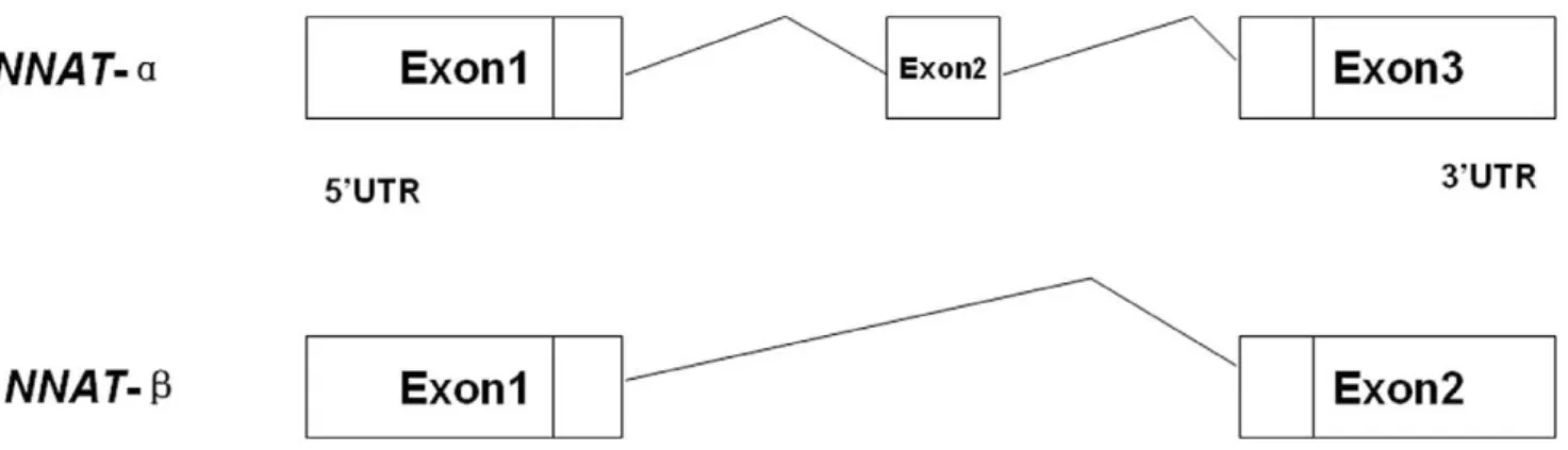
From analysis of the human and mouse NNAT sequences, together with the porcine EST analysis, the structure of porcine
NNAT was determined, as shown in Fig. 1. Two transcripts of NNAT existed in porcine placenta, and both of them were expressed in the placentas of Yorkshire and Meishan pigs at different developmental stages (Fig. 2). The expression level was analyzed by software Quality one. The expression ofNNATband
NNATa were decreased in developing placentas in both breeds (Table 1).
Monoallelic Expression of theNNATGene in the Porcine Placenta
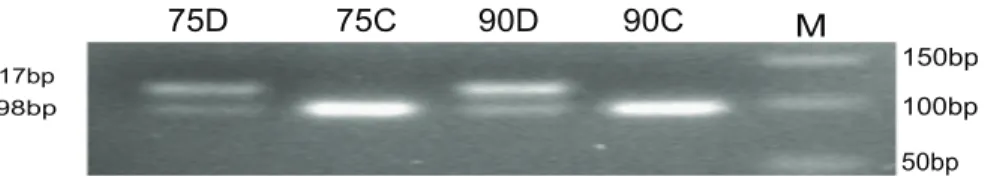
A SNP in 39UTR, c.*711C.T was used to detect the biallelic expression status ofNNAT. When a C was present at the c.*711 position,Hinf- I can cut the 117 bp product into two fragments (98 and 19 bp, C-allele), while when a T was present,Hinf-I did not cut the PCR product (117 bp, T-allele) (Fig. 3). The genotypes of two pigs (on day75 and d90 of gestation) were CT, while the cDNA expressed only the C allele. This result showed thatNNAT
was also monoallelically expressed in the placentas, besides other reported tissues.
High Expression of NNAT in the Luminal Epithelium and Epithelial Glands of Early Yorkshire and Meishan Pig Placentas
To detect the location of NNAT protein in the placenta, we conducted IHC analysis in the placentas of Meishan and Yorkshire pigs. The luminal epithelium, endometrial fold and fetal chorion of Yorkshire pigs were larger than those of Meishan
Figure 1. Two transcripts of porcineNNAT.NNAT-ahad three exons, whileNNAT-blacked the second exon, which consisted of 81 base pairs. doi:10.1371/journal.pone.0043325.g001
Figure 2. RT-PCR analysis ofNNATmRNA in different placentas of different breeds and at different developmental stages.The 280 base pairNNAT-asequence and the 199 base pairNNAT-bsequence were both detected in all samples. From left to right, the lane contents were as follows: 500 base pair DNA ladder (Takara), products amplified from the placentas of Yorkshire pigs on day26 and d50 of gestation, Meishan pigs on day26 and day50 of gestation, Yorkshire pigs on day75 and d90 of gestation, and Meishan pigs on day75 and d90 of gestation.
doi:10.1371/journal.pone.0043325.g002
Table 1.Ratios betweenNNATa/b.
Y26 Y50 M26 M50 Y75 Y90 M75 M90
NNATa 7499 11992 6816 15540 21838 21768 16594 13984
NNATb 4595 5526 5938 6874 10012 6746 6183 6246
pigs. Strong NNAT protein signal was detected in luminal and glandular epithelia in Meishan and Yorkshire pig placentas on day26 and d50 of gestation (Fig. 4, dark brown color).
Gene-interaction Network Construction and qPCR Detection of the mRNA Level in Placentas
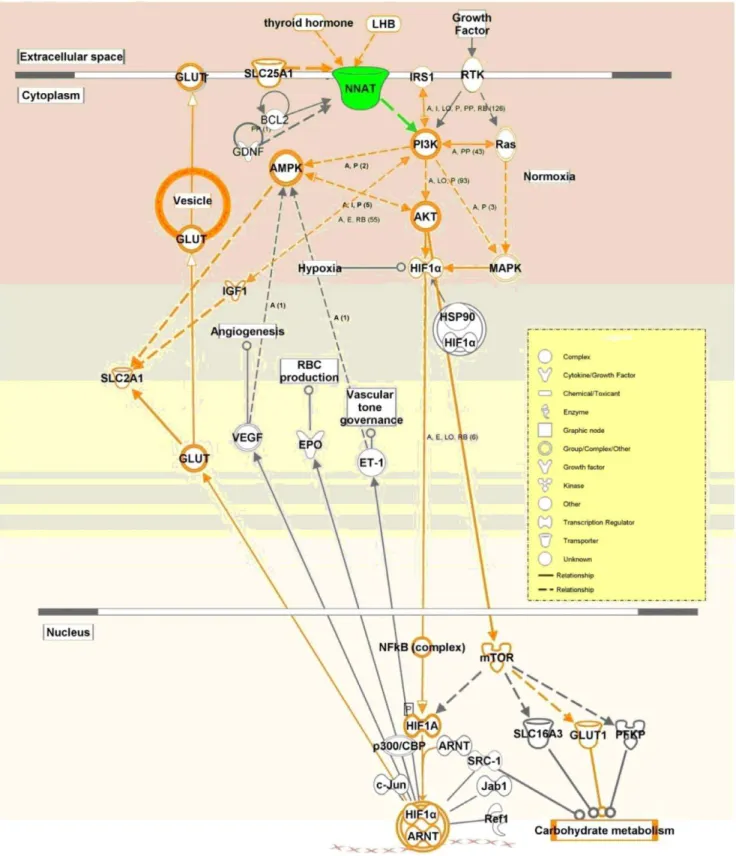
Based on the KEGG prediction and on published papers, a pathway includingGLUT1, GLUT3, PI3K, AKT, HIF1A, mTOR, and IRS genes that may be affected byNNAT in placentas was drawn by Ingenuity Pathway Analysis (IPA) (Fig. 5). qPCR using placental RNAs from the Meishan and Yorkshire pig breeds on day75 and day90 of gestation showed thatNNATexpression varied with the same pattern as genes in the predicted pathway, indicating thatNNATmay regulate the transportation of glucose through thePI3K-AKTpathway (Figs. 6 and 7).
Promoter Analysis
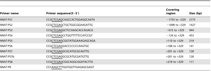
We cloned a 1766 bp fragment upstream of the transcription starting point using the primers NNAT-PS1 and NNAT-PR (Table 2). To determine the transcriptional control regions in theNNATpromoter, a series of reporter vectors consisting of firefly luciferase regulated by a consecutively truncated promoter were
constructed and transfected into porcine kidney 15 cells (PK-15) and the human placental choriocarcinoma cell line JEG3.
We found that 59 deletions in a region from +201 to +218 upstream of the promoter (NNAT-PS7 to NNAT-PS8) almost eliminated its transcription in both the cell lines (Fig. 8). When two base pairs in the+201 to+218 regions were mutated from CA into AT (NNAT-PS7b), the transcriptional activity was reduced to a level similar to the basic control. Table 2 showed the mutated base pairs in primersNNAT-PS7 andNNAT-PS7b in square box.
Dual luciferase promoter activity detection demonstrated that the crucial promoter region ofNNATcontained an E-box family member binding sequence (CANNTG) located at+210 to +215 was sufficient and necessary for transcription.
Discussion
NNAT was an important gene in regulating mouse brain and adipocyte differentiation and insulin secretion in response to glucose in mouse pancreatic cells [7,8,9]. NNATwas paternally expressed in human and mouse brains. In pigs, it was reported to be imprinted in eleven tissues. However, there is no study focused on the function of NNAT in placentas. We detected NNAT
Figure 4. IHC analysis of porcine NNAT in the placenta using rabbit anti-NNAT.Stroma (S) represented the maternal side and fetal chorion (C) represented the fetal side. L: Luminal epithelium; G: glandular epithelium. All images were obtained at 406magnification. Scale bars represented 50mm.
expression in placentas. It was of great interest to illustrate possible roles ofNNATin the regulation of glucose transportation based on the known roles played by most imprinted genes on fetal and placental development. As indicated by the classic parent-offspring conflict theory, higher expression of paternally expressed NNAT
would result in a larger fetus in Yorkshire pigs, as the opposite happened in the Mest knockout mice [1]. Our study demonstrated thatNNATwas monoallelically expressed in porcine placentas and may regulate glucose transportation though the PI3K-AKT
pathway. The results led us to conclude that NNAT was an important imprinted gene in the placenta.
The results of the IHC experiments showed that NNAT was highly expressed in porcine uterine luminal and glandular epithelia. NNAT was expressed in two types of exocrine cells, indicating that this gene may regulate transportation of nutrients. Carbohydrate- and lipid-rich glandular secretions were an important source of nutrients for the fetus when maternal arterial supply to the placenta has not been formed during the early stages of fetal development, and the uterine luminal epithelium mediates transportation between the embryo and mother at this time [13]. The two transcripts ofNNAThad different functions.NNATwas significantly increased in the adipose tissue of ZDF (fat) rats and ectopic expression of NNATa augmented adipocyte by increase adipogenic transcription factors in 3T3-L1 cell line [8]. Study in pancreas showed that NNATa and NNATb increased insulin
indicatingNNATmay affect or be co-regulated by the insulin and glucokinase pathways in the placenta.
Further, we investigated crucial transcription regions of the
NNAT gene. Dual luciferase activity detection and basic muta-tional analysis confirmed that an E-box binding site in the+210 to
+215 position of the NNAT promoter region was the crucial transcriptional binding site. This was similar to results in the mouse in which ChIP and EMSA shift experiments in thebTC3 cell line confirmed the existence of an E-box binding site at the
2644 to2366 position [14]. These results indicated that the E-box transcription factor was conserved in cell lines from mice, pigs, and humans, but the binding site was species specific.
This is the first report that NNAT is expressed in porcine placenta and involved in regulating glucose transportation.
Materials and Methods
Tissue Collection
A total of 12 placentas from Meishan and Yorkshire pigs on day75 and d90 of gestation were obtained from a Wen’s Company pig farm. 12 porcine placentas at different stages of gestation (3 each of Meishan and Yorkshire pig placentas on day26 and day50 of gestation) were collected from the swine farm of Huazhong Agricultural University. All sample tissues were either fixed in 4% paraformaldehyde followed by paraffin embedding or stored at
280uC or RNA extraction. All animals involved in this study were conducted according to the regulation (No. 5 proclaim of the Standing Committee of Hubei People’s Congress),which was approved by the Standing Committee of Hubei People’s Congress, and the ethics committee of Huazhong Agricultural University, P. R. China. The approved permit number for this study is ‘‘HBAC20091138’’.
Two Different Transcripts of NNAT Expressed on Porcine Placenta
Total RNA was extracted using the RNAprep pure Tissue kit (Tiangen biotech co., Ltd, Beijing, China) and quantified by a Nanodrop ND1000 spectrophotometer. Two micrograms of RNA was reverse-transcribed using M-MLV Reverse Transcriptase (Invitrogen, San Diego, CA) according to the manufacturer’s instructions. RT-PCR was used to detect mRNA of NNAT in porcine placentas using the primer pairNNATpig2, as shown in Table 2.
Biallelic Expression Status Analysis ofNNATGenes
cDNA of the porcineNNATgene from three Meishan and three Yorkshire were amplified by PCR and then blasted by NCBI Blast and a SNP in 39 end was detected. As there is no available
Figure 6. qPCR was used to investigate the mRNA profile of
NNATin Yorkshire and Meishan porcine placentas on day75 of
gestation (Y75 and M75) and 90 (Y90 and M90). NNAT was significantly lower expressed in M75 compared with M90 and Y75 by SAS Anova test. The different superscript alphanumeric characters indicated a statistically significant difference at p,0.05.
commercial enzyme for this SNP, a pair of mutant primers (NNAT-imprint in Table 1) that created aHinf- I enzyme site by replacing a C with an A was designed to amplify all individuals’ genomic DNA. The cDNA from the heterozygous samples was amplified and enzyme cut to detect the biallelic expression status. Sequencing was conducted to confirm the genotype.
Immunohistochemistry (IHC)
IHC to determine NNAT expression in placentas from different pig breeds at 2 gestational time points was performed by standard IHC procedures as described [15]. Three sections of each different sample (5mm) were deparaffinized and rehydrated in xylene and ethanol. Slides were boiled in citrate buffer (10 mM citrate sodium, 10 mM citric acid, pH = 6.0) in a microwave oven and then cooled to room temperature twice to retrieve antigen. Sections were then incubated with 3% H2O2 in methanol for
10 min to quench endogenous peroxidase and then in a normal goat serum blocking solution for 20 min. Sections were incubated with the polyclonal primary antibody against NNAT (Abcam Inc., MA, USA) at 4uC overnight and then in biotinylated secondary antibody. Sections were counterstained with hematoxylin and mounted. For each sample, a negative control was performed by replacing the primary antibody with PBS buffer.
Quantitative RT-PCR (qPCR) Analysis
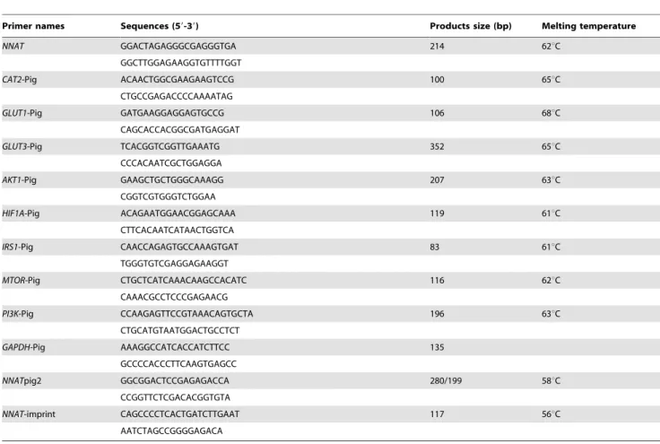
qPCR was carried out on the LightCycler 480 Real-Time PCR machine (Roche Diagnostics Ltd., Forrentrasse, Switzerland) using Thunderbird SYBR qPCR Mix (Toyobo co., ltd, Osaka, Japan). PCR conditions were as follows: single cycle of 5 min at 95uC, followed by 40 cycles of 30 sec at 95uC, 20 sec at 62uC, and 15 sec at 72uC. The primers were designed by the Primer 6.0 program. The primer sequences, melting temperature and product size were shown in Table 1. TheGAPDHgene was used as a control. The ANOVA test in the SAS 8.1 program was used to perform statistical analysis.
pGL3-NNAT-PS Series Reporter Construction, Transfection, and Dual-luciferase Reporter Assay
A total of 16 pGL3-NNATluc+vectors were constructed and transfected into PK-15 and JEG-3 cell lines as described previously [16]. A successive series of fragments covering21766 to+329 of the 59-flanking promoter of the porcineNNATgene were amplified using the primers in Table 3 and the high fidelity DNA polymerase KOD-Plus (Toyobo) and then inserted into the XhoI/HindIII sites of the pGL3-Basic Vector (Promega, Madison, USA) to construct the pGL3-NNATluc+reporters. These vectors were transfected into PK-15 cells and the human placental choriocarcinoma cell line JEG3 by Lipofectamine 2000
(Invitro-Figure 7. qPCR was used to investigate the mRNA profile of genes in thePI3Kpathway in Yorkshire and Meishan pig placentas on day75 of gestation (Y75 and M75) and 90 (Y90 and M90).The expression levels were analyzed by SAS Anova test. The different superscript alphanumeric characters indicated a statistically significant difference at p,0.05.
Figure 8. Identification of crucial transcriptional regions ofNNAT.Three replicates were used for each vector in each transfection. When two base pairs in the region were mutated from CA into AT (NNAT-PS7b) in square box, the transcriptional activity was reduced to a level similar to the basic control.
doi:10.1371/journal.pone.0043325.g008
CCCACAATCGCTGGAGGA
AKT1-Pig GAAGCTGCTGGGCAAAGG 207 63uC
CGGTCGTGGGTCTGGAA
HIF1A-Pig ACAGAATGGAACGGAGCAAA 119 61uC
CTTCACAATCATAACTGGTCA
IRS1-Pig CAACCAGAGTGCCAAAGTGAT 83 61uC
TGGGTGTCGAGGAGAAGGT
MTOR-Pig CTGCTCATCAAACAAGCCACATC 116 62uC
CAAACGCCTCCCGAGAACG
PI3K-Pig CCAAGAGTTCCGTAAACAGTGCTA 196 63uC
CTGCATGTAATGGACTGCCTCT
GAPDH-Pig AAAGGCCATCACCATCTTCC 135
GCCCCACCCTTCAAGTGAGCC
NNATpig2 GGCGGACTCCGAGAGACCA 280/199 58uC
CCGGTTCTCGACACGGTGTA
NNAT-imprint CAGCCCCTCACTGATCTTGAAT 117 56uC
AATCTAGCCGGGGAGACA
gen). The activity of the promoter vectors was detected by a Dual-Luciferase Reporter Assay System (Promega).
Acknowledgments
We thank Hongmei Wang at the State Key Laboratory of Reproductive Biology, Institute of Zoology, Chinese Academy of Science for providing
the JEG-3 cell line. We also thank Xiang-Dong Liu, Lu Jing, Chu-xin Liu and Ze-tan Li for their technical help.
Author Contributions
Conceived and designed the experiments: SHZ QYZ CCL. Performed the experiments: TG XS QYZ YD. Analyzed the data: TG. Contributed reagents/materials/analysis tools: SHZ XYL MY. Wrote the paper: TG.
References
1. Moore T, Haig D (1991) Genomic imprinting in mammalian development: a parental tug-of-war. Trends Genet 7: 45–49.
2. Lefebvre L, Viville S, Barton SC, Ishino F, Keverne EB, et al. (1998) Abnormal maternal behaviour and growth retardation associated with loss of the imprinted gene Mest. Nat Genet 20: 163–169.
3. Charalambous M, Smith FM, Bennett WR, Crew TE, Mackenzie F, et al. (2003) Disruption of the imprinted Grb10 gene leads to disproportionate overgrowth by an Igf2-independent mechanism. Proc Natl Acad Sci U S A 100: 8292–8297. 4. Bazer FW, Thatcher WW, Martinat-Botte F, Terqui M (1988) Conceptus
development in large white and prolific Chinese Meishan pigs. J Reprod Fertil 84: 37–42.
5. Wilson ME, Biensen NJ, Youngs CR, Ford SP (1998) Development of Meishan and Yorkshire littermate conceptuses in either a Meishan or Yorkshire uterine environment to day 90 of gestation and to term. Biol Reprod 58: 905–910. 6. Zhou QY, Fang MD, Huang TH, Li CC, Yu M, et al. (2009) Detection of
differentially expressed genes between Erhualian and Large White placentas on day 75 and 90 of gestation. BMC Genomics 10: 337.
7. Joseph R, Dou D, Tsang W (1994) Molecular cloning of a novel mRNA (neuronatin) that is highly expressed in neonatal mammalian brain. Biochem Biophys Res Commun 201: 1227–1234.
8. Suh YH, Kim WH, Moon C, Hong YH, Eun SY, et al. (2005) Ectopic expression of Neuronatin potentiates adipogenesis through enhanced phosphor-ylation of cAMP-response element-binding protein in 3T3-L1 cells. Biochem Biophys Res Commun 337: 481–489.
9. Joe MK, Lee HJ, Suh YH, Han KL, Lim JH, et al. (2008) Crucial roles of neuronatin in insulin secretion and high glucose-induced apoptosis in pancreatic beta-cells. Cell Signal 20: 907–915.
10. Evans HK, Wylie AA, Murphy SK, Jirtle RL (2001) The neuronatin gene resides in a ‘‘micro-imprinted’’ domain on human chromosome 20q11.2. Genomics 77: 99–104.
11. Kagitani F, Kuroiwa Y, Wakana S, Shiroishi T, Miyoshi N, et al. (1997) Peg5/ Neuronatin is an imprinted gene located on sub-distal chromosome 2 in the mouse. Nucleic Acids Res 25: 3428–3432.
12. Cheng HC, Zhang FW, Deng CY, Jiang CD, Xiong YZ, et al. (2007) NNAT and DIRAS3 genes are paternally expressed in pigs. Genet Sel Evol 39: 599– 607.
13. Burton GJ, Jauniaux E, Charnock-Jones DS (2007) Human early placental development: potential roles of the endometrial glands. Placenta 28 Suppl A: S64–69.
14. Chu K, Tsai MJ (2005) Neuronatin, a downstream target of BETA2/NeuroD1 in the pancreas, is involved in glucose-mediated insulin secretion. Diabetes 54: 1064–1073.
15. Fu JJ, Lin P, Lv XY, Yan XJ, Wang HX, et al. (2009) Low molecular mass polypeptide-2 in human trophoblast: over-expression in hydatidiform moles and possible role in trophoblast cell invasion. Placenta 30: 305–312.
16. Chen H, Cheng L, Yang S, Liu X, Liu Y, et al. Molecular characterization, induced expression, and transcriptional regulation of porcine S100A12 gene. Mol Immunol 47: 1601–1607.
Table 3.Primers used inNNATpromoter serious deletion analysis.
Primer name Primer sequence(59-39)
Covering
region Size (bp)
NNAT-PS1 CCGCTCGAGCAGCCACTGGAGGCAATA 21755 to+329 2175
NNAT-PS2 CCGCTCGAGCTGCTGGCGGAAGATTG 21098 to+329 1427
NNAT-PS3 CCGCTCGAGGCTCCAAGCACCAGACG 2615 to+329 944
NNAT-PS4 CCGCTCGAGCCTGGTTTTTCCATCCGT 2124 to+329 453
NNAT-PS5 CCGCTCGAGCGGTATGGAAAGAGCAGA +110 to+329 219
NNAT-PS6 CCGCTCGAGGACCCCCCCAAGTGC +188 to+329 141
NNAT-PS7 CCGCTCGAGGCGCATGCGCAGTTG +201 to+329 128
NNAT-PS7b CCGCTCGAGGCGCATGCGATGTTG +201 to+329 128
NNAT-PS8 CCGCTCGAGCGGCAGGCGGATACTTA +218 to+329 111
NNAT-PR CCCAAGCTTTGGTGGTTGAGAGCGAGT