Abstract — A variety of medical applications require the proper characterization of the thermomechanical behaviors of skin tissue. However, few quantitative studies have been conducted on its temperature-dependent mechanical properties. This paper reports the tensile, compressive and viscoelastic behaviors of skin tissue under different temperatures, focusing on the influence of temperature and the corresponding dermal collagen denaturation. Differential scanning calorimetry (DSC) was used to detect dermal collagen denaturation and to assess its thermal stability. The DSC results under various heating rates were used to derive the Arrhenius parameters for burn damage integration, which were subsequently used to calculate the degree of denatured collagen in the skin. The results of hydrothermal tests show that temperature has a great influence on both the tensile and compressive properties of skin, but the mechanisms are different. The variation of skin tensile properties is caused by the thermal denaturation of collagen with increasing temperature, while the variation of skin compressive behavior is likely caused by hydration change with thermal denaturation. Temperature tests were also performed using a dynamic mechanical analyzer (DMA) to evaluate changes in skin viscoelastic properties as a function of collagen damage,; specifically, changes in skin storage modulus (E') and loss factor (tanδ) were assessed. The results show remarkable changes in E', due possibily to the release of water, but there is no significant influence of collagen damage on
tanδ . These results suggest that at fixed frequency the denaturation of collagen molecules has little effect on skin viscoelasticity.
Index Terms—Skin tissue, thermal denaturation, compressive behavior, tensile behavior, viscoelastic behavior.
I. INTRODUCTION
Skin, consisted in general of three distinct layers (epidermis, dermis, and subcutaneous tissue), plays a variety of important roles including sensory, thermoregulation and host defense etc. Advances in laser, microwave and similar technologies have
Manuscript received September 13, 2007. This work is supported by the Overseas Research Studentship (ORS) and Overseas Trust Scholarship of Cambridge University, the National Natural Science Foundation of China (10572111, 10632060), the National 111 Project of China (B06024), and the National Basic Research Program of China (2006CB601202).
F. Xu is with the Engineering Department, Cambridge University, Cambridge, CB2 1PZ UK (e-mail: [email protected]).
K. A. Seffen is with the Engineering Department, Cambridge University, Cambridge, CB2 1PZ UK (e-mail: [email protected]).
T. J. Lu is with the MOE Key Laboratory of Strength and Vibration, School of Aerospace, Xi’an Jiaotong University, Xi’an 710049 P.R. China (phone: 0086-29-82665600; fax: 0086-29-83234781; e-mail: [email protected])
led to recent developments in thermal treatments of diseased and injured skin tissue such as skin cancer and skin burn. The aim is to induce thermal injury precisely within skin tissue several millimeters below the surface but without affecting the surrounding, healthy tissue. In spite of the widespread use of heating therapies in dermatology, these nonetheless do not draw upon the detailed understanding of the bio-thermo-mechanical behavior of skin tissue, for none exists to date, even though each behavioral facet has been well established and understood. It is proposed therefore that a detailed understanding of the coupled biological-mechanical response under thermal loading will contribute to the design, characterization and optimization of strategies for delivering better treatment.
Since it is technically very difficult to measure experimentally the bio-thermo-mechanical behavior of skin tissue in physiological conditions, analytical and numerical simulations are often used, for which the quantification of skin mechanical properties is an essential step towards building reliable computer simulations. So far, skin mechanical properties under normal conditions have been studied experimentally in vivo and in vitro ever since the study of Langer in 1861. However, few studies have focused on skin mechanical properties under hyperthermia conditions as well as on the coupling effects between thermal and mechanical behaviors.
A. Experimental characterization of skin mechanical properties
Tests on skin mechanical behaviors can be broadly divided into two categories, in vitro (ex vivo) and in vivo, where in vitro tests involve testing the skin tissue after removal from the living body, whereas in vivo tests examine the skin while it is still on the living body. The only truly reliable method to determine skin properties is through in vivo testing, since the deformation may be strongly dependent on active processes. In comparison, during in vitro testing, the skin is away from its influential surroundings such as blood perfusion, lymphatic drainage, metabolism, nervous and hormonal controls. For example, during compressing, the resulting deformation is largely a function of fluid interchange with the surrounding unstressed skin [1]. The epidermal tissue used for in vitro experiments was not perfused and thus lacked restorative fluidic pressures, which could potentially have aided in reducing the visible compression of the tissue after application of pressure [2]. However, in vivo measurements are affected by
Temperature-Dependent Mechanical
Behaviors of Skin Tissue
F. Xu, K. A. Seffen, and T. J. Lu
both skin tissue itself and other structures it is attached to, which means it is technically very difficult to obtain a uniform strain field in the sample and control boundary conditions when performing, thus in vitro tests are also widely used as in this study.
Most in vitro skin experiments use a tensile method, and it is now accepted that a biaxial mechanical test can better mimic the sorts of deformation that in vivo skin experiences. However, there exist many difficulties in performing biaxial tensile experiments on soft biological tissues [3]. In view of these problems, an unconfined compression test through the thickness direction has been suggested as an approximation which gives an averaged in-plane tensile response [4, 5]. If the skin deforms with a constant volume, then the compression test will have a similar effect to a biaxial test in the plane of the skin (through thickness compression results in lateral in-plane expansions). For example, Shergold [6] found that the Ogden model calibrated from his compression tests of pig skin provided a reasonable approximation to the tensile orthotropic constitutive behavior measured by Ankerson et al. [7]. However, there are also problems associated with unconfined compression testing, the critical one being the friction between skin sample and compression plates, which will limit lateral expansion. Fortunately, several methods can be adopted to circumvent this issue, such as covering the moving platen with a polytetrafluoroethylene (PTFE) sheet [8], using platens of polished stainless steel [9], removing the epidermis layer from skin sample [4], coating smooth compression plates with lubricant or grease (margarine) [4, 5], and using lubricated squeezing flow [10].
The extension of the results in compression to biaxial tension, however, should be carefully made. Mechanically, skin is a complex nonlinear composite, with collagen fibers serving as the reinforcements. These fibers are very stiff in stretch, but carry little load in compression. Under tension, the majority of the load is carried by collagen fibers; while under compressive loading, most of the load is carried by the tissue matrix - the collagen fibers embedded in the tissue matrix are comparable to hard inclusions in a composite in this case. Due to its complex micro-structures, skin is expected to exhibit asymmetric behavior in tension and compression [11].For example, it was found that the long-term stiffness of the skin is approximately two times higher in compression [11] than that in tension [12-14].
As for viscoelastic behavior, previous studies on skin tissue reveal that its stress-strain relationship depends on strain rate, loading rate, the period of loading and on the preconditioning stress historyI; it has also been established that skin tissue exhibits considerable hysteresis in cyclic tests, as well as stress relaxation under constant strain [15-17]. Most of these studies were performed under quasi-static experimental conditions: only a few studies have investigated the dynamic viscoelastic properties of skin tissue [18, 19].
B. Thermal denaturation of skin collagen
Collagen is the major component of skin, which accounts for
approximately 75% of skin dry weight and 18-30% of dermis volume [20], and provides the principal structural and mechanical support in skin tissue. Under thermal loading, with the increase of skin temperature, the heat-labile intramolecular crosslinks in collagen are gradually broken, and the collagen undergoes a transition from a highly organized crystalline structure to a random, gel-like state [21]. This process is termed ‘thermal denaturation’, which appears accordingly as thermal shrinkage. Skin also contains a small mount of elastin (0.1% of skin dry weight), which is thermally very stable [22], and capable of surviving in boiling water for hours with no apparent change. Therefore, when discussing the mechanical behaviors of skin tissue under different thermal loadings, only collagen will be addressed.
With the thermal denaturation of collagen, there are not only structural changes, but also changes in collagen hydration which may involveliberation of water initially and, absorption of water later. Not surprisingly, thermal denaturation of a collagenous tissue can lead to remarkable changes in the mechanical, thermal, electrical, and optical properties. However, despite the skin dermis being mainly composed of collagen, there are comparatively few studies on skin thermal denaturation [23-26], and there is no published study on the temperature-dependent skin viscoelasticity, although other collagenous tissues have been studied, for example, cartilage [27, 28] and bone [29, 30].
The shrinkage of collagen due to macro scale thermal denaturation can be used as a convenient continuum metric of thermal damage [31, 32]. Diller & Pearce [32] pointed out that the dimensionless indicator of damage (Ω) is, in fact, the logarithm of the relative concentration of “reactants” (un-denatured collagen) in the collagen denaturation process:
(0) ( ) ln
( )
C t
C t
⎛ ⎞
Ω = ⎜ ⎟
⎝ ⎠
(1)
where C(0) is the initial concentration and C t( ) is the concentration of un-denatured collagen remaining at time t. Then, the degree of thermal denaturation Deg t( ), defined as the fraction of denatured collagen, can be calculated as:
(
)
(0) ( )( ) 1 exp ( )
(0)
C C t
Deg t t
C
−
= = − −Ω
(2)
As for the calculation of thermal damage, the Arrhenius burn integration proposed by Henriques & Moritz [33] is widely used. They proposed that skin damage could be represented as a chemical rate process, which could be calculated by using a first order Arrhenius rate equation, whereby damage is related to the rate of protein denaturation (k) and exposure time (t) at a given absolute temperature (T ). The measure of thermal injury Ω was introduced and its rate k was postulated to satisfy:
( ) d exp Ea
k T A
dt RT
Ω ⎛ ⎞
= = ⎜− ⎟
⎝ ⎠ (3)
or, equivalently:
0 exp
t
a E
A dt
RT
⎛ ⎞
Ω = ⎜− ⎟
⎝ ⎠
∫
(4)where t is the time after the starting of heating, A is a material parameter (frequency factor), Ea is the activation energy, and
8.314 J/mol K
R= is the universal gas constant. In the present study, Arrhenius parameters (Ea, A) for pig skin are derived experimentally from DSC results in Section III.
C. Aim and scope
This study aims to test the hypothesis that collagen is the key determinant of skin mechanical properties as well as thermally induced changes in these properties. For this purpose, differential scanning calorimetry (DSC) measurements is used to detect the denaturation of collagen and to measure its thermal stability. The integrity of the collagen network is then assessed using the thermal damage integration method. Hydrothermal tensile and compressive experiments are subsequently performed. In addition, in order to characterize the viscoelastic properties of skin tissue as a function of temperature and collagen denaturation, a dynamic mechanical analysis versus temperature (DMA) are carried out.
II. MATERIALS AND METHODS
A. Samples
The ethical and immunological issues associated with testing human skin demand a proper substitute. Pig skin is chosen due to its high degree of structural and functional similarities to human skin [5]. In addition, repetitive tests can be realized for the same animal because of its large size, which reduces the variation in results [34]. In the present series of experiments, skins of UK domestic pigs were used, which were obtained from a local slaughter house near Cambridge at Dalehead Foods, Linton.
B. Samples preparation
Samples of pig skin to a depth of subcutaneous fat at different body sites were procured daily, ten minutes post mortem by block dissection. They were fast-chilled following the standard organ procurement protocol to 4°C in a pre-gassed (95%O2, 5%CO2) Krebs-Henseleit Ringer (KHR, pH7.4). In the laboratory, samples were separated from the subcutaneous fat by wet/fast-dissection on a bed of ice and KHR at 4°C. The skin samples were tested within a few hours from slaughter, in order to minimize degradation of the tissue structure. Before each tensile or compressive test, the skin sample was preconditioned so that a reproducible response could be
obtained from nominally identical tests. The preconditioning was completed in KHR solution at 37 °C.
C. Experimental system and test procedure
Differential Scanning Calorimetry (DSC)
Differential scanning calorimetry has been used extensively to characterize the thermal behavior of collagenous tissues [35, 36]. DSC detects thermodynamic changes by measuring the flow of heat between a sample and a reference, from which the effect of thermal history on the heat capacity can be determined, enabling different parameters to be interrogated, such as the thermal denaturation temperatures of skin collagen, reaction kinetics, etc. In this study, the thermal stability of collagen in pig skin was assessed with a TA Instruments DSC of type Q1000 Tzero, scanning from 20 to 100 °C at four different heating rates (2, 5, 10, 20 °C/min).
Tensile and Compressive Tests
These tests were performed using purpose-built hydrothermal tensile and compressive testing systems, as shown schematically in Fig. 1 and Fig. 2. Details of the test procedure have been reported elsewhere [37, 38], and hence only a brief introduction is given below. A specimen was put inside the test chamber and the liquid in the high temperature reservoir was heated to a chosen temperature between 30 and 100 °C, while the low temperature reservoir contained liquid at room temperature. A valve in the test chamber was opened to allow the hot (or cold) liquid to fill the test chamber and the sample was fully immersed in the liquid, while a thermocouple near the sample was used to inform a software, via a PC, the exact time at which the experiment started. A mechanical load was then applied to the skin sample. Temperature and applied displacement, strain, and loads were recorded subsequently.
Dynamic Mechanical Analysis (DMA)
Various techniques have been used to characterize engineering viscoelastic materials, including Dynamic Mechanical Analysis (DMA), resonant method [39, 40], non-resonant method [41, 42] and wave propagation method [43-45]. Amongst these, DMA is the most commonly used due to its commercial availability and suitability for investigating the viscoelastic properties of polymers in a wide range of temperatures and mechanical loading frequencies [46, 47].
In the present study, the dynamic viscoelasticity of pig skin was therefore investigated by DMA Q800. The principle of DMA is based on the fact that strain (stress) in a viscoelastic material in response to stress (strain) has a time course. In the case of a sinusoidal oscillation, the response of the material is of the same form as the input (sinusoidal with the same frequency) but has different amplitudes and a phase shift due to viscous behavior.
based on displacement amplitudes within the linear viscoelastic region to ensure that the material behavior is independent of the magnitude of deformation [28]. In this study, the flexural storage moduli were measured within the linear elastic region. The deformation of a single cantilevered beam specimen produces a combination of both flexural and shear forces. In short, thick specimens, shear forces are dominant while in long, thin specimens, flexural deformation is the dominant mode. In our tests, the length to thickness ratio of skin samples was greater than 10 and, at this ratio level, the deformation mode is purely flexural [28]. It should be noted here that all the tests were performed in a test chamber filled with air.
Before testing, the length, width and thickness of each sample were measured using a micrometer. The specimen was clamped between the movable and stationary knife-edges, and then enclosed in the thermal chamber. The frequency, amplitude, and a temperature range appropriate for the material being tested were specified. The Analyzer applied an oscillation of fixed frequency of 1 Hz to the specimen while slowly moving through the specified temperature range. Although it is expected that the loading frequency may also play a role, the study of the possible influence of frequency is beyond the scope of the present study. Each loading condition was tested three times and the averaged values of test data were used.
Fig. 1 Schematic of hydrothermal tensile testing system
Fig. 2 Schematic of hydrothermal compressive testing system
III. RESULTS AND DISCUSSION
A. DSC results
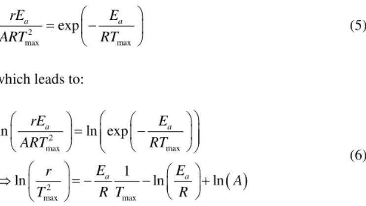
A typical DSC thermogram of a pig back skin sample is shown in Fig. 3. The measured specific heat capacity, cp, compares well with existing results [33, 35]. For a stress-free skin sample, denaturation is characterized by a sudden increase in the energy absorption endotherm, starting at about 60 °C: with further heating, the endotherm reaches its maximum value at the denaturation temperature of 67.39 °C and then decreases. It has also been established that the denaturation endotherm characteristics are heating rate dependent, although the results are not shown here for they offer a similar trend.
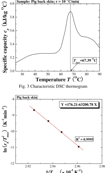
In this study, the collagenous tissue was heated at a constant heating rate, r . According to thermodynamics and the Arrhenius equation, the following relation between the Arrhenius parameters, Ea and A, and the peak temperature of thermal denaturation, Tmax, can be obtained as [48-50]:
2
max max
exp
a a
rE E
RT ART
⎛ ⎞
= ⎜− ⎟
⎝ ⎠
(5)
which leads to:
( )
2
max max
2
max max
ln ln exp
1
ln ln ln
a a
a a
rE E
RT ART
E E
r
A
R T R
T
⎛ ⎞
⎛ ⎞ ⎛ ⎞
= ⎜ − ⎟
⎜ ⎟ ⎜ ⎜ ⎟⎟
⎝ ⎠
⎝ ⎠ ⎝ ⎠
⎛ ⎞ ⎛ ⎞
⇒ ⎜ ⎟= − − ⎜ ⎟+
⎝ ⎠
⎝ ⎠
(6)
The activation energy Ea can be obtained from the slope of the plot of
(
2)
max
ln r T versus 1Tmax , while the material constant A can be derived from the intercept. A characteristic plot for pig back skin sample is given in Fig. 4, whilst the Arrhenius parameters obtained from separate tests with belly, flank, ear and face skin are presented in Table I. As shown in an earlier study [51], there exists a linear relationship between
( )
ln A and Ea, given by (upon curve fitting):
21149.324 2688.367 ln( )
a
E = + A
(7)
30 40 50 60 70 80 90 3.0
3.2 3.4 3.6 3.8
4.0 Sample: Pig back skin; r = 10 O
C/min
Tmax=67.39 OC
S
p
ecif
ic cap
a
cit
y
c
p(
kJ/
k
g
O
C
)
Temperature
T
(
OC
)
Fig. 3 Characteristic DSC thermogram
2.92 2.94 2.96 2.98
-12 -11 -10 -9 -8
Y =176.21-63200.78 X
Pig back skin
ln
(
r
h/
T
2 max
)
(
K
-1
min
-1)
1/
T
max
(
×10
-3
K
-1)
R2 = 0.9995
Fig. 4 Characteristic plot of
(
2)
maxln r T versus 1Tmax
100 150 200 250 300
3 4 5 6 7 8
Eq.(7)
Back
Belly
Ear
Face
Flank
E
a(
10
5
J/
mol
)
ln(A)
(
ln
(
s
-1))
Fig. 5 Comparison of measured Arrhenius parameters (Ea, A) with Eq. (7)
Table I Experimental results of Arrhenius parameters (Ea, A)
Sample
(
5)
10 J mol
a
E
( )
-1s
A
Back skin 5.255 2.126 × 1081 Belly skin 3.935 1.151 × 1061 Ear skin 5.867 5.240 × 1091 Face skin 4.710 4.575 × 1072 Flank skin 4.012 1.501 × 1061
B. Tensile behavior
Uniaxial tensile tests of ear skin under different temperatures have been performed; the corresponding stress versus strain curves are presented in Fig. 6. Note that there exixt two distinct regimes for all the curves. In the first regime, where ε<50%, the curves corresponding to different temperatures almost overlap. In this low modulus region, skin deformation is accommodated principally by the stretching of elastin, which is thermally stable and invariant to small temperature changes. The differences amongst the curves can be attributed to the gradual straightening of an increasing fraction of wavy collagen fibers. In the second region, ε>50% , the stress increases almost linearly with strain, and the gradient of the stress-strain curve decreases with increasing temperature. This is caused by the stretching and slippage of collagen molecules within crosslinked collagen fibers as well as collagen fibril slippage; with increasing temperature, the highly organized crystalline structure of collagen changes to a random, gel-like state, which results in a decreasing stiffness. Another possible effect stems from collagen dehydration which can be linked to thermal damage. For example, Fig. 7 and Fig. 8 show that, when T ≥60 C° , collagen in the dermis is fully denaturized almost instantaneously, whilst at T = 45 and 50 ºC, denaturation is slow. Thus, this divergence in behavior around
60
T= °C seems to fit well with experimental observations.
0 20 40 60 80 100
0.0 0.2 0.4 0.6 0.8 1.0
T (OC)
37 45 50 60 70 80
Stress
σ
(MP
a
)
Strain
ε(%)
Pig ear skin, strain rate: 12.5%/s
Fig. 6 Hydrothermal tensile stress-strain relation at different temperatures
0 20 40 60 80 100 10-9
10-6 10-3 100
103 106
T (OC) 37 45 50 60 70 80
Th
ermal d
a
mage
Ω
Strainε (%)
Pig ear skin, strain rate: 12.5%/s
Fig. 7 Hydrothermal tensile stress-thermal damage relation at different temperatures
0 20 40 60 80 100
10-10 10-8 10-6 10-4 10-2 100
T (OC) 37 45 50 60 70 80
Th
ermal damage degree
Deg
Strainε (%)
Pig ear skin, strain rate: 12.5%/s
Fig. 8 Hydrothermal tensile stress-thermal damage degree relation at different temperatures
C. Compressive behavior
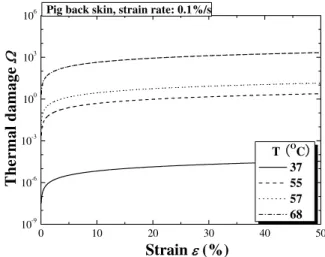
The hydrothermal compressive stress-strain curves of pig back skin are plotted in Fig. 9 for different temperatures. Similar to tensile response, all the curves display two distinct regions: a toe region of low stiffness at low strain, and a region of high stiffness at high strain, with the transition from low to high stiffness occurring around strain levels of 0.1~0.3. Results similar to those of Fig. 9 have been reported for the compressive behavior of skin tested at room temperature [52]. It is interesting to note that, contrary to the tensile tests, the compressive stiffness increases with increasing temperature, although both the thermal damage and the degree of thermal denaturation also increase, as shown in Fig. 10 and Fig. 11. One key difference is that the compressive tests were performed through the thickness of skin samples, in a direction perpendicular to the principal orientation of collagen and elastin fibers. Even though there may exist dehydration effects and denaturation of collagen, similar to tensile response, the
compressive behavior is governed by the mechanical properties of the gel-like ground substance, within which the fibers are embeded [53, 54]. Very little is known about the mechanical properties of the ground substance, which therefore should be the subject of future work; at present one can only speculate that its stiffness increases with increasing temperatures.
0 10 20 30 40 50
0.0 0.3 0.6 0.9 1.2
1.5 Pig back skin, strain rate: 0.1%/s
T
(
OC
)
37
55
58
67
Stre
ss
σ
(
MP
a
)
Strain
ε
(%)
Fig. 9 Hydrothermal compressive stress-strain relations at different temperatures
0 10 20 30 40 50
10-9 10-6 10-3 100 103 106
T (OC) 37 55 57 68
T
h
ermal damage
Ω
Strainε (%)
Pig back skin, strain rate: 0.1%/s
Fig. 10 Hydrothermal compressive stress-thermal damage relations at different temperatures
0 10 20 30 40 50 10-10
10-8 10-6 10-4 10-2 100
T (OC) 37 55 57 68
Th
ermal d
a
mage
d
e
gree
Deg
Strainε (%)
Pig ear skin, strain rate: 0.1%/s
Fig. 11 Hydrothermal compressive stress-thermal damage degree relations at different temperatures
D. DMA results
The viscoelastic properties of skin were obtained from DMA tests. During each DMA experiment, the storage modulus ( E'), loss modulus ( E") and loss factor (tanδ ), were measured as a function of the temperature history. The storage modulus is related to the stored elastic energy in the viscoelastic cycle, and can be considered analogous to a stiffness measurement of a monotonic test; the loss factor yields the ratio between the mechanical energy lost and stored during a given cycle, tanδ =E" E' , effectively measuring the damping performance of the sample.
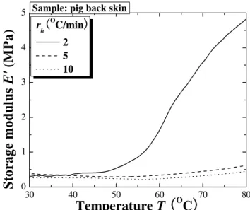
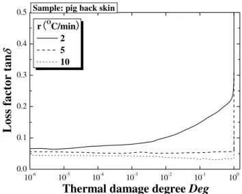
The storage modulus and loss factor are given separately in Fig. 12 and Fig. 13 as a function of temperature. The storage modulus increases with increasing temperature: the skin becomes stiffer at higher temperatures, with the rate increasing with heating rates. The loss factor, however, is insensitive to temperature change but increases marginally with heating rate. In other words, temperature has little effect on the viscous and damping property of skin tissue. The measured value of the loss factor is about 0.2, similar to the result measured with the method of surfacewave propagation at room temperature [55].
It has been generally accepted that, under quasi-static conditions, collagen is responsible for the viscoelastic properties and stiffness of skin [56], suggesting that collagen plays an important role in determining the overall mechanical properties of skin. Thus, it seems reasonable to assume that collagen plays a major role in the viscoelastic nature of skin tissue under thermal loadings. In order to investigate this hypothesis, the storage modulus and loss factor are plotted as a function in Figs. 14 to 17 of thermal damage and thermal denaturation degree. These results show that the effects of thermal damage and thermal denaturation degree on skin viscoelastic behaviour are similar to that of temperature above. In other words, changes in the interval structure of collagen during thermal denaturation do not affect storage modulus and loss factor at the test frequency used in this study. It has been
shown elsewhere that the mechanical changes observed using dynamic mechanical analysis are caused by water loss and changes in the intrinsic molecular structure of the material with heating [28]. Furthermore, weight loss of skin samples has also been observed during the test. Thus, the variations observed in our tests may be attributed to the release of water due to denaturation of collagen molecules during heating.
30 40 50 60 70 80
0 1 2 3 4
5 Sample: pig back skin
rh
(
OC/min)
2 5 10
St
orage mod
u
lu
s
E'
(MPa
)
Temperature
T
(
OC
)
Fig. 12 Storage modulus plotted as a function of temperature for selected heating rates
30 40 50 60 70 80
0.0 0.1 0.2 0.3
0.4 Pig back skin
rh(OC/min)
2 5 10
Temperature
T
(
OC
)
Los
s factor tan
δ
Fig. 13 Loss factor plotted as a function of temperature for selected heating rates
10-6 10-4 10-2 100 102 104 106 0
1 2 3 4
5 Sample: pig back skin
r
(
OC/min)
2 5 10
Thermald damage
Ω
Sto
r
ag
e mo
dul
u
s
E'
(MP
a
)
Fig. 14 Storage modulus plotted as a function of thermal damage for selected heating rates
10-6 10-4 10-2 100 102 104 106
0.0 0.2 0.4 0.6 0.8
1.0 Sample: pig back skin
r(OC/min) 2 5 10
Loss f
a
ctor tan
δ
Thermal damage
Ω
Fig. 15 Loss factor plotted as a function of thermal damage for selected heating rates
10-6 10-5 10-4 10-3 10-2 10-1 100
0 1 2 3 4
5 Sample: pig back skin
r(OC/min) 2 5 10
Thermal damage degree
Deg
Sto
r
a
g
e
mo
dul
us
E'
(MPa)
Fig. 16 Storage modulus plotted as a function of thermal damage degree for selected heating rates
10-6 10-5 10-4 10-3 10-2 10-1 100
0.0 0.1 0.2 0.3 0.4
0.5 Sample: pig back skin r(OC/min)
2 5 10
Lo
ss fac
tor
tan
δ
Thermal damage degree
Deg
Fig. 17 Loss factor plotted as a function of thermal damage degree for selected heating rates
IV. CONCLUSION
The tensile, compressible and viscoelastic behaviors of pig skin tissue were measured under different temperatures. To our best knowledge, this is the first systematical study to characterize the effect of temperature and the corresponding thermal denaturation of skin collagen on skin mechanical properties. The main findings are: 1) DSC results show that the transition temperature of collagen in our skin samples is about 66.8 ºC. 2) Under tensile loading, the stiffness of skin decreases with increasing temperature due to thermal denaturation of skin collagen. 3) Under compressive loading, the stiffness of skin tissue decreases with increasing thermal damage degree, with strain rate sensitivity observed at different damage levels. However, the denaturation of collagen should not be the main mechanism, since the compressive tests are performed through the thickness of skin and similar strain sensitivity has been observed at all damage levels. The rate sensitivity and decreased stiffness with thermal damage are caused mainly by hydration changes. 4) As for dynamic skin viscoelasticity, DMA results show that the storage modulus is highly temperature dependent, due to the release of loosely and strongly bound water. However, the loss factor does not exhibit a significant dependency on either temperature or thermal denaturation. These results suggest that, at fixed frequency, the denaturation of collagen triple-helical molecules may have little effect on skin viscoelasticity.
More experiments are needed to better understand these phenomena and to quantify the variation of skin properties with temperature and collagen denaturation, so that these properties can be considered reliably in future models.
REFERENCES
[1] C. H. Daly, "Biomechanical properties of dermis," J.Invest Dermatol., vol. 79 Suppl 1, 1982, pp. 17s-20s.
[2] C. M. A. Bonilla, A. R. F. Massanet, and N. V. Almodóvar, "Mechanics of Biomaterials: Skin Repair and Grafts," in Proceedings of
Applications of Engineering Mechanics in Medicine, University of
Puerto Rico - Mayaguez, 2005.
[3] M. S. Sacks and W. Sun, "Multiaxial mechanical behavior of biological materials," Annu. Rev. Biomed. Eng., vol. 5, 2003, pp. 251-284. [4] D. G. Hepworth, A. Steven-fountain, D. M. Bruce, and J. F. Vincent,
"Affine versus non-affine deformation in soft biological tissues, measured by the reorientation and stretching of collagen fibres through the thickness of compressed porcine skin," J Biomech, vol. 34, Mar 2001, pp. 341-6.
[5] O. A. Shergold, N. A. Fleck, and D. Radford, "The uniaxial stress versus strain response of pig skin and silicone rubber at low and high strain rates," International Journal of Impact Engineering, vol. 32, Sep 2006, pp. 1384-1402.
[6] O. A. Shergold, "The mechanics of needle-free injection," in Department
of Engineering. vol. PhD Thesis Cambridge: University of
Cambridge, 2004.
[7] J. Ankersen, A. E. Birkbeck, R. D. Thomson, and P. Vanezis, "Puncture resistance and tensile strength of skin simulants," Proc Inst Mech
Eng [H], vol. 213, 1999, pp. 493-501.
[8] K. Miller and K. Chinzei, "Constitutive modelling of brain tissue: Experiment and theory," Journal of Biomechanics, vol. 30, 1997, pp. 1115-1121.
[9] J. E. Miller-Young, N. A. Duncan, and G. Baroud, "Material properties of the human calcaneal fat pad in compression: experiment and theory," Journal of Biomechanics, vol. 35, 2002, pp. 1523-1531. [10] S. Nasseri, L. Bilston, and R. Tanner, "Lubricating squeezing flow: a
useful method for measuring the viscoelastic properties of soft tissues," Biorheology vol. 40, 2003, pp. 545-551.
[11] J. Z. Wu, R. G. Dong, W. P. Smutz, and A. W. Schopper, "Non-linear and viscoelastic characteristics of skin under compression: experiment and analysis," Biomed. Mater. Eng., vol. 13, 2003, pp. 373-385. [12] Y. Lanir and Y. C. Fung, "Two-dimensional mechanical properties of
rabbit skin. II. Experimental results," J.Biomech., vol. 7, 1974, pp. 171-182.
[13] A. W. Wan, "Biaxial tension test of human skin in vivo," Biomed. Mater. Eng., vol. 4, 1994, pp. 473-486.
[14] M. G. Dunn, F. H. Silver, and D. A. Swann, "Mechanical analysis of hypertrophic scar tissue: structural basis for apparent increased rigidity," J Invest Dermatol, vol. 84, Jan 1985, pp. 9-13.
[15] Y. C. Fung, Biomechanics: Mechanical Properties of Living Tissues. New York: Springer-Verlag, 1993,
[16] Y. Lanir, "Biaxial stress relaxation in skin," Ann. Biomed. Eng., vol. 4, 1976, pp. 250-270.
[17] T. Hermanns-Le, I. Uhoda, S. Smitz, and G. E. Pierard, "Skin tensile properties revisited during ageing. Where now, where next?," J.
Cosmet. Dermatol., vol. 3, 2004, pp. 35-40.
[18] E. K. Dawes-Higgs, M. V. Swain, R. J. E. D. Higgs, R. C. Appleyard, and S. Kossard, "Accuracy and reliability of a dynamic biomechanical skin measurement probe for the analysis of stiffness and viscoelasticity," Physiol. Meas., vol. 25, 2004, pp. 97-105.
[19] C. Jacquemoud, K. Bruyere-Garnier, and M. Coret, "Methodology to determine failure characteristics of planar soft tissues using a dynamic tensile test," Journal of Biomechanics, vol. 40, 2007, pp. 468-475.
[20] F. Ebling, R. Eady, and I. Leigh, "Anatomy and organization of human skin," in Textbook of Dermatology, 5th ed, R. H. Champion, J. L. Burrington, and F. J. G. Ebling, Eds. New York: Blackwell Scientific Publications, 1992.
[21] P. J. Flory and R. R. Garrett, "Phase transition in collagen and gelatin systems," J. Am. Chem. Soc., vol. 80, 1958, pp. 4836-4845. [22] J. M. Davidson, M. Giro, M. Sutcli.e, O. Zoia, Q. D., J. Liu, J. M., E.
Perkett, B. Meryck, K. N. Broadley, S. Russell, and G. C. Sephel, "Regulation of elastin synthesis," in Elastin: Chemical and
Biological Aspects, A. M. Tamburro and J. M. Davidson, Eds.
Galatina, Italy: Congedo Editore, 1990.
[23] M. Le Lous, L. Cohen-Solal, J. C. Allain, J. Bonaventure, and P. Maroteaux, "Age related evolution of stable collagen reticulation in human skin," Connect Tissue Res., vol. 13, 1985, pp. 145-155. [24] M. Melling, W. Pfeiler, D. Karimian-Teherani, M. Schnallinger, G. Sobal,
C. Zangerle, and E. J. Menzel, "Differential scanning calorimetry, biochemical, and biomechanical analysis of human skin from
individuals with diabetes mellitus," The Anatomical Record, vol. 59, 2000, pp. 327-333.
[25] R. Reihsner, M. Melling, W. Pfeiler, and E. J. Menzel, "Alterations of biochemical and two-dimensional biomechanical properties of human skin in diabetes mellitus as compared to effects of in vitro non-enzymatic glycation," Clinical Biomechanics, vol. 15, 2000, pp. 379-386.
[26] M. C. Pierce, R. L. Sheridan, B. H. Park, B. Cense, and J. F. De Boer, "Collagen denaturation can be quantified in burned human skin using polarization-sensitive optical coherence tomography," Burns, vol. 30, 2004, pp. 511-517.
[27] K. K. H. Chao, M. A. Burden, and B. J. F. Wong, "Dynamic changes in the elastic modulus of lagomorph nasal septal cartilage during Nd:YAG (λ=1.32 μm) laser irradiation," San Jose, CA, 2001, pp. 247-254.
[28] Y. Chae, G. Aguilar, E. J. Lavernia, and B. J. Wong, "Characterization of temperature dependent mechanical behavior of cartilage," Lasers
Surg. Med., vol. 32, 2003, pp. 271-278.
[29] J. Yamashita, B. R. Furman, H. R. Rawls, X. Wang, and C. M. Agrawal, "Collagen and bone ciscoelasticity: a dynamic mechanical analysis,"
J. Biomed. Mater. Res. (Appl. Biomater.), vol. 63, 2002, pp. 31-36.
[30] J. F. Mano, "Viscoelastic properties of bone: Mechanical spectroscopy studies on a chicken model," Materials Science and Engineering C, vol. 25, 2005, pp. 145-152.
[31] Y. C. Fung, "Biomechanics: Motion, Flow, Stress, and Growth," New York: Springer-Verlag, 1990.
[32] K. R. Diller and J. A. Pearce, "Issues in modeling thermal alterations in tissues," Annals New York Academy of Science, vol. 888, 1999, pp. 153-164.
[33] F. C. Henriques and A. R. Moritz, "Studies of thermal injury, 1. The conduction of heat to and through skin and the temperatures attained therein. A theoretical and an experimental investigation," A. J. Pathol., vol. 23, 1947, pp. 531-549.
[34] E. Middelkoop, A. J. Van den Bogaerdt, E. N. Lamme, M. J. Hoekstra, K. Brandsma, and M. M. W. Ulrich, "Porcine wound models for skin substitution and burn treatment," Biomaterials, vol. 25, 2004, pp. 1559-1567.
[35] G. S. Young, "Thermodynamic Characterization of Skin, Hide and Similar Materials Composed of Fibrous Collagen," Studies in
Conservation, vol. 43, 1998, pp. 65-79.
[36] R. Schiller, A. P. Funke, and C. Gunther, "DSC measurements on full thickness mice skin: An additional tool to investigate permeation enhancement of highly lipophilic drugs," Journal of Thermal
Analysis and Calorimetry, vol. 77, 2004, pp. 497-510.
[37] F. Xu, "Thermomechanical analysis of skin tissue, PhD First Year Report," Cambridge, UK Engineering Department, Cambridge University, 2005.
[38] F. Xu, "Skin biothermomechanics and thermal pain, PhD Second Year Report," Engineering Department, Cambridge University, 2006. [39] W. M. Madigosky and G. F. Lee, "Improved resonance technique for
materials characterization," J. Acous. Soc. Am., vol. 73, 1983, pp. 1374-1377.
[40] S. O. Oyadiji and G. R. Tomlinson, "Determination of the complex moduli of viscoelastic structural elements by resonance and non-resonance methods," J. Sound Vib., vol. 101, 1985, pp. 277-298.
[41] T.-K. Ahn and K.-J. Kim, "Sensitivity Analysis For Estimation Of Complex Modulus Of Viscoelastic Materials By Non-resonance Method," J. Sound Vib., vol. 176, 1994, pp. 543-561.
[42] L. F. Nielsen, N. J. Wismer, and S. Gade, "Improved method for complex modulus estimation," Sound Vib. , vol. 34, 2000, pp. 20-24. [43] J. M. Pereira, J. M. Mansour, and B. R. Davis, "Dynamic Measurement of
the Viscoelastic Properties of Skin," Journal of Biomechanics, vol. 24, 1991, pp. 157-162.
[44] K. B. Arbogast, K. L. Thibault, B. Scott Pinheiro, K. I. Winey, and S. S. Margulies, "A high-frequency shear device for testing soft biological tissues," Journal of Biomechanics, vol. 30, 1997, pp. 757-759.
[45] M. Mossberg, L. Hillström, and T. Söderström, " Non-parametric identification of viscoelastic materials from wave propagation experiments," Automatica vol. 37, 2001, pp. 511-521.
[47] N. G. McCrum, B. E. Read, and G. Williams, Anelastic and Dielectric
Effects in Polymer Solids. New York: Dover Publications, 1991,
[48] T. Ozawa, "Kinetic analysis of derivatives curves in thermal analysis,"
Journal of Thermal Analysis and Calorimetry, vol. 2, 1970, pp.
301-324.
[49] C. A. Miles, "Kinetics of collagen denaturation in mammalian lens capsules studied by differential scanning calorimetry," Int. J. Biol.
Macromol., vol. 15, 1993, pp. 265-271.
[50] C. A. Miles, T. V. Burjanadze, and A. J. Bailey, "The kinetics of the thermal denaturation of collagen in unrestrained rat tail tendon determined by differential scanning calorimetry," J.Mol.Biol., vol. 245, 1995, pp. 437-446.
[51] F. Xu, T. Wen, K. A. Seffen, and T. J. Lu, "Biothermomechanics of Skin Tissue," Journal of the Mechanics and Physics of Solids, 2007, [52] J. Z. Wu, R. G. Cutlip, M. E. Andrew, and R. G. Dong, "Simultaneous
determination of the nonlinear-elastic properties of skin and subcutaneous tissue in unconfined compression tests," Skin
Research and Technology, vol. 13, Feb 2007, pp. 34-42.
[53] R. T. Tregear and P. Dirnhuber, "Viscous flow in compressed human and rat skin," J. Invest. Dermatol., vol. 45, 1965, pp. 119-125. [54] S. Dikstein and A. Hartzshtark, "What does low-pressure indentometry
measure," Arzt. Kosmetol., vol. 16, 1983, pp. 327-328.
[55] S. J. Kirkpatrick, D. D. Duncan, and L. Fang, "Low-frequency surface wave propagation and the viscoelastic behavior of porcine skin,"
Proceedings of SPIE vol. 5474, 2004, p. 127
[56] F. H. Silver, J. W. Freeman, and D. DeVore, "Viscoelastic properties of human skin and processed dermis," Skin Res.Technol., vol. 7, 2001, pp. 18-23.