A Versatile Vector for
Monitoring of
Type I Interferon Induction and Signaling
Estanislao Nistal-Villan1,3¤, Joanna Poutou1,3, Estefania Rodríguez-Garcia1,3, Maria Buñuales1,3, Beatriz Carte-Abad1,3, Jesus Prieto1,3, Gloria Gonzalez-Aseguinolaza1,3, Ruben Hernandez-Alcoceba1,3
*, Esther Larrea2,3
*
1Gene Therapy and Regulation of Gene Expression Program, Center for Applied Medical Research (CIMA), University of Navarra, Pamplona, Spain,2Instituto de Salud Tropical, University of Navarra, Pamplona, Spain,3IdiSNA Navarra Institute for Health Research, Pamplona, Spain
¤ Current address: Microbiology Section, Department of Pharmaceutical and Health Science, Faculty of Pharmacy, University CEU San Pablo, Campus Montepríncipe, Madrid, Spain
*elarrea@unav.es(EL);rubenh@unav.es(RHA)
Abstract
Development of reporter systems forin vivoexamination of IFN-βinduction or signaling of type I interferon (IFN-I) pathways is of great interest in order to characterize biological responses to different inducers such as viral infections. Several reporter mice have been developed to monitor the induction of both pathways in response to different agonists. How-ever, alternative strategies that do not require transgenic mice breeding have to date not been reported. In addition, detection of these pathwaysin vivoin animal species other than mice has not yet been addressed. Herein we describe a simple method based on the use of an adeno-associated viral vector (AAV8-3xIRF-ISRE-Luc) containing an IFN-βinduction and signaling-sensitive promoter sequence controlling the expression of the reporter gene luciferase. This vector is valid for monitoring IFN-I responsesin vivoelicited by diverse sti-muli in different organs. Intravenous administration of the vector in C57BL/6 mice and Syr-ian hamsters was able to detect activation of the IFN pathway in the liver upon systemic treatment with different pro-inflammatory agents and infection with Newcastle disease virus (NDV). In addition, intranasal instillation of AAV8-3xIRF-ISRE-Luc showed a rapid and tran-sient IFN-I response in the respiratory tract of mice infected with the influenza A/PR8/34 virus lacking the NS1 protein. In comparison, this response was delayed and exacerbated in mice infected with influenza A/PR/8 wild type virus. In conclusion, the AAV8-3xIRF-ISRE-Luc vector offers the possibility of detecting IFN-I activation in response to different stimuli and in different animal models with no need for reporter transgenic animals.
Introduction
The interferon (IFN)-βinduction pathway and type I IFN (IFN-I) signaling are two related pathways culminating in the induction of critical antiviral and immuno-stimulatory genes [1]. The IFN-βinduction pathway activates IFN regulatory factor (IRF) 3 and 7, which can OPEN ACCESS
Citation:Nistal-Villan E, Poutou J, Rodríguez-Garcia E, Buñuales M, Carte-Abad B, Prieto J, et al. (2016) A Versatile Vector forIn VivoMonitoring of Type I Interferon Induction and Signaling. PLoS ONE 11(3): e0152031. doi:10.1371/journal.pone.0152031
Editor:Mirco Schmolke, University of Geneva, SWITZERLAND
Received:October 15, 2015
Accepted:March 8, 2016
Published:March 23, 2016
Copyright:© 2016 Nistal-Villan et al. This is an open access article distributed under the terms of the
Creative Commons Attribution License, which permits unrestricted use, distribution, and reproduction in any medium, provided the original author and source are credited.
Data Availability Statement:All relevant data are within the paper.
bind specific IRF genomic DNA elements called IRF-E and stimulate the transcription of sev-eral genes [2]. Type I IFNs, including IFN-β, bind IFN-I receptor and trigger the IFN-I sig-naling cascade, activating STAT1, STAT2 and IRF-9. These three transcription factors form the so-called IFN-stimulated gene factor 3 (ISGF3) complex. ISGF3 binds DNA elements named IFN stimulated response elements or ISRE [3], triggering transcription of IFN-stimu-lated genes (ISGs) and the subsequent activation of cellular pathways associated with IFN stimulation.
IRF-E elements present a consensus sequence:WWNNRAAANNGAAA) [4], where W can be
A/T, R can be G/A and N any nucleotide. The ISRE consensus sequence isGAAANNGAAACT
[5]. The similarity of both consensus sequences supports the fact that many genes can be acti-vated by both signaling pathways, such asMx2[6] orISG15[7]. Schmidt et al. [7] completed a
thorough manipulation of ISG15 IRF-E and ISRE elements and found a sequence with opti-mized IRF-7 and ISGF3 binding properties (TCGGGAAACCGAAACT). This sequence is named
the IRF-ISRE element throughout this text.
Several groups have generated transgenic mice expressing reporter genes under the control of IFN-β[8,9], IFN-α6 [10] or Mx2 promoters [6] to detect IFN induction or IFN-I activityin vivo. In addition, the IFN-specific responsive promoter Mx1 has been used to control the
expression of Cre recombinase in mice [11] in order to generate conditional knock out models. All reporter systems for IFN-βinduction or type I IFN signaling described to date require the use of animal transgenesis and husbandry. These methods are expensive, time-consuming and restricted to a particular mouse strain. In addition, many viral infections have other animal models different from mice. Monitoring IFN-I responsesin vivoin these other
animal species is not an option, and detection of the IFN-I signature requires other invasive methods.
In the present study, we explored the possibility of developing an adeno-associated virus (AAV) reporter vector which enables live monitoring of IFN-I signature in different organs and animal species. AAV is a small, nonpathogenic parvovirus extensively used in gene transfer approaches. AAV-based vectors allow long-term expressionin vivowithout virus replication,
and can transduce different organs/tissues depending on the serotype and/or the route of administration [12]. AAV vectors based on serotype 8 (rAAV8) can transduce the liver with high efficiency [13] when injected intravenously (iv) and the upper respiratory tract when inoc-ulated through the nasal route [14]. The ability of this vector to deliver inducible expression systems has been previously demonstrated [15].
We describe here an AAV vector carrying an IRF-ISRE inducible sequence that controls the expression of firefly luciferase reporter gene (AAV8-3xIRF-ISRE-Luc). We have tested its abil-ity to respond to different stimuli in different organs in C57BL/6 mice and in the liver of Syrian Hamsters.
Materials and Methods
Cells lines
The human cell lines HuH-7 (JCRB Genebank, Japan), Hep2 (ATCC CCL-23) and HepG2 (ATCC HB-8065), mouse cells Hepa1.6 (ATCC CRL-1830) and B16-OVA (courtesy of Dr. P. Sarobe, CIMA, Spain) [16] and the Syrian hamster cell line H2T (courtesy of Dr. C.M. Town-send, University of Texas, Galveston, TX, USA) [17] were cultured in Dulbecco’s modified Eagle’s medium supplemented with 10% heat-inactivated fetal bovine serum, 2 mM L-gluta-mine, 50μg/ml penicillin/streptomycin (all culture reagents from Invitrogen). All cells were grown at 37°C in a 5% CO2incubator.
Reagents
The following reagents were used throughout the experiments performedin vitroorin vivo:
human IFN-α(Sicor Biotech), human IFN-β, mouse IFN-αand mouse IFN-β(PBL). R848 (InvivoGen), CpG and DEAE Dextran (Sigma) and poly I:C (Oncovir).
Reporter assays
All cell culture reporter assays were performed using the indicated IFN-βinduction or IFN-I signaling sensitive DNA elements cloned into the pGL4.17 Firefly luciferase reporter plasmid (Promega). pRL-TK plasmid was used in all cases in order to normalize pGL4.17 reporter plas-mid induction. Cell transfection with the appropriate plasplas-mids was performed as described [18]. A dual-luciferase reporter assay kit (Promega) was used to measure reporter activities fol-lowing the manufacturer’s instructions.
Plasmid construction
Plasmids containing artificial IRF-ISRE or MxA ISRE sequences fused to a minimal MxA pro-moter sequence were synthesized by GenScript. The regions of interest were subcloned into a luciferase reporter plasmid pGL4.17[luc2/Neo]. Reporter plasmids bearing multiple copies of
IRF-ISRE were obtained by inserting a DNA element composed of two complementary oligo-nucleotide sequences containing the IRF-ISRE element. This insert was flanked by PacI restric-tion sites that were used to insert multiple repetirestric-tions of this insert into a PacI site between the original IRF-ISRE and MxA promoter sequences. AAV-MCS vector (Cell Biolabs) was used to insert the promoter and reporter elements. CMV promoter was removed and the 3xIRF-IS-RE-MxA-luciferase sequence was subcloned into the AAV multi cloning site.
Viruses
AAV8-3xIRF-ISRE-Luc rescue was performed as described previously [19]. Recombinant rNDV-F3aa-GFP LaSota, Sendai Cantell virus and influenza viruses used to infect mice and hamsters have been described before [14]. Viral doses per animal are expressed as viral genomes (vg) for AAV vectors and infectious units (iu) for NDV and influenza viruses.
Animal handling and treatments
Six week old female C57BL/6 mice and Syrian (Golden) hamsters (Mesocricetus Auratus; HSD
HAN: AURA, 5 weeks of age) were purchased from Harlan Laboratories. Animals were main-tained under pathogen-free conditions in the CIMA’s animal facility. All of the animal proce-dures were performed in accordance with institutional guidelines and were approved by the Animal Experimentation Ethics Committee of the University of Navarra (Permit number: CEEA-153-14). All animal manipulations were performed under isofluorane inhalatory anes-thesia (Abbott labs). Hydrodynamic plasmid injection in mice was performed as previously described [20], using 20μg of reporter plasmids. IFN-βwas administered intraperitoneally at the indicated doses. The IFN-agonists: poly I:C (50μg), CpG DNA (50μg) and imiquimod-R848 (100μg) were administered intravenously. For intranasal administration, viruses were formulated in 50μl saline solution and instilled drop by drop during at least 1 minute in anes-thetized animals.
Bioluminescence imaging (BLI)
of 100 and 300μl D-luciferin potassium-salt substrate (Promega), respectively, dissolved in PBS at a final concentration of 30 mg/ml. Light emission was measured using a Photon Imager device (Biospace Lab). Photon counts were acquired 8 minutes after substrate administration during 1 minute. Light surface images were obtained immediately after each photon counting session to provide an anatomical view of the animal. Image processing and signal intensity quantifications were performed using M3 Vision software (Biospace Lab). Images are displayed as a pseudo-color photon count image, superimposed on a gray scale anatomic white-light image, allowing assessment of both bioluminescence intensity and its anatomical source. The number of photons emitted per second per square centimeter per steradian was calculated as a measure of luciferase activity utilizing a constant region of interest.
Analysis of gene expression
Peripheral blood mononuclear cells were purified using Ficoll-Paque (GE Bioscience). Total RNA from cells or liver tissue was extracted using the automated MagMax Express 96 system (Applied Biosystems) using the Magmax-96 total RNA isolation kit (Life Technologies). Reverse transcription was performed as previously reported [21]. Real-time polymerase chain reactions were performed with iQ SYBR Green supermix (Bio-Rad) in a CFX96 system from Bio-Rad, using specific primers for each gene [14]. The amount of each transcript was expressed by the formula: 2ct(β-actin)−ct(gene), with ct being the point at which the fluorescence rises appreciably above the background fluorescence.
Quantification of neutralizing antibodies
The reporter virus NDV-GFP was serially diluted in a 1:2 series. A virus dilution corresponding to the last dose presenting near 100% GFP signal in Hep2 cells was later used for incubation at 25°C for 2 hours with mouse or hamster sera. Ab neutralizing titers were obtained in quadru-plicate using the Reed-Muench method to calculate the inhibitory ED50 also in Hep2 cells.
Measurement of type I IFNs by bioassay and ELISA
The amount of type I IFN present in serum from hamsters was analyzed by measuring its abil-ity to protect hamster H2T cells against the cytopathic effect of encephalomyocarditis virus. The assay was performed in a 96-well microtiter plate. First, 2x10⁴H2T cells per well were seeded in 150μl of medium containing serial dilutions of serum. After incubation for 24 hours, cells were infected with encephalomyocarditis virus (5x10⁶pfu per well), and 24 hours later, the cytopathic effect was measured by staining with crystal violet dye solution (0.5% in 1/4 v/v methanol/water). The optical density was read at 595 nm. At the same time, serial dilutions of human IFN-αwere tested to obtain a standard curve. Results are calculated interpolating the optical density of each sample in the standard curve and are expressed as units/ml.
The levels of IFN-αand IFN-βprotein in serum samples from mice were quantified by ELISA (PBL assay science) following the manufacturer΄s instructions.
Statistical analysis
Results
In vitro
characterization of IFN-I reporters
Although several plasmids containing ISRE-driven reporter elements have been generated, few studies have undertaken the task of improving such reporter plasmids and analyzing their pos-sible use for thein vivomonitoring of IFN-I signature. We aimed to build an artificial
opti-mized DNA element that responds to IFN-βinduction and type I IFN signaling pathways. We hypothesized that a promoter containing such an element controlling expression of the lucifer-ase reporter gene should be able to monitor IFN-I signaturein vivo. In order to generate the
reporter system, the IRF-ISRE enhancing element was fused to a minimal human MxA pro-moter. A series of reporter plasmids bearing different numbers of IRF-ISRE repeats was obtained (Fig 1A). These constructs were transfected in HuH-7 cells together with the pRL-TK plasmid in order to normalize specific firefly luciferase reporter activity. Cells were then stimu-lated with 500 units/ml of human IFN-αfor 24 hours. A plasmid containing a tandem of the previously described ISRE sequence was included for comparison. The new IRF-ISRE sequence was more efficient than the ISRE in the response to IFN-α, even when only one repeat of this sequence was included in the plasmid (Fig 1B). Addition of multiple repeats of this element increased the potency of the reporter plasmid. The promoter containing three elements (3xIR-F-ISRE) was chosen for further characterization because it showed the maximal potency with-out increasing baseline activity (Fig 1C). This construct also responds to the IFN-βinduction pathway stimulation, such as Sendai Cantell virus infection in Hepa 1.6 cells [22] (Fig 1D). Fur-thermore, the 3xIRF-ISRE sequence can be induced by IFN-αin human (HuH-7 and HepG2) and mouse (Hepa 1.6 and B16.OVA) cell lines (Fig 1E) and is able to respond to different
IFN-βor IFN-I signaling pathway stimuli such as IFN-α, IFN-β, Poly I:C and Poly I:C plus DEAE (Fig 1F). The kinetics of luciferase activity in response to IFN-αin vitroshow stimulation 24
hours after induction and progressive decay, returning to baseline three days after stimulation (Fig 1G).
In vivo
characterization of 3xIRF-ISRE-Luc reporter plasmid delivered to
mouse liver by hydrodynamic injection
The ability of the 3xIRF-ISRE-Luc reporter plasmid to respond to murine IFN-βin vivowas
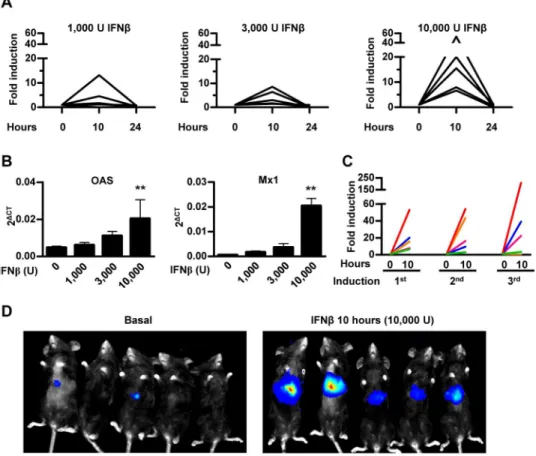
analyzed using hydrodynamic plasmid injection through the tail vein in mice. Using this method of gene delivery, plasmids penetrate hepatocytes in high amounts. An intense initiation of transgene expression soon after injection is usually followed by a rapid reduction with fur-ther stabilization 3–4 weeks later [23]. Once the luciferase signal was stable (four weeks after hydrodynamic injection, data not shown), animals in three different groups received 1,000, 3,000 or 10,000 units of murine IFN-βintraperitoneally.In vivobioluminescent imaging after
administration of the substrate D-luciferin revealed an induction of reporter gene expression 10 hours after murine IFN-βtreatment. We observed that, at the maximum dose of IFN-β
(10,000 U) all animals responded consistently, while at doses of 1,000 and 3,000 U of IFN-β
some of the animals showed no increase in transgene expression. Luciferase activity returned to baseline levels after 24 hours (Fig 2A). Activation of the IFN-I pathway was verified by quan-tifying mRNA expression of ISGs such asOASandMx1in peripheral blood leukocytes 10
hours after treatment (Fig 2B). A good correlation was observed between activation of IFN-β
In vivo
activity of AAV8-3xIRF-ISRE-Luc in response to IFN-agonist
We constructed a rAAV8 vector carrying the 3xIRF-ISRE-Luc cassette (Fig 3A). A dose of 3x10¹⁰viral genomes/mouse of the AAV8-3xIRF-ISRE-Luc vector was injected intravenously in C57BL/6 mice. Baseline luciferase activity was measured until it stabilized approximately 2 weeks after injection, as expected for this type of vector [19]. We detected no elevations of type I IFN protein in sera from mice at days 1 and 7 after AAV8-3xIRF-ISRE-Luc vector inoculation (data not shown). Four groups of mice received different IFN-agonists: poly I:C, CpG DNA, Imiquimod (R848) or 10,000 units of murine IFN-β. Administration of agonists was repeated every week for 3 times. In all cases, an increase in luciferase signal was observed following each round of stimulation (Fig 3B). Light emission was localized mainly in the liver (Fig 3C). Fig 1.In vitrocharacterization of IFN-I reporters.A) Schematic representation of reporter plasmid constructs. B) Dual luciferase reporter assay in HuH-7 cells transfected with the indicated plasmids in response to 500 units/ml of human IFN-αfor 24 hours. C) Luciferase activity (fold induction) in HuH-7 cells transfected with the indicated reporter plasmids and treated with 500 units/ml of human IFN-αfor 24 hours. D) Dual luciferase reporter assay in Hepa 1.6 cells transfected with the indicated plasmids in response to Sendai Cantell virus (20 hemagglutination units, 24 hours). E) Fold induction of luciferase activity in cells transfected with the 3xIRF-ISRE reporter plasmids in the indicated cell lines treated during 24 hours with 500 units/ml of the corresponding species-specific IFN-α. F) Luciferase activity fold induction in murine cell lines transfected with the 3xIRF-ISRE reporter plasmid and treated with 500 units/ml murine IFN-α, 500 units/ml murine IFN-β, 50μg/ml poly I:C or 1μg/ml poly I:C mixed with 250μg/ml DEAE-Dextran for 24 hours. G) Time course of luciferase activity in HuH-7 cells transfected with the 3xIRF-ISRE-luc reporter plasmids and treated with 500 units/ml of human IFN-α. These data are from one experiment representative of four.***p<0.001, ns: not significant. TK: Thymidine kinase.
In vivo
activity of AAV8-3xIRF-ISRE-Luc in mice in response to
intravenous NDV-GFP administration
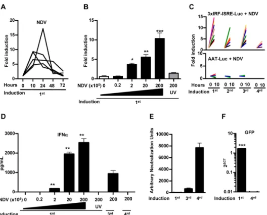
It has been previously shown that the intravenous administration of NDV induces IFN-I in the mouse liver [10]. In order to validate the activity of our reporter vectorin vivoin response to
NDV, mice were treated with AAV8-3xIRF-ISRE-Luc and then received an iv administration of NDV-F3AA-GFP LaSota (2x10⁷iu). A consistent and specific stimulation of luciferase signal was observed as early as 10 hours after virus exposure and the activity returned to baseline lev-els 48–72 hours after NDV infection (Fig 4A). Further experiments demonstrated a dose-dependent activation of the reporter system in response to NDV (Fig 4B), whereas inactivation of the virus by freeze/thaw and UV irradiation abolished the increase in luciferase signal. Lucif-erase expression was consistently re-induced every week for at least 3 times after administra-tion of the same dose of NDV (Fig 4C). No increase in luciferase signal was observed when the same dose of NDV-F3AA-GFP LaSota virus was administered to mice previously transduced with an AAV8 vector expressing luciferase under the control of the constitutive liver-specific Fig 2.In vivocharacterization of 3xIRF-ISRE-Luc reporter plasmid delivered to mouse liver by hydrodynamic injection.A) Mice received a hydrodynamic injection with 20μg of 3xIRF-ISRE-Luc reporter plasmid through the tail vein. Once luciferase activity stabilized (one month after injection), mice were treated intraperitoneally with the indicated doses of murine IFN-β. Light emission was quantified by BLI 10 and 24 hours after treatment. Values correspond to fold luciferase activity, using baseline (pre-induction) activity as a reference. B) Quantitative RT-PCR ofOASandMx1genes in peripheral blood lymphocytes of animals treated for 24 hours with different doses of recombinant murine IFN-β. C) Reporter activity re-induction in mice determined every week by intraperitoneally administration of 10,000 units of IFN-β. Each line represents an individual mouse. D) Representative BLI images of mice before and 10 hours after administration of 10,000 U of murine IFN-β. These data are from one experiment representative of three.**p<0.01vs3,000, 1,000 and 0 IFN-βunits.
alpha-1 anti-trypsin promoter (AAV8-AAT-Luc) [24] (Fig 4C). Interestingly, repeated activa-tion of the AAV8-3xIRF-ISRE-Luc reporter during the first month correlated with the stimula-tion of the IFN-I response evidenced by the increase in serum IFN-αafter each viral infection (Fig 4D). The reporter activation was drastically diminished when the NDV virus was adminis-tered again at later times (Fig 4C), coinciding with the lack of IFN-I elevation in serum after 24 hours (Fig 4D), the appearance of high titers of anti-NDV neutralizing antibodies (Fig 4E) and the inability to detect the virus infection in the liver (Fig 4F). These results indicate that the AAV8-3xIRF-ISRE-Luc reporter is able to detect specifically the activation of IFN-I responses in the liver resulting from infection by NDV.
In vivo
activity of AAV8-3xIRF-ISRE-Luc in hamster in response to
intravenous NDV-GFP administration
In order to study the ability of AAV8-3xIRF-ISRE-Luc reporter vector to detect IFN-I signature in animals other than mice, we used Syrian hamsters as a model. These animals are suitable for BLI [25] and have been used to study different viral infections due to their broad permissive-ness [26–30]. After iv AAV8-3xIRF-ISRE-Luc vector administration, baseline luciferase activ-ityin vivowas monitored until stabilization. As observed previously in mice, the main
luciferase signal in hamsters originated from the liver (data not shown), which indicates a pref-erential hepatic transduction of the AAV8 vector. Intravenous administration of
NDV-F3AA-GFP LaSota induced the luciferase reporter activity in the liver (Fig 5A and 5B), with kinetics similar to those observed in mice treated with the same virus (Fig 5A). A second Fig 3.In vivoactivity of AAV8-3xIRF-ISRE-Luc in response to IFN-agonist.A) Schematic representation of the AAV8-3xIRF-ISRE-Luc vector (not drawn to scale). B) Mice were inoculated iv with 3x10¹⁰vg of the vector. Two weeks later, animals were divided in different groups and received the following iv stimuli: poly I: C, CpG DNA, Imiquimod (R848) or murine IFN-β. Treatments were repeated weekly for 3 weeks. Luciferase activity was quantified by BLI. Each line represents the fold luciferase activity of each individual mouse. C) Representative BLI images of AAV8-3xIRF-ISRE-Luciferase transduced mice before and 10 hours after murine IFN-βadministration. These data are from one experiment representative of two.
stimulation of the reporter in response to NDV injection was observed 6 weeks later (Fig 5C). This is consistent with a peak of IFN-I in serum (Fig 5D). Under these experimental condi-tions, the titer of anti-NDV neutralizing antibodies is still moderate in hamsters (Fig 5E), which may explain the preservation of the IFN-I response.
In vivo
reporter activity of AAV8-3xIRF-ISRE-Luc in the respiratory tract
Although not very efficiently, AAV8 can also transduce the respiratory tract after intranasal instillation. We employed this route to test if our reporter vector could deliver the 3xIRF-ISRE-driven luciferase reporter in the respiratory tract. This should allow detection of IFN-I induc-tion elicited by airway infecinduc-tion with viruses displaying different pathogenicity. These viruses include a non-pathogenic virus such as NDV-F3AA-GFP LaSota (107iu/mouse) or mouse-adapted influenza viruses: the attenuated mutant A/PR8/34-ΔNS1 (107iu/mouse) and the Fig 4.In vivoactivity of AAV8-3xIRF-ISRE-Luc in mice in response to intravenous NDV-F3AA-GFP LaSota administration.Mice were inoculated iv with 3x10¹⁰vg of the AAV8-3xIRF-ISRE-Luc vector or a
control vector AAV8-AAT-Luc, in which luciferase expression is controlled by a constitutive liver-specific promoter. Two weeks later, animals started to receive iv administrations of NDV-F3AA-GFP LaSota virus at different doses and schedules A)In vivoluciferase activity stimulation in the AAV8-3xIRF-ISRE-Luc transduced animals at 10, 24, 48 and 72 hours after iv administration of the NDV virus at 2x10⁷iu. Each line represents an individual mouse. B) Mice transduced with AAV8-3xIRF-ISRE-Luc received the indicated doses of NDV-F3AA-GFP LaSota virus or UV-inactivated virus (UV), and the stimulation of luciferase activity was determined 10 hours later (indicated as average fold induction for each group). C) Mice transduced with AAV8-3xIRF-ISRE-Luc (upper panel) or AAV8-AAT-Luc (lower panel) received repeated iv inoculations of 2x10⁷iu NDV one week apart. Stimulation of luciferase activity was determined 10 hours after every NDV
injection. Each line represents an individual mouse. D) Concentration of IFN-αin the serum of mice, determined by ELISA 24 hours after each NDV administration E) NDV neutralizing antibodies in serum of mice, determined one day after each round of NDV administration. F) A sub-group of mice was sacrificed after the first or fourth NDV-GFP administration, and expression of virally encoded GFP was determined by qRT-PCR in liver samples. These data are from one experiment representative of three.
corresponding wild type (Wt) A/PR8/34 (2x10²iu /mouse). Our results indicate that the mouse immune system reacted against NDV, as shown by a potent activation of the reporter element circumscribed to the upper respiratory tract, which lasted approximately 3 days (Fig 6A). A similar result was observed after infection with A/PR8/34-ΔNS1, which was well-toler-ated despite the high dose used (Fig 6A). This is compatible with an apparent localized infec-tion of the upper respiratory tract and efficient limitainfec-tion of viral spread by the strong innate immune response. To allow monitorization of Wt A/PR8/34 infection over time, a sublethal dose of the virus was intranasally inoculated in mice. In contrast to NDV or the attenuated A/ PR8/34-ΔNS1 influenza virus, a robust IFN-I signature was detected in the upper and lower respiratory tracts of animals, starting 3–4 days after infection (Fig 6A and 6B). This result is Fig 5.In vivoactivity of AAV8-3xIRF-ISRE-Luc in hamster in response to intravenous NDV-F3AA-GFP LaSota administration.Syrian hamsters were inoculated iv with 1x10¹¹vg of the AAV8-3xIRF-ISRE-Luc
vector. Three weeks later, animals received an iv administration of 1x10⁹iu NDV-F3AA-GFP LaSota. A)In
vivoluciferase activity was monitored at 10, 24, 48 and 72 hours after iv administration of the NDV virus. Each
line represents an individual hamster. B) Image of a representative hamster before (basal) and 10 hours after the first NDV administration. C) Luciferase activity stimulation in hamsters receiving two doses of
NDV-F3AA-GFP LaSota 6 weeks apart one from the other. D) Type I IFN activity in serum of hamsters before and 24 hours after second NDV administration, measured by bio-assay. E) NDV neutralizing antibodies in serum of hamsters before and 6 weeks after the first NDV administration. These data are from one experiment representative of two.
consistent with active replication of the Wt A/PR8/34 virus in the lung as well as the activation of a strong inflammatory process which is type I IFN-dependent as previously reported [31,32]. The effect of virus replication on the reporter activation in the mouse upper and lower respiratory tracts can be clearly visualized 120 hours after infection (Fig 6B).
Discussion
We describe here a novel technique to monitor IFN-Iin vivowithout the need for animal
trans-genesis by the administration of a non-replicating AAV vector. We demonstrate the utility of this systemin vivoby characterizing different factors capable of enhancing IFN-I expression or
signaling. A vector such as AAV8-3xIRF-ISRE-Luc is a novel and valid biotechnological tool to follow IFN-I signature in mice and hamsters and most likely in many more animal species since the IRF and ISRE promoter sequences are conserved across vertebrates [33].
In the present study, we have been able to monitor IFN-I signature after administration of NDV-GFP, a chicken-adapted virus, with limited replication in mammals [34]. A detailed characterization in mice indicated that the activation of the reporter system in the liver was dose-dependent and correlated with the presence of IFN-αin serum (Fig 4). Interestingly, the system was able to detect repeated activations of the IFN-I response upon NDV re-administra-tion, until the titer of neutralizing antibodies was high enough to block liver infection. At this point, a residual reporter signal was present in the absence of detectable IFN-αin serum. This may reflect a higher sensitivity of the AAV8-3xIRF-ISRE-Luc vector compared to the IFN-α
ELISA or the detection of NDV-encoded GFP by qRT-PCR. Alternatively, it is possible that the IFN-I response is activated in these circumstances in the absence of liver infection. Although Fig 6.In vivoreporter activity of AAV8-3xIRF-ISRE-Luc in the respiratory tract.Mice received an intranasal instillation of AAV8-3xIRF-ISRE-Luc (1x10¹¹viral genomes/mouse) and were then divided in 4
groups, according to the following intranasal stimuli: Saline solution (Mock); 2x10⁷ iu NDV-F3AA-GFP LaSota; 2x10⁷iu influenza A/PR8/34-ΔNS1 or 2x10²iu Wt A/PR8/34. A) Luciferase activity was measured by
BLI in the upper and lower respiratory tract at the indicated times. B) Representative images of mice before (baseline) and 120 hours after Wt PR8 infection. Each line represents an individual mouse. These data are from one experiment representative of two.
the mechanism is still speculative, one possibility is that antibody-neutralized NDV immune complex could be uptaken by a particular phagocytic cell in the liver. This neutralized virus genetic material could be detected and trigger type I IFN in a process similar to the one that triggers the nucleic acid-antibody immune-complexes responsible for type I IFN stimulation in systemic lupus erythematosus [35]. In addition, although not described for NDV, it has been proposed that some virus infections, specifically in the entry process, could be enhanced by virus opsonization [36]. Alternatively, a small fraction of NDV could escape from antibody-driven phagocytosis and be detected by previously described FcγR negative monocytes (CD64-or CD64 low) which could trigger the production of type I IFN [37]. Further work will deter-mine if the same pattern of response is observed in different species. We provide evidence that the reporter can be re-induced by NDV infection in Syrian hamsters when the titer of antibod-ies is still relatively low and there is new peak of IFN-I in serum, but long-term follow-up in this and other animal models will expand our knowledge about innate immune response against this virus.
The study performed with the influenza viruses, A/PR8/34-ΔNS1 and Wt A/PR8/34 high-lights the relevance and usefulness of the reporter vector presented here and its possible adap-tation to different organs, in this case the respiratory tract. Our results indicate that influenza induces a mild and early IFN-I response capable of controlling viral infection when the virus lacks the NS1 protein, whereas this response is not detected when the virus expresses NS1. In fact, the NS1-expressing virus induces a delayed but very strong IFN-I response clearly detected by the reporter system in the lower respiratory tract. This signal correlates with a robust immune response compatible with an effective replication of influenza virus in these mice. Equivalent studies can be carried out to identify new pathogenic factors related to the IFN-I responsein vivoafter infection with a variety of viruses and animal models where AAV could
be employed as the delivery vector.
In vivobioluminescence detection requires specialized instrumentation that may not be
adapted easily to large animals. In these situations an IFN-I signature could be detectedin vivo
by replacing the luciferase gene with a soluble reporter gene easily detected in blood, such as secreted alkaline phosphatase (SEAP) [38] or others. This system could bypass the requirement for specie-specific type I IFN ELISA kits. The inducible sequence could also be moved to other viral vectors or organ-specific nanoparticles that would be useful to monitor specific activation of type I IFN upon different stimuli.
Alternatively the reporter element described here could be inserted into the genome of dif-ferent nuclear DNA replicating viruses. Indeed, activation of this reporter in infected cells may be useful to study kinetics of the type I IFN responsein vivoin the infected cell.
Finally, a potential application of this tool in the field of gene therapy would involve the use of the IFN-responsive promoter to control the expression of a therapeutic gene. Delivery of such construct in the target organ would achieve activation of transgene expression in response to endogenous IFN responses. Alternatively, recombinant type I IFN could be used as an inducer.
Conclusions
We present here a novel tool to deliver an IFN-I responsive reporter system to the livers or the respiratory tract that could be extended to other organs by changing the vector, AAV serotype and/or the route of administration. This convenient method allows sustained monitoring of IFN-I inductionin vivowithout the need for breeding transgenic animals. Furthermore, this
available, increasing the opportunities of studying IFN-I induction by a wide number of viruses or agents.
Acknowledgments
We acknowledge Carlos Alfaro Alegría, David Corbacho and Marianna DiScala from the CIMA at the University of Navarra for their help with reagents and technical issues and Paul Miller for language editing.
Author Contributions
Conceived and designed the experiments: EL RHA ENV GGA J. Prieto. Performed the experi-ments: EL ENV J. Pouto ERG MB BCA. Analyzed the data: EL ENV GGA J. Prieto RHA. Wrote the paper: EL ENV RHA.
References
1. Geiss G, Jin G, Guo J, Bumgarner R, Katze MG, Sen GC. A comprehensive view of regulation of gene expression by double-stranded RNA-mediated cell signaling. J Biol Chem. 2001; 276:30178–30182.
PMID:11487589
2. Grandvaux N, Servant MJ, tenOever B, Sen GC, Balachandran S, Barber GN, et al. Transcriptional pro-filing of interferon regulatory factor 3 target genes: direct involvement in the regulation of interferon-stimulated genes. J Virol. 2002; 76:5532–5539. PMID:11991981
3. Ivashkiv LB, Donlin LT. Regulation of type I interferon responses. Nat Rev Immunol. 2014; 14:36–49.
doi:10.1038/nri3581PMID:24362405
4. Tanaka N, Kawakami T, Taniguchi T. Recognition DNA sequences of interferon regulatory factor 1 (IRF-1) and IRF-2, regulators of cell growth and the interferon system. Mol Cell Biol. 1993; 13:4531–
4538. PMID:7687740
5. Stark GR, Kerr IM, Williams BR, Silverman RH, Schreiber RD. How cells respond to interferons. Annu Rev Biochem. 1998; 67:227–264. PMID:9759489
6. Pulverer JE, Rand U, Lienenklaus S, Kugel D, Zietara N, Kochs G, et al. Temporal and spatial resolu-tion of type I and III interferon responses in vivo. J Virol. 2010; 84:8626–8638. doi: 10.1128/JVI.00303-10PMID:20573823
7. Schmid S, Mordstein M, Kochs G, Garcia-Sastre A, Tenoever BR. Transcription factor redundancy ensures induction of the antiviral state. J Biol Chem. 2010; 285:42013–42022. doi:10.1074/jbc.M110. 165936PMID:20943654
8. Lienenklaus S, Cornitescu M, Zietara N, Lyszkiewicz M, Gekara N, Jablonska J, et al. Novel reporter mouse reveals constitutive and inflammatory expression of IFN-beta in vivo. J Immunol. 2009; 183:3229–3236. doi:10.4049/jimmunol.0804277PMID:19667093
9. Scheu S, Dresing P, Locksley RM. Visualization of IFNbeta production by plasmacytoid versus conven-tional dendritic cells under specific stimulation conditions in vivo. Proc Natl Acad Sci U S A. 2008; 105:20416–20421. doi:10.1073/pnas.0808537105PMID:19088190
10. Kumagai Y, Takeuchi O, Kato H, Kumar H, Matsui K, Morii E, et al. Alveolar macrophages are the pri-mary interferon-alpha producer in pulmonary infection with RNA viruses. Immunity. 2007; 27:240–252.
PMID:17723216
11. Kuhn R, Schwenk F, Aguet M, Rajewsky K. Inducible gene targeting in mice. Science. 1995; 269:1427–1429. PMID:7660125
12. Chenuaud P, Larcher T, Rabinowitz JE, Provost N, Joussemet B, Bujard H, et al. Optimal design of a single recombinant adeno-associated virus derived from serotypes 1 and 2 to achieve more tightly reg-ulated transgene expression from nonhuman primate muscle. Mol Ther. 2004; 9:410–418. PMID: 15006608
13. Paneda A, Collantes M, Beattie SG, Otano I, Snapper J, Timmermans E, et al. Adeno-associated virus liver transduction efficiency measured by in vivo [18F]FHBG positron emission tomography imaging in rodents and nonhuman primates. Hum Gene Ther. 2011; 22:999–1009. doi:10.1089/hum.2010.190
PMID:21320035
Models Deficient in Type I IFN Response. J Innate Immun. 2015; 7:466–481. doi:10.1159/000375262
PMID:25966783
15. Vanrell L, Di Scala M, Blanco L, Otano I, Gil-Farina I, Baldim V, et al. Development of a liver-specific Tet-on inducible system for AAV vectors and its application in the treatment of liver cancer. Mol Ther. 2011; 19:1245–1253. doi:10.1038/mt.2011.37PMID:21364542
16. Aranda F, Llopiz D, Diaz-Valdes N, Riezu-Boj JI, Bezunartea J, Ruiz M, et al. Adjuvant combination and antigen targeting as a strategy to induce polyfunctional and high-avidity T-cell responses against poorly immunogenic tumors. Cancer Res. 2011; 71:3214–3224. doi: 10.1158/0008-5472.CAN-10-3259PMID:21402711
17. Simmons DJ, Seitz PK, Cooper CW, Krukowski M, Townsend CM Jr. Antitumor effect of positively charged resin in the hamster cheek pouch model. J Biomed Mater Res. 1997; 34:393–400. PMID: 9086409
18. Larrea E, Riezu-Boj JI, Aldabe R, Guembe L, Echeverria I, Balasiddaiah A, et al. Dysregulation of inter-feron regulatory factors impairs the expression of immunostimulatory molecules in hepatitis C virus genotype 1-infected hepatocytes. Gut. 2014; 63:665–673. doi:10.1136/gutjnl-2012-304377PMID: 23787026
19. Paneda A, Vanrell L, Mauleon I, Crettaz JS, Berraondo P, Timmermans EJ, et al. Effect of adeno-asso-ciated virus serotype and genomic structure on liver transduction and biodistribution in mice of both genders. Hum Gene Ther. 2009; 20:908–917. doi:10.1089/hum.2009.031PMID:19419275
20. Fioravanti J, Gonzalez I, Medina-Echeverz J, Larrea E, Ardaiz N, Gonzalez-Aseguinolaza G, et al. Anchoring interferon alpha to apolipoprotein A-I reduces hematological toxicity while enhancing immu-nostimulatory properties. Hepatology. 2011; 53:1864–1873. doi:10.1002/hep.24306PMID:21425312
21. Larrea E, Aldabe R, Molano E, Fernandez-Rodriguez CM, Ametzazurra A, Civeira MP, et al. Altered expression and activation of signal transducers and activators of transcription (STATs) in hepatitis C virus infection: in vivo and in vitro studies. Gut. 2006; 55:1188–1196. PMID:16120756
22. Rajsbaum R, Albrecht RA, Wang MK, Maharaj NP, Versteeg GA, Nistal-Villan E, et al. Species-specific inhibition of RIG-I ubiquitination and IFN induction by the influenza A virus NS1 protein. PLoS Pathog. 2012; 8:e1003059. doi:10.1371/journal.ppat.1003059PMID:23209422
23. Berraondo P, Gonzalez-Aseguinolaza G, Troconiz IF. Semi-mechanistic pharmacodynamic modelling of gene expression and silencing processes. Eur J Pharm Sci. 2009; 37:418–426. doi:10.1016/j.ejps. 2009.03.013PMID:19491033
24. Unzu C, Sampedro A, Mauleon I, Alegre M, Beattie SG, de Salamanca RE, et al. Sustained enzymatic correction by rAAV-mediated liver gene therapy protects against induced motor neuropathy in acute porphyria mice. Mol Ther. 2011; 19:243–250. doi:10.1038/mt.2010.210PMID:20877347
25. Bunuales M, Garcia-Aragoncillo E, Casado R, Quetglas JI, Hervas-Stubbs S, Bortolanza S, et al. Eval-uation of monocytes as carriers for armed oncolytic adenoviruses in murine and Syrian hamster models of cancer. Hum Gene Ther. 2012; 23:1258–1268. doi:10.1089/hum.2012.043PMID:22985305
26. Morrey JD, Day CW, Julander JG, Olsen AL, Sidwell RW, Cheney CD, et al. Modeling hamsters for evaluating West Nile virus therapies. Antiviral Res. 2004; 63:41–50. PMID:15196819
27. Li G, Duan T, Wu X, Tesh RB, Soong L, Xiao SY. Yellow fever virus infection in Syrian golden hamsters: relationship between cytokine expression and pathologic changes. Int J Clin Exp Pathol. 2008; 1:169–
179. PMID:18784801
28. Sbrana E, Xiao SY, Popov VL, Newman PC, Tesh RB. Experimental yellow fever virus infection in the golden hamster (Mesocricetus auratus) III. Clinical laboratory values. Am J Trop Med Hyg. 2006; 74:1084–1089. PMID:16760525
29. Tesh RB, Guzman H, da Rosa AP, Vasconcelos PF, Dias LB, Bunnell JE, et al. Experimental yellow fever virus infection in the Golden Hamster (Mesocricetus auratus). I. Virologic, biochemical, and immu-nologic studies. J Infect Dis. 2001; 183:1431–1436. PMID:11319679
30. Paessler S, Aguilar P, Anishchenko M, Wang HQ, Aronson J, Campbell G, et al. The hamster as an ani-mal model for eastern equine encephalitis—and its use in studies of virus entrance into the brain. J
Infect Dis. 2004; 189:2072–2076. PMID:15143475
31. Garcia-Sastre A, Durbin RK, Zheng H, Palese P, Gertner R, Levy DE, et al. The role of interferon in influenza virus tissue tropism. J Virol. 1998; 72:8550–8558. PMID:9765393
32. Manicassamy B, Manicassamy S, Belicha-Villanueva A, Pisanelli G, Pulendran B, Garcia-Sastre A. Analysis of in vivo dynamics of influenza virus infection in mice using a GFP reporter virus. Proc Natl Acad Sci U S A. 2010; 107:11531–11536. doi:10.1073/pnas.0914994107PMID:20534532
34. Kumar R, Tiwari AK, Chaturvedi U, Kumar GR, Sahoo AP, Rajmani RS, et al. Velogenic newcastle dis-ease virus as an oncolytic virotherapeutics: in vitro characterization. Appl Biochem Biotechnol. 2012; 167:2005–2022. doi:10.1007/s12010-012-9700-1PMID:22644640
35. Ronnblom L, Eloranta ML, Alm GV. The type I interferon system in systemic lupus erythematosus. Arthritis Rheum. 2006; 54:408–420. PMID:16447217
36. Iankov ID, Pandey M, Harvey M, Griesmann GE, Federspiel MJ, Russell SJ. Immunoglobulin g anti-body-mediated enhancement of measles virus infection can bypass the protective antiviral immune response. J Virol. 2006; 80:8530–8540. PMID:16912303
37. Grage-Griebenow E, Flad HD, Ernst M. Fc gamma receptor I (CD64)-negative human monocytes are potent accessory cells in viral antigen-induced T cell activation and exhibit high IFN-alpha-producing capacity. J Leukoc Biol. 1996; 60:389–396.