Simultaneous overexpression of GH and STAT5b genes inhibits the STAT5
signalling pathway in tilapia (
Oreochromis niloticus
) embryos
Luis Fernando Marins
2, Arati Iyengar
1, Norman Maclean
1, Jose A. Levy
2and Frédéric Sohm
11
School of Biological Sciences, Biomedical Sciences Building, University of Southampton,
Bassett Crescent East, Southampton, UK.
2
Fundação Universidade Federal do Rio Grande, Departamento de Química,
Laboratório de Bioquímica Marinha, Rio Grande, RS, Brazil.
Abstract
In this study, we describe the use of a STAT5 responsive element (LHRE) reporter gene to monitor the activity of the growth hormone (GH) transduction pathway following expression of heterologous fish GH and rat STAT5b in tilapia embryos and fish fibroblast cells. Our results indicate that both GH and STAT5b are able to activate the LHRE at high levels when transferred separately, demonstrating the substantial level of conservation of the GH signal transduction pathways between fish and mammals. Unexpectedly, co-expression experiments show a strong inhibition of the GH-dependent activation, suggesting that simultaneous GH and STAT5b overexpression can counteract effects of GH expression in tilapia embryos.
Key words: teleost, growth hormone, signal transduction, cytokine, trangenesis signal transducers and activators of transcription. Received: April 6, 2002; accepted: July 23, 2002.
Introduction
Growth is the result of complex molecular interac-tions in which growth hormone plays a major role in all ver-tebrates. Although the physiological effects of GH result from transcriptional regulation by several signalling path-ways, the most direct involves the JAK/STAT signalling pathway (Moutoussamy et al., 1998). Fish GH receptor (GHR) cDNAs have been recently cloned and encode pro-teins of approximately 600 amino acids (Calduch-Gineret al., 2001; Leeet al., 2001). The GHR signal transduction follows a pattern shared amongst the receptors of the class I cytokine receptor superfamily. Briefly, the interaction of the hormone with the receptors induces receptor homodi-merisation which in turn activates the GHR-associated Ja-nus Kinase 2 (JAK2). The subsequent phosphorylation of the receptor tyrosines allows the recruitment of a class of highly related cytoplasmic proteins called signal transduc-ers and activators of transcription or STATs. The GHR-bound STATs are themselves phosphorylated by JAK2. Once phosphorylated, the cytoplasmic STATs form dimers, translocate into the nucleus where they bind to a palindromic DNA motif present in STAT inducible gene promoters and activate the transcription of target genes
(Kopchick and Andry, 2000; Takeda and Akira, 2000). GH activates STAT1, STAT3 and STAT5 (Moutoussamy et al., 1998). In mammals, GH responsive elements have been identified within several gene promoters and some of them have been investigated using reporter genes (Sotiropoulos
et al., 1996; Liuet al., 1997; Storzet al., 1999; von Laueet al., 2000; Ehretet al., 2001).
In fish, growth enhancement has been achieved by transgenic overexpression of GH in several lines, while no enhancement has been observed in others, including a recent report of lack of growth enhancement in genetically selected strains of trout (Duet al., 1992; Devlinet al., 1994; de la Fuenteet al., 1999; Rahman and Maclean, 1999; Devlinet al., 2001). Additionally, better growth enhancement has been obtained with relatively low levels of circulating GH (de la Fuenteet al., 1999). Even though several genes from the JAK/STAT signal transduction pathway have been iden-tified in fish (Rycyzynet al., 1998; Baudleret al., 1999; Oateset al., 1999a, 1999b; Leuet al., 2000), little is known about fish GH signal transduction pathways and just recently the GH receptor itself has been cloned in this group of verte-brates (Calduch-Gineret al., 2001; Leeet al., 2001).
In the present study, we describe the use of LHRE (lactogenic hormone responsive element), a STAT5 re-sponsive element (Sotiropouloset al., 1996) spliced to a lu-ciferase reporter gene, to monitor the activation of the JAK/STAT signalling pathway in response to heterologous Send correspondence to F. Sohm. Department of Biology and
Bio-chemistry, University of Bath, Bath, BA2 7AY U.K. E-mail: [email protected].
GH and STAT5b overexpression in fish embryos. To ex-amine this question we have used vectors expressing the marine silverside (Odontesthes argentinensis) fish GH and the rat STAT5b. We show that the GH and the STAT5b are able to induce the expression of a luciferase gene under control of a STAT5 responsive element both in fish fibroblasts and tilapia embryos. Moreover, we show that the overexpression of both GH and STAT5b inhibits the ex-pression of the LHRE element.
Materials and Methods
Plasmids
pP3PA-cβActin/lacZ (hereafter referred to as pcβA/ lacZ) consists of the lacZ gene under the control of the carp
β-actin promoter and pCMV/STAT5b-GFP expresses a STAT5b-GFP fusion protein under the control of the cyto-megalovirus promoter (Alamet al., 1996; Herringtonet al., 1999). pLHRE-TK/luc and pTK/luc consist of the luciferase gene under the control of the thymidine kinase minimal pro-moter with and without a six times repeat of the lactogenic re-sponsive element respectively (Sotiropouloset al., 1996). The pcβA/msGH was constructed by replacing the lacZ sequence of pcβA/lacZ by aNcoI/EcoRI fragment containing the cDNA of the growth hormone gene of the marine silverside fish (Genbank/EMBL accession number AF236091). The inser-tion was confirmed by sequencing. The vector pP3PA-cβAcin/GFP was constructed as the pcβA/msGH but the cDNA coding for the green fluorescent protein was used in-stead of the msGH (S. Brooks, unpublished).
Cell culture and transfections
The BF-2 cell line (Bluegill sunfish fibroblasts, Ref. 87032603, ECACC, Bath, UK) were cultured in L-15 me-dium, supplemented with 10% foetal calf serum (ICN), 2 mM glutamine (Sigma) and antibiotic/antimycotic solu-tion (Sigma) at 26 °C without CO2. For transient
trans-fection 300,000 cells per well (12-well plates) were seeded and incubated overnight. Transfections were performed at approximately 60% confluence using FuGENE 6 trans-fection reagent (Roche Molecular Biochemicals, UK). Prior to transfection, the cells were washed to remove the serum. For the transfection, 400µL of medium without se-rum plus 100µL of DNA-FuGENE 6 mixture were added to each well in all experiments. The total amount of DNA transfected per well was always 1µg. Cells were incubated for 24 h at 26 °C without CO2and then analysed for
lucifer-ase expression.
The dose response experiment was carried out by transfecting increasing quantities of the pcβA/msGH con-struct (0 to 200 ng) together with the pLHRE-TK/luc re-porter construct (330 ng), and pcβA/lacZ (330 ng). The transfections were completed with the pcβA/GFP construct to keep the amount of transfected DNA (1µg) constant. In
order to confirm GH secretion into the medium, we also re-covered, centrifuged and filtered the supernatant of BF-2 cells previously transfected with pcβA/msGH and utilised this to replace the medium of cells transfected without pcβA/msGH. In a second set of experiments, in order to verify the effect of the heterologous STAT5b gene expres-sion on the JAK/STAT signalling pathway in fish fibroblast cells, 100 ng of the vector expressing the rat STAT5b (pCMV/STAT5b-GFP) were co-transfected with pLHRE-TK/luc and pcβA/lacZ, with or without pcβA/msGH (100 ng). For all these tests, control experiments with the pTK/luc instead of the pLHRE-TK/luc were carried out un-der the same conditions. Every transfection experiment was performed at least three times in triplicate.
Tilapia egg microinjections
Transgenic tilapias were generated by co-injecting 3x10⁵copies of GH expression vector (pcβA/msGH) and either pLHRE-TK/luc or pTK/luc into the cytoplasm of one-cell stage fertilised eggs. A second experiment was carried out using STAT5b expressing vector instead of the GH expressing vector. Groups of embryos were injected with either pLHRE-TK/luc or pTK/luc and used as con-trols. Total DNA injected was kept at 106 copies using pcβA/GPF whenever necessary. A total number of 562 em-bryos were reared until hatching and immediately frozen in liquid nitrogen and stored at -70 °C until the luciferase as-say.
Luciferase andβ-Galactosidase assay
The Luciferase Assay System (Promega, UK) was used to analyse both cells and tilapia embryos. Briefly, 24 h after transfection or 4 h after the change of medium, the cells were washed twice with PBS and lysed in 600µL of the 1X Cell Culture Lysis solution. 20µL of the lysate was transferred into scintillation vials and counted after addi-tion of 100µL luciferin solution. The samples were mea-sured in a liquid scintillation counter (LKB-Wallac). Frozen tilapia embryos were thawed at room temperature and homogenised in 300µL of 1X Cell Culture Lysis solu-tion and 100µL of the lysate was used for counting.
β-Galactosidase assays were performed as described in Rahmanet al. (2000). After normalisation of the lucifer-ase activity by theβ-Galactosidase activity in each well, data are presented as the mean fold-induction ± S.E. Cell culture data were analysed using t-test, while the data for embryo were analysed using the Mann-Whitney U-test.
Results and Discussion
Activation of the LHRE by msGH overexpression
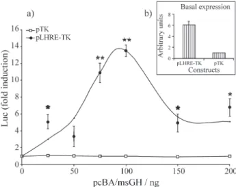
quantities of the GH expressing construct pcβA/msGH led to increased expression of the pLHRE-TK/luc in the BF-2 cells, while none of the pcβA/msGH concentrations tested affected the luciferase induction of the control pTK/luc vector (Figure 1a). A basal activity of STAT5 is likely to be present in BF-2 cells since luciferase expression was found to be higher in cells transfected with the LHRE reporter construct alone when compared to cells transfected with the pTK/luc (Figure 1b). The luciferase activity of the pLHRE-TK/luc increased steadily with transfected concen-tration of the pcβA/msGH construct up to a maximum of 13.5 fold induction. Further addition of the GH expressing construct reduced the LHRE-TK promoter induction there-after. The bell-shaped activation curve suggests that, in fish as in mammals, STAT5b induction is dependent on dimerisation of the GH receptor (Ilondoet al., 1994). To demonstrate the secretion of the msGH, cells were transfected without msGH but exposed for 4 h to the supernatant from cells transfected with pcβA/msGH (0 to 200 ng DNA). This supernatant was indeed able to signifi-cantly induce luciferase expression to levels up to 70% of the pcβA/msGH transfected cell, (p < 0.05, t-test, data not shown). This expression cannot be explained by leftover DNA/FuGENE 6 mixture in the supernatant since 4 h is not sufficient to observe any transfection in BF-2 cells (data not shown).
We next injected the pcβA/msGH construct together with the pLHRE-TK/luc into one-cell stage tilapia eggs. As shown in Figure 2, our results demonstrate a highly signifi-cant increase of luciferase induction (10.3-fold) in a group of fish co-injected with pcβA/msGH and pLHRE-TK/luc when compared to the control group (p < 0.001, Mann-Whitney U-test). Again, no significant induction of luciferase expression was observed in embryos injected with the pTK/luc vector. These observations are in agree-ment with the data obtained fromex vivoexperiments and prove that the marine silverside GH driven by the carpβ-actin promoter (pcβA/msGH) is able to induce the GH signalling pathway to high levels both in fish fibroblast cells and in tilapia embryos.
Activation of the LHRE by rat STAT5b
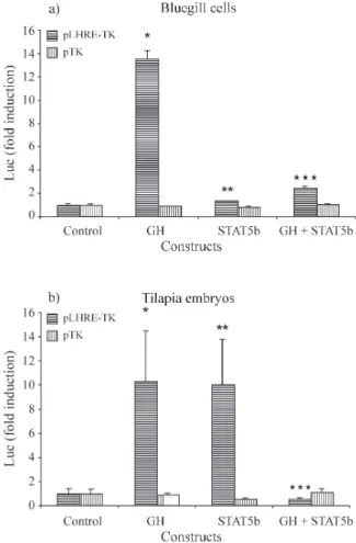
Overexpression of STAT5b induces a slight but sig-nificant increase in luciferase expression in LHRE-TK/luc transfected cells (1.4-fold induction, p < 0.05, t-test, Figure 3a). The overexpression of STAT5b in fish embryos pro-duced a highly significant 10-fold induction of luciferase over the control batches of injected embryos (p < 0.001, Mann-Whitney U-test, Figure 3b). This high level of induc-tion observed in tilapia embryos suggests that rat STAT5b is activated by endogenous fish growth factors and is fully functional in fish. To the best of our knowledge, this is the first demonstration that a mammalian STAT5b is func-tional in a teleost. Taken together with the results on
activa-tion of the JAK/STAT signal transducactiva-tion pathway in mammalian cells by the tilapia prolactin receptor (Sohmet al., 1998) or the zebrafish STAT1 (Oates et al., 1999b),
Figure 2- Luciferase expression induced by the GH expressing vector in tilapia embryos. Control: pLHRE-TK/luc or pTK/luc vector injected alone. GH: pcβA/msGH was co-injected either with the pLHRE-TK/luc vector or the pTK/luc vector. Fold induction, see Figure 1. The asterisk in-dicates a significant difference with the group which was injected with the corresponding reporter vector alone:*= p < 0.001, Mann-Whitney U-test, n = 32 to 78.
these results indicate that JAK/STAT transduction path-ways are remarkably similar in fish and in mammals, al-though we know now that the overall homology between teleost GHRs and GHRs of other species is in fact rather low (≈40 % in aa identity; Calduch-Gineret al., 2001; Lee
et al., 2001).
Co-expression of msGH and STAT5b
When the pcβA/msGH and pCMV/STAT5b-GFP constructs were co-transfected in BF-2 cells an induction of 2.4-fold over the pLHRE-TK/luc control (p < 0.01, t-test) and 1.8-fold over STAT5b alone was observed, (p < 0.05, t-test), but there was a 5-fold inhibition of luciferase ex-pression (p < 0.005, t-test) when compared to the msGH
transfected cells (Figure 4a). These results are in contrast to the observations made in mammalian fibroblasts where GH and STAT5a were found to act synergistically (von Laueet al., 2000). Interestingly, tilapia embryos co-injected with both GH and STAT5b expression vectors demonstrate an even stronger inhibition (≈20x) of the LHRE-TK/luc ex-pression when compared to the pcβA/msGH injected em-bryos (p < 0.001, Figure 4b). In mammals, STAT5a/b interact and are regulated by several growth factors and hormones that can down regulate their signalling (Yoshi-mura, 1998; Favre-Young et al., 2000; von Laue et al., 2000). However, the most likely candidates for the down regulation of STAT5b signalling in the presence of GH are the suppressors of cytokine signalling or SOCS proteins which have been shown to regulate the signal transduction of GH receptors (Adamset al., 1998; Favreet al., 1999). Moreover, in mice, STAT5 is implicated in the
Figure 3- Luciferase expression induced by the STAT5b expressing vec-tor in fish fibroblast cells (a) and in tilapia embryos (b). Control: pLHRE-TK/luc or ppLHRE-TK/luc vector transfected alone. STAT5b: pCMV/STAT5b co-transfected with the pLHRE-TK/luc vector or the pTK/luc vector. The fold induction and asterisks are defined as in Figure 1.*= p < 0.05, t-test, n = 3,**= p < 0.001, Mann-Whitney U-test.
GH-dependent transcription of SOCS-3, which in turn in-hibits GH ability to induce the transcription of the Spi 2.1 promoter (Adamset al., 1998). Our results clearly show that the overexpression of STAT5b can counteract effects of GH overexpression in fish, but the precise mechanism of this down regulation of STAT5 responsive elements by overexpressed-STAT5b remains to be clarified.
This down regulation of the GH signalling could have important consequence in fish. Recently, Devlin and col-laborators have shown that in fast growing selected strains of trout, growth hormone overexpression frequently did not improve growth and could even have deleterious effects on the health of the fish (Devlinet al., 2001). It is likely that genetic selection may act to increase plasma GH levels but may also modify GH signal transduction pathways. In re-gard to our results, it is probable that GH-dependent STAT5 signal transduction pathways are at least partially responsible for the deleterious effects of GH overexpres-sion observed in genetically selected fish.
At a time when increasing concern regarding trans-genesis is being expressed, a proper understanding of the GH transduction pathway in fish is needed. The production of fish lineages carrying reporter gene constructs from the best characterised mammalian transduction signal path-ways could already bring more understanding of GH trans-duction signal in fish. STAT responsive elements seem good candidates to start with and would allow further in-vestigations of GH, prolactin and cytokines transduction signal pathwaysin vivoin teleosts.
Acknowledgements
The authors wish to thank Dr. Christin Carter-Su (University of Michigan Medical School, Michigan, USA) and Dr. Li-yuan Yu-Lee (Baylor College of Medicine, Houston, USA) who kindly provided the pCMV/STAT5b-GFP, Dr. Jöelle Finidori (Hôpital Necker-Enfants Malades, Paris, France) for the gift of the pLHRE-TK/luc and the pTK/luc plasmids, and Dr. Suzanne Brooks (University of Southampton, UK) for providing the plasmid pP3PA-cβActin/GPF. We are also grateful to Rosemary Bell for her help with the scintillation counter. L.F. Marins was sup-ported by a CAPES scholarship of the Brazilian Ministry of Education.
References
Adams TE, Hansen JA, Starr R, Nicola NA, Hilton DJ and Billestrup N (1998) Growth hormone preferentially induces the rapid, transient expression of SOCS-3, a novel inhibitor of cytokine receptor signaling. J Biol Chem 273:1285-1287. Alam MS, Lavender FL, Iyengar A, Rahman MA, Ayad HH,
Lathe R, Morley SD and Maclean N (1996) Comparison of the activity of carp and rat b-actin gene regulatory sequences in tilapia and rainbow trout embryos. Mol Reprod Dev 45:117-122.
Baudler M, Schartl M and Altschmied J (1999) Specific activation of a STAT family member inXiphophorusmelanoma cells. Exp Cell Res 249:212-220.
Calduch-Giner J, Duval H, Chesnel F, Boeuf G, Perez-Sanchez J and Boujard D (2001) Fish growth hormone receptor: mo-lecular characterization of two membrane-anchored forms. Endocrinology 142:3269-3273.
de la Fuente J, Guillén I, Martínez R, and Estrada MP (1999) Growth regulation and enhancement in tilapia: basic re-search findings and their applications. Genet Anal 15:85-90. Devlin RH, Biagi CA, Yesaki TY, Smailus DE and Byatt JC
(2001) Growth of domesticated transgenic fish. Nature 409:781-782.
Devlin RH, Yesaki TY, Biagl CA, Donaldson EM, Swanson P and Chan W-K (1994) Extraordinary salmon growth. Nature 371:209-210.
Du S-J, Gong Z, Fletcher GL, Shears MA, King MJ, Idler DR and Hew CL (1992) Growth enhancement in transgenic Atlantic salmon by the use of an “all fish” chimeric growth hormone gene construct. Bio-Technol 10:176-181.
Ehret GB, Reichenbach P, Schindler U, Horvath CM, Fritz S, Nabholz M and Bucher P (2001) DNA binding specificity of different STAT proteins: comparison ofin vitrospecificity with natural target sites. J Biol Chem 276:6675-6688. Favre H, Benhamou A, Finidori J, Kelly PA and Edery M (1999)
Dual effects of suppressor of cytokine signaling (SOCS-2) on growth hormone signal transduction. FEBS Lett 453:63-66.
Favre-Young H, Dif F, Roussille F, Demeneix BA, Kelly PA, Edery M and de Luze A (2000) Cross-talk between signal transducer and activator of transcription (Stat5) and thyroid hormone receptor-β1 (TRβ1) signaling pathways. Mol Endocrinol 14:1411-1424.
Herrington J, Rui L, Luo G, Yu-Lee L and Carter-Su C (1999) A functional DNA binding domain is required for growth hor-mone-induced nuclear accumulation of Stat5b. J Biol Chem 274:5138-5145.
Ilondo MM, Damholt AB, Cunningham BA, Wells JA, Meyts P and Shymko RM (1994) Receptor dimerization determines the effects of growth hormone in primary rat adipocytes and cultured human IM-9 lymphocytes. Endocrinology 6:2397-2403.
Kopchick JJ and Andry JM (2000) Growth hormone (GH), GH re-ceptor, and signal transduction. Mol Genet Metab 71:293-314.
Lee LT, Nong G, Chan YH, Tse DL and Cheng CH (2001) Molec-ular cloning of a teleost growth hormone receptor and its functional interaction with human growth hormone. Gene 270:121-129.
Leu JH, Yan SJ, Lee TF, Chou CM, Chen ST, Hwang PP, Chou CK and Huang CJ (2000) Complete genomic organization and promoter analysis of the round-spotted pufferfish JAK1, JAK2, JAK3, and TYK2 genes. DNA Cell Biol 19:431-446. Liu N, Mertani HC, Norstedt G, Törnell J and Lobie PE (1997)
Mode of the autocrine/paracrine mechanism of growth hor-mone actions. Exp Cell Res 237:196-206.
Moutoussamy S, Kelly PA and Finidori J (1998) Growth-hormone-receptor and cytokine-receptor-family signaling. Eur J Biochem 5:1-11.
AF (1999a) Gene duplication of zebrafish JAK2 homologs is accompanied by divergent embryonic expression patterns: only jak2a is expressed during erythropoiesis. Blood 94:2622-2636.
Oates AC, Wollberg P, Pratt SJ, Paw BH, Johnson SL, Ho RK, Postlethwait JH, Zon LI and Wilks AF (1999b) Zebrafish stat3 is expressed in restricted tissues during embryogenesis and stat1 rescues cytokine signaling in a STAT1-deficient human cell line. Dev Dynam 215:352-370.
Rahman MA, Hwang G-L, Razak SA, Sohm F and Maclean N (2000) Copy number related transgene expression and mo-saic somatic expression in hemizygous and homozygous transgenic tilapia (Oreochromis niloticus). Transgenic Res 9:417-427.
Rahman MA and Maclean N (1999) Growth performance of transgenic tilapia containing an exogenous piscine growth hormone gene. Aquaculture 173:333-346.
Rycyzyn MA, Wilson MR, Bengten E, Warr GW, Clem LW and Miller NW (1998) Mitogen and growth factor-induced acti-vation of a STAT-like molecule in channel catfish lymphoid cells. Mol Immunol 35:127-136.
Sohm F, Pezet A, Sandra O, Prunet P, de Luze A and Edery M (1998) Activation of gene transcription by tilapia prolactin variants tiPRL188 and tiPRL177. FEBS Lett 438:119-123. Sotiropoulos A, Moutoussamy S, Renaudie F, Clauss M, Kayser
C, Gouilleux F, Kelly PA and Finidori J (1996) Differential activation of Stat3 and Stat5 by distinct regions of the growth hormone receptor. Mol Endocrinol 10:998-1009. Storz P, Döppler H, Horn-Müller J, Groner B, Pfizenmaier K and
Müller G (1999) A cellular reporter assay to monitor insulin receptor kinase activity based on STAT5-dependent lucifer-ase gene expression. Anal Biochem 276:97-104.
Takeda K and Akira S (2000) STAT family of transcription fac-tors in cytokine-mediated biological responses. Cytokine Growth FR 11:199-207.
von Laue S, Finidori J, Maamra M, Shen X-Y, Justice S, Dobson PRM and Ross RJM (2000) Stimulation of endogenous GH and interleukin-6 receptors selectively activates different Jaks and Stats, with a Stat5 specific synergistic effect of dexamethasone. J Endocrinol 165:301-311.