IgE expression on the surface of B1 and
B2 lymphocytes in experimental murine
schistosomiasis
1Departamento de Histologia e Embriologia, Instituto de Ciências Biomédicas, 2Programa Avançado de Biologia Celular Aplicada à Medicina,
Hospital Universitário Clementino Fraga Filho,
Universidade Federal do Rio de Janeiro, Rio de Janeiro, RJ, Brasil F.L. Oliveira1,
A.M.Aguiar1,
R. Borojevic1,2 and
M.C. El-Cheikh1
Abstract
In a previous study we monitored the distribution and phenotype expression of B1 cells during the evolution of experimental murine schistosomiasis mansoni and we proposed that the B1 cells were heterogeneous: a fraction which originated in the spleen and followed the migratory pathway to mesenteric ganglia, while the other was the resident peritoneal B1-cell pool. In the present study, we have ad-dressed the question of whether these two B1-lymphocyte populations are involved in the production of the late Ig isotype IgE, which is present in high levels in schistosomal infection. Lymphocyte expres-sion of surface markers and immunoglobulins were monitored by immunofluorescence flow cytometry. Both in the spleen and mesen-teric ganglia, the B1 and B2 cells were induced to switch from IgM to IgE in the early Th2-dominated phase of the disease, with an increase of IgE in its later phases. Conversely, peritoneal B1-IgM+ switched to
the remaining IgE+ present in high numbers in the peritoneal cavity
throughout the disease. We correlated the efficient induction of the expression of late Ig isotypes by B1 cells with high levels of inflam-matory cytokines due to the intense host response to the presence of worms and their eggs in the abdominal cavity. In conclusion, B1 cells have a different switch behavior from IgM to IgE indicating that these cell sub-populations depend on the microenvironment.
Correspondence M.C. El-Cheikh Caixa Postal 68021 21941-970 Rio de Janeiro, RJ Brasil
Fax: +55-21-2562-6483 E-mail:
Presented at the XI Congresso Brasileiro de Biologia Celular, Campinas, SP, Brazil, July 15-18, 2004.
Research supported by PRONEX, FINEP, CNPq, FAPERJ, and the Associação Técnico-Científica Paul Ehrlich.
Received June 24, 2004 Accepted March 11, 2005
Key words
•Schistosomiasis •B lymphocytes •Immunoglobulin switch
Introduction
B1 lymphocytes are defined as a subset of B cells that express the surface pan-T marker CD5 together with the B-cell surface markers B220lo, IgMhi, and IgDlo. In contrast
to standard B cells (B2 lymphocytes), they are CD23-. When located in celomic
cavi-ties, which are the major site of their homing
and proliferation, they are Mac1+. In
addi-tion to their phenotypic differences, B1 cells are distinguished from the major B-lympho-cyte population, the B2 cells, by their differ-ent origin, localization and immunoglobulin repertoires, presumably reflecting different functions in the immune system (reviewed in Refs. 1-3).
are the major if not the sole B-cell popula-tion in the fetal liver and omentum. In adult mice they predominate in the pleural and peritoneal cavities. They are frequent in the lamina propria, but represent only a small percentage of B cells in the spleen, and are essentially absent from lymph nodes and Peyer’s patches. B1 cells are maintained by self-replenishment, i.e., by division of fully mature cells. In contrast, B2 cells are replen-ished throughout life by proliferation and differentiation of immature progenitors in bone marrow (1). In addition to these appar-ently separate life histories and characteris-tics of the B1 and B2 lineages, it has been shown that CD5 expression can be induced in mature B2 cells, and the alternative origin of B1 cells among the cells of the B2 lineage has been subsequently proposed (4,5). At present, heterogeneous properties of the B-cell compartment with a double origin have been suggested, depending on the quality of interaction with antigens and with other cells engaged in the immune response (6).
B1 cells produce antibodies that are fre-quently polyreactive and of low affinity. These “natural antibodies” produced spon-taneously under low immunization condi-tions are often autoreactive, such as anti-bodies for phosphatidyl choline, DNA and immunoglobulins, or react with common bacterial carbohydrate antigens like phos-phorylcholine. Most B1 cells produce IgM.
In vitro studies have indicated the inability of B1 cells to produce more mature isotypes, but in vivo the B1 population as a whole can produce all Ig isotypes (1). The two subsets of B1 cells, the B1a and B1b cells, are distin-guished by the presence of CD5 in B1a and its absence in B1b cells, but despite their similar origin and characteristics they switch to different Ig isotypes. In response to IL-5, B1a cells tend to produce IgG2a, IgG2b and IgG3, while B1b cells produce IgG1 and IgE in response to IL-9 (7,8).
In view of their early appearance and broad reactivity, as well as their strategic
submucosal location, B1 cells are consid-ered to be part of the natural resistance to the intestinal flora (9). They can also participate in defense against viral infections (10), and in protection or recovery from experimental infections with large parasites such as Brugia
perivascular inflammation of the mesenteric venous system gives rise to plasmacytogenic foci, where immature plasmacytes are ob-served close to the blood vessel walls and mature ones are located on the surface of the mesenterium, predominantly secreting IgG and IgM (19,20).
In a previous study, we monitored the distribution and phenotype expression of B1 cells during the evolution of experimental murine schistosomiasis mansoni (21). We found no evidence of an increase of B1 cells in the peritoneum, nor a mobilization of B1 cells expressing the peritoneal phenotype (CD5lo, IgMhi) in the tissues involved by the
infection, despite the systemic increase of the B-cell response and overall hyperplasia of peritoneal cells. Conversely, we detected in the spleen a subpopulation of B lympho-cytes characterized as CD5hi and IgMlo, which
appeared subsequently in Peyer’s patches and mesenteric ganglia, with a progressive acquisition of Mac-1 expression. We pro-posed that the B1-cell population may be heterogeneous in schistosomiasis: a fraction is originated in the spleen and follows the migratory pathway to mesenteric ganglia, while the other is the resident peritoneal pool that is apparently not increased in situ. In the present study, we have addressed the question of whether these two lymphocyte populations (the splenic and the peritoneal pools) are involved in the production of IgE antibodies, which are produced in high lev-els in schistosomal infection.
Material and Methods
Schistosomal infection
Inbred C3H/HeN mice of both sexes were obtained from the colony bred at the Federal University of Rio de Janeiro and infected by transcutaneous penetration of 40 S. mansoni
cercariae (BH strain, Oswaldo Cruz Insti-tute, Rio de Janeiro, RJ, Brazil). Mice were studied after 45-50 and 90-95 days of
infec-tion, corresponding to the acute phase and the beginning of the chronic phase of the disease, respectively (20). Uninfected age-and sex-matched mice were used as con-trols.
Preparation of cell suspensions
Cell suspensions from normal and in-fected mice were obtained ex vivo by stand-ard mechanical dissociation of spleen, mes-enteric ganglia and Peyer’s patches. The peritoneal cavity was washed with phos-phate-buffered saline (PBS) and red blood cells were lysed with Gey’s solution. After lysis, the cell suspensions were washed twice with PBS, pH 7.2, containing 3% fetal bo-vine serum, and cell concentration was ad-justed to 1 x 10⁶ cells/ml. In order to saturate
the Fc receptors, the cells were incubated with normal mouse serum for 10 min before adding the specific monoclonal antibodies.
Immunofluorescence
The presence of cell surface markers on lymphoid cells was monitored with the fol-lowing monoclonal antibodies for three-color analysis: fluorescein isothiocyanate-conju-gated anti-IgM (Sigma, St. Louis, MO, USA) and anti-IgE (Pharmingen, San Diego, CA, USA); phycoerythrin-conjugated anti-B220 (Pharmingen), and quantum red-conjugated anti-CD5 (Sigma). Staining was done sepa-rately for each monoclonal antibody for 30 min on ice, followed by washing with cold PBS.
Data acquisition and analysis
com-pensation was performed using single-color-stained cells from each organ separately. Cell debris and dead cells were excluded according to the light scatter parameters, and the gate including the lymphocyte popu-lation was named R1. Data are reported as dot plots and histograms.
Statistical analysis
Data were analyzed statistically by the Tukey multiple comparison test, with the level of significance set at P < 0.05.
Results
Expression of IgE on the surface of B
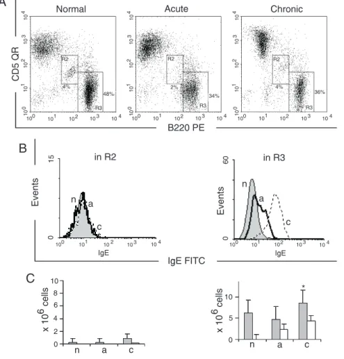
cells was monitored in the mesenteric gan-glia, Peyer’s patches, spleen, and peritoneal cavity of normal and schistosome-infected mice during the evolution of the disease from the acute to the chronic phase (Figures 1 to 4). B1, B2 and T lymphocytes were defined according to differential expression of B220 and CD5 (Figures 1-5A).
The labeling pattern of the cells that did not produce IgE was recorded in all experi-ments (Figures 1B to 4B) and pooled, and compared to the pattern of cells that reacted with anti-IgE antibodies. In agreement with reported levels of Th2 cytokines (14), mes-enteric ganglia showed increased IgE+ cell
numbers from the acute to the chronic phase of the disease (Figure 1B). The median
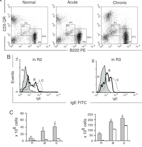
ues were moderately increased in diseased animals, suggesting that rare cells presented low to intermediate IgE fluorescence inten-sity on their surface. This low or intermedi-ate reactivity is not suggestive of immuno-globulin secretion, but indicates that this B-cell subpopulation has the genetic machin-ery able to perform all the gene recombina-tions necessary to switch from IgM to IgE. Due to the low number of events, the differ-ence between the groups was only sugges-tive (P = 0.08). Conversely, in Peyer’s patches, only B2 lymphocytes were involved in the switching from IgM to IgEisotype (Figure 2B and C), with a progressive in-crease from the acute to the chronic phase indicative of switching from the IgM isotype. In schistosomiasis, the spleen receives a permanent stimulation of the immune
sys-tem, evolving to splenomegaly associated with an increase in B-cell numbers and their polyclonal activation (13). In normal spleens, IgE+ cells were very rare in all the B-cell
subsets studied (Figure 3B), but their num-ber increased in the acute and chronic phases of the disease. The number of B1-IgE+ cells
remained stable in infected mice (Figure 3C). In contrast, the total B2 and B2-IgE+
cells increased during the evolution of the disease (Figure 3B and C). In agreement with the results shown for mesenteric gan-glia, B1-IgE+ cells were found in the spleen,
participating with B2 lymphocytes in the production of IgE, present in high concen-trations in serum of mice during both the acute and chronic phases of the disease.
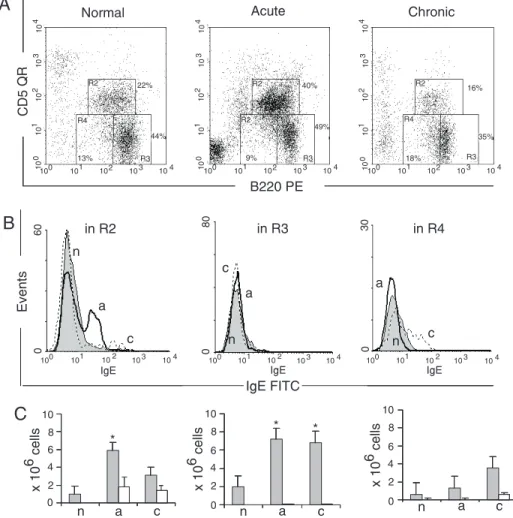
The peritoneal cavity is the major site of maintenance and production of B1 cells. In
Figure 2. Surface expression of B220, CD5 and IgE on cells from Peyer’s patches. A, Dot plot of cells in the lymphocyte gate. The R2 and R3 regions represent the B1 and B2 cells, respectively. B, Intensity of IgE on the B-cell sur-face. C, Total cell numbers (filled columns) and IgE+ cells (open columns). Data are reported as means ± SEM and are repre-sentative of five independent ex-periments, each carried out in 2 normal mice (n), 5 mice with acute infection (a) and 5 with chronic infection (c). *P < 0.05 for B-IgE+ cells compared to to-tal B cells (Tukey multiple com-parison test).
normal adult mice B1 lymphocytes repre-sent up to 30% of peritoneal cells. B1a cells were increased during the acute phase of the disease, and in the chronic phase their rela-tive number decreased compared to normal mice. According with previously published data (21), the total number of peritoneal B1 cells remained constant throughout the dis-ease as a consequence of the incrdis-eased num-ber of peritoneal cells. B1a-IgE+ cells were
negligible in normal mice (Figure 4B and C). Mice with acute infection had an in-creased B1a-IgE+ cell fraction,which
re-mained at the same level during chronic infection. B1b cells (Figure 4B) were nega-tive in normal mice and in mice with acute infection, but in the chronic phase, they were split into two clearly distinct IgE+ and IgE
-subsets (Figure 4B and C). The conventional
B2 lymphocytes present in the peritoneal cavity were not involved in IgE production in the studied groups (Figure 4B and C). The increase of IgE+ cells was thus due to the
appearance of a new increasing IgE+ cell
population in the peritoneal cavity of in-fected mice, which did not exist in normal mice.
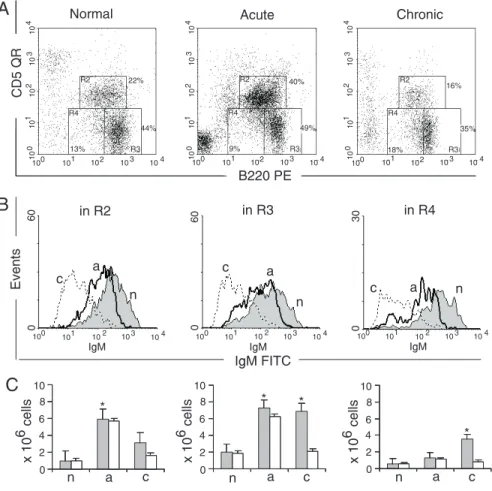
We have addressed the question of the presence of IgM+ cells in this cavity in order
to monitor the kinetics of the switch of IgM+
cells toIgE+ cells (Figure 5A and B). IgM+
cells were the vast majority in the entire peritoneal B-cell population of normal mice (Figure 5B). The evolution of the disease was concomitant with a progressive decrease of IgM+ cells in all B-cell populations.
Moni-toring IgM expression identified two sub-populations among IgM+ cells: a very bright
and another one with slightly lower IgM expression (Figure 5B). We understand that the latter population corresponds to the IgM cells switching to the expression of IgE. In the acute phase, an increase of IgMlo cells
was concomitant with the appearance of IgE+
cells (Figures 4B and 5B) indicating the isotype switching. In the chronic phase, a larger part of B1 cells were IgM- (Figure
5B), indicating a full switch to more mature Ig isotypes, as demonstrated by the presence of B1a and B1b-IgE+ cells (Figure 4B). A
smaller IgMlo population potentially
corre-sponds to the cells switching to other iso-types. It is notable that no IgMhi cells were
found in this phase of infection, indicating that self-replenishment of IgM-producing cells did not occur. Low quantities of IgM+
cells in this phase (Figure 5C) were
appar-ently engaged in switching to more mature isotypes, while the constant presence of IgE-producing cells in the peritoneal cavity may correlate with the permanent granulomatous and diffuse inflammation of the viscera in response to the long-lasting presence of worms and their eggs in the tributaries of the mesenteric venous system. In conclusion, we understand that IgE+ cell kinetics in the
peritoneal cavity has different patterns and is controlled by distinct molecular mechan-isms.
Discussion
B1 cells can produce IgM and IgG3 spon-taneously in the absence of T cells. This production is considered to be part of the natural resistance to exogenous antigens,
and can participate in a short-term response of the immune system to infectious agents (22-24). T cells can increase the production of these isotypes, presumably in an antigen-dependent manner, as part of the adaptive immune response. The production of IgG1 by B1 cells is totally dependent on T cells.
Production of other immunoglobulins by B1 cells can be either independent or T-dependent, and these antibodies strongly contribute to the IgA-secreting plasma cells of the intestinal lamina propria (25,26). A significant difference between the response of splenic and peritoneal B cells to IL-5 has been reported (27-29). We observed simul-taneous generation of B1-IgE+ cells in the
spleen and in the peritoneal cavity during the acute phase of the disease when the overall pattern of the immune response is
Th2-domi-nated. While the great majority of splenic cells that switched to IgE+ were B2, in the
peritoneal cavity IgE+ cells apparently
origi-nated in the B1-IgM+-cell population,
sug-gesting the generation and expansion of a new cell population in response to the infec-tion. Schistosome soluble egg antigens are known to elicit a very strong Th2 response, with high levels of IL-4 and IL-5 (15,30).
In vitro, the induction of IgE switch in B1 cells has been shown to be difficult, requir-ing very high levels of cytokines (29), in studies based on the production of IgM, IgG1, IgE, and IgA by B1 (CD23-) and B2
lymphocytes (CD23+). Both subpopulations
were obtained by sorting from the spleen and peritoneal cavity and cultured with lipo-polysaccharide and IL-4. Only the B1 and B2 cells purified from spleen were capable
References
1. Herzenberg LA, Baumgarth N & Wilshire JA (2000). B-1 cell origins and VH repertoire determination. Current Topics in Microbiology and Immunology, 252: 3-13.
2. Hayakawa K, Shinton SA, Asano M & Hardy RR (2000). B-1 cell definition. Current Topics in Microbiology and Immunology, 252: 15-22.
3. Fagarasan S, Watanabe N & Honjo T (2000). Generation, expan-sion, migration and activation of mouse B1 cells. Immunological Reviews, 176: 205-215.
4. Wortis HH (1992). Surface markers, heavy chain sequences and B cell lineages. International Review in Immunology, 8: 235-246. 5. Haughton G, Arnold LW, Whitmore AC & Clarke SH (1993). B1 cells
are made, not born. Immunology Today, 14: 84-87.
6. Potter M & Melchers F (2000). Opinions on the nature of B1 cells and their relationship to B cell neoplasia. Current Topics in Microbi-ology and ImmunMicrobi-ology, 252: 307-324.
7. Takatsu K, Takaki S, Hitoshi Y, Mita S, Katoh S, Yamaguchi N & Tominaga A (1992). Cytokine repertoires on Ly-1 B cells. IL-5 and its receptor system. Annals of the New York Academy of Sciences, 651: 241-258.
8. Vink A, Warnier G, Brombacher F & Renauld JC (1999). Interleukin 9-induced in vivo expansion of the B1 lymphocyte population. Jour-nal of Experimental Medicine, 189: 1413-1423.
9. Murakami M, Tsubata T, Shinkura R, Nisitani S, Okamoto M, Yoshioka H, Usui T, Miyawaki S & Honjo TJ (1994). Oral administra-tion of lipopolysaccharides activates B1 cells in the peritoneal cavity and lamina propria of the gut and induces autoimmune symptoms in an autoantibody transgenic mouse. Journal of Experimental Medi-cine, 180: 111-121.
10. Ochsenbein AF, Fehr T, Lutz C, Suter M, Brombacher F, Hengartner H & Zinkernagel RM (1999). Control of early viral and bacterial distribution and disease by natural antibodies. Science, 286: 2156-2159.
11. Paciorkowski N, Porte P, Shultz LD & Rajan TV (2000). B1 B lymphocytes play a critical role in host protection against lymphatic filarial parasites. Journal of Experimental Medicine, 191: 731-736. 12. Gaubert S, Costa AV, Maurage CA, Lima ECS, Fontaine J, Lafitte S,
Minoprio P, Capron A & Grzych JM (1999). X-linked immunodefi-ciency affects the outcome of Schistosoma mansoni infection in the murine model. Parasite Immunology, 21: 89-101.
13. El-Cheikh MC, Dutra HS, Minóprio P & Borojevic R (1994). Increase of B-lymphocyte number and activity during experimental murine schistosomiasis mansoni. Brazilian Journal of Medical and Biologi-cal Research, 27: 1605-1617.
14. Grzych JM, Pearce E, Cheever A, Caulada ZA, Caspar P, Heiny S, Lewis F & Sher A (1991). Egg deposition is the major stimulus for
of producing all the tested isotypes, includ-ing high levels of IgE. Here we demon-strated in vivo the presence of B1-IgE+ in
infected mice. Although B1-IgE+ were
rela-tively rare compared to B2-IgE+ cells, they
showed intermediate to high fluorescence intensity, suggesting that both cell types par-ticipated in the production of IgE, known to be high in this disease (31). In vitro, perito-neal B1a cells were able to produce IgG1, but only rarely switched further to the IgE isotype, indicating that this second step re-quires additional stimuli (32). Moreover, the rare B1a cells that were reactive for IgE on the surface did not produce the antibody to the supernatant. In our model, the peritoneal environment was able to fulfill these re-quirements, generating a considerable quan-tity of IgE-producing B1 cells, particularly so during the acute phase of the disease, consistently with the high level of Th2 cyto-kines in this phase.
Our observation of the relatively large number of B1 cells switching to IgE+ cells in vivo potentially corresponds to the
associa-tion between high cytokine levels and ap-propriate presentation of antigens in the granulomatous and diffuse inflammatory reactions in the abdominal cavity, and in the downstream sites of the antigen circulation in the spleen. The presence of B-IgE+ cells in
all the compartments studied agrees with the high concentration of IgE in the serum of infected mice in the late chronic phase of the disease (normal mice = 1.07 ± 1.01 µg/ml, versus infected mice = 69.4 ± 43.3 µg/ml; El-Cheikh MC, unpublished data).
In the experimental model employed here, the peritoneal B1-IgE+ cells did not decrease
during the evolution of the disease, indicat-ing that their migration and homindicat-ing are dif-ferent from those observed for IgA+ cells
the production of Th2 cytokines in murine schistosomiasis mansoni. Journal of Immunology, 146: 1322-1327.
15. Sher A, McIntyre S & Lichtenberg F (1977). Schistosoma mansoni: kinetics and class specificity of hypergammaglobulinemia induced during murine infection. Experimental Parasitology, 41: 415-422. 16. Bout D, Rousseaux R, Calier Y & Capron A (1980). Kinetics of
classes and sub-classes of total immunoglobulins and specific anti-bodies to Schistosoma mansoni during murine infection. Parasitol-ogy, 80: 247-256.
17. Jarret E & Bazin H (1974). Elevation of total serum IgE in rats following helminth parasite infection. Nature, 251: 613-614. 18. Weinberg DF, Baldo-Correa E, Lenzi HL & Borojevic R (1992).
Schistosoma mansoni: peritoneal plasmacytogenesis and polypoid transformation of mesenteric milky spots in infected mice. Experi-mental Parasitology, 74: 408-416.
19. Lenzi HL, Oliveira DN, Pelajo-Machado M, Borojevic R & Lenzi JA (1996). Coelom-associated lymphomyeloid tissue (milky spots): site of lymphoid and myelomonocytic cell generation. Brazilian Journal of Medical and Biological Research, 29: 19-24.
20. Borojevic R, Nicola MH & Santos-da-Silva C (1984). Modulation of macrophage and polymorphonuclear granulocyte inflammatory re-actions in experimental murine Schistosomiasis mansoni. Cellular and Molecular Biology, 30: 37-42.
21. El-Cheikh MC, Bonomo AC, Rossi MID, Pinho MFB & Borojevic R (1998). Experimental murine schistosomiasis mansoni: modulation of the B1 lymphocyte distribution and phenotype expression. Immu-nobiology, 199: 51-62.
22. Murakami M & Honjo T (1995). Involvement of B1 cells in mucosal immunity and autoimmunity. Immunology Today, 16: 534-539. 23. Martin F & Kearney JF (2001). B1 cells: similarities and differences
with other B cell subsets. Current Opinion in Immunology, 13: 195-201.
24. Thurnheer MC, Zuercher AW, Cebra JJ & Bos NA (2003). B1 cells
contribute to serum IgM, but not to intestinal IgA, production in gnotobiotic Ig allotype chimeric mice. Journal of Immunology, 170: 4564-4571.
25. Taki S, Schmitt M, Tarlinton D, Forster I & Rajewsky K (1992). T cell-dependent antibody production by Ly-1 B cells. Annals of the New York Academy of Sciences, 651: 328-335.
26. Whitmore AC, Haughton G & Arnold LW (1992). Isotype switching in CD5 B cells. Annals of the New York Academy of Sciences, 651: 143-151.
27. Coffman RL, Shrader B, Carty J, Mossmann TR & Bond MW (1987). A mouse T cell product that preferentially enhances IgA production. I. Biologic characterization. Journal of Immunology, 139: 3685-3690. 28. Sher A, Coffman RL, Hieny S & Cheever AW (1990). Ablation of eosinophil and IgE responses with anti-IL-5 or anti-IL-4 antibodies fails to affect immunity against Schistosoma mansoni in the mouse. Journal of Immunology, 145: 3911-3916.
29. Foy TM & Waldschmidt TJ (1993). Switching capacity of Fcε RIII-positive and negative murine B cells. European Journal of Immunol-ogy, 23: 3208-3216.
30. Erickson LD, Foy TM & Waldschmidt TJ (2001). Murine B1 B cells require IL-5 for optimal T cell-dependent activation. Journal of Im-munology, 166: 1531-1539.
31. Prevost-Rousseaux R, Bazin H & Capron A (1977). IgE in experi-mental schistosomiasis. I. Serum IgE levels after infection by Schis-tosoma mansoni in various strains of rats. Immunology, 33: 501-505.
32. Siebenkotten G, Esser C, Wabl M & Radbruch A (1992). The murine IgG1/IgE class switch program. European Journal of Immunology, 22: 1827-1834.