Glomerular Immune Deposits Are Predictive
of Poor Long-Term Outcome in Patients with
Adult Biopsy-Proven Minimal Change
Disease: A Cohort Study in Korea
Sung Woo Lee1,2, Mi-Yeon YU2, Seon Ha Baek2, Shin-Young Ahn1,2, Sejoong Kim2, Ki Young Na2,3, Dong-Wan Chae2,3, Ho Jun Chin1,2,3*
1Department of Immunology, Seoul National University Postgraduate School, Seoul, Korea,2Division of Nephrology, Department of Internal Medicine, Seoul National University Bundang Hospital, Seongnam, Korea,3Department of Internal Medicine, Seoul National University College of Medicine, Seoul, Korea
Abstract
Background and Objectives
There has been little published information on risk factors for poor long-term outcome in adult biopsy-proven minimal change disease (MCD).
Methods
Data from sixty-three adult, biopsy-proven primary MCD patients treated at a tertiary univer-sity hospital between 2003 and 2013 were analyzed. Baseline clinical and pathologic fac-tors were assessed for the associations with composite outcome of creatinine doubling, end stage renal disease, or all-cause mortality.
Results
During a median (interquartile) 5.0 (2.8–5.0) years, the composite outcome occurred in 11.1% (7/63) of patients. The rate of glomerular immune deposits was 23.8% (15/63). Patients with glomerular immune deposits showed a significantly lower urine protein creati-nine ratio than those without deposits (P= 0.033). The rate of non-responders was signifi-cantly higher in patients with glomerular immune deposits than in those without deposits (P= 0.033). In patients with deposits, 26.7% (4/15) developed the composite outcome, while only 6.3% (3/48) developed the composite outcome among those without deposits (P= 0.049). In multivariate Cox proportional hazards regression analysis, the presence of glomerular immune deposits was the only factor associated with development of the com-posite outcome (hazard ratio: 2.310, 95% confidence interval: 1.031–98.579,P= 0.047).
OPEN ACCESS
Citation:Lee SW, YU M-Y, Baek SH, Ahn S-Y, Kim S, Na KY, et al. (2016) Glomerular Immune Deposits Are Predictive of Poor Long-Term Outcome in Patients with Adult Biopsy-Proven Minimal Change Disease: A Cohort Study in Korea. PLoS ONE 11(1): e0147387. doi:10.1371/journal.pone.0147387
Editor:Jeff M Sands, Emory University, UNITED STATES
Received:November 4, 2015
Accepted:January 4, 2016
Published:January 22, 2016
Copyright:© 2016 Lee et al. This is an open access article distributed under the terms of theCreative Commons Attribution License, which permits unrestricted use, distribution, and reproduction in any medium, provided the original author and source are credited.
Data Availability Statement:All relevant data are within the paper and its Supporting Information files.
Funding:The authors have no support or funding to report.
Conclusion
Glomerular immune deposits were associated with increased risk of a composite outcome in adult MCD patients. The higher rate of non-responders in patients with deposits might be related to the poor outcome. Future study is needed.
Introduction
Minimal change disease (MCD) accounts for approximately 10% and 30% of cases of adult pri-mary glomerulonephritis (GN) and nephrotic syndrome, respectively [1–3]. MCD is generally
thought to show excellent long-term patient and renal survival [4]. Lee et al. analyzed 1941 patients with GN. The proportion of MCD was 9.6% (187/1941), and 5.9% (11/187) of all-cause mortality was identified [2]. Chou et al. reviewed 580 patients with GN, and MCD accounted for 18.8% (109/580). Among the 109 MCD patients, dialysis dependency and all-cause mortality were identified in 0.9% (1/109) and 3.7% (4/109), respectively [5]. Because of the excellent long-term outcome, there has been little attention focused on the risk factors for poor long-term outcome in patients with MCD.
The pathogenesis of MCD remains unclear. The most accepted explanation is the presence of soluble factors, such as interleukin-13 [6], angiopoietin-like 4 [7] and CD80 [8] which initi-ate glomerular changes in MCD [9]. However, a smaller group shows evidence of humoral antibody or a complement response, predominantly immunoglobulin (Ig) M [10,11] or C1q [12]. These subsets of MCD tend to be resistant to treatment [13,14] and have poor long-term outcomes [13,15]. Previous studies have mainly focused on clinicopathologic features of GN with mesangial IgM [15–19] or C1q deposits [14,20–22], but other humoral factors could also
play a role in the pathogenesis of MCD. Moreover, there has been no report on the clinical sig-nificance of glomerular immune deposits solely in MCD patients. Therefore, we performed the current study to determine whether glomerular immune deposits are associated with poor long-term outcome in adult patients with biopsy-proven MCD.
Materials and Methods
Patients
Between 2003 and 2013, 82 patients were diagnosed with biopsy-proven MCD at Seoul National University Bundang Hospital, a tertiary care hospital. A total of 63 patients were eligi-ble for the study (Fig A inS1 Fig), after excluding those aged<15 years (n = 4), and those with
the diagnosis of focal segmental glomerulosclerosis (FSGS) in a subsequent biopsy (n = 1), inadequate specimen for immunofluorescence (IF) staining (n = 1), and less than 1 year of fol-low-up after the start of the study (n = 13). The study protocol complied with the Declaration of Helsinki and received full approval from the Seoul National University Bundang Hospital institutional review board (IRB number: B-1410/272-119). The need for informed consent was waived because the study did not infringe on patient privacy or health status.
Definitions and measurements
start. ESRD development was determined from the registry database of the Korean Society of Nephrology, and all-cause mortality was determined from the database of Korean Statistics. The first date of admission for kidney biopsy was the start of our study, and the end of the study was a later date between the date of creatinine doubling, ESRD, or death. The study out-come was the composite of creatinine doubling, ESRD, or all-cause mortality.
Body mass index was calculated as weight (kg) per square of height (m2). Complete remis-sion was defined as urine protein creatinine ratio (UPCR)<0.3 g/g or<1+ on urine dipstick
for 3 consecutive measurements. Non-responders were defined as patients without complete remission throughout the treatment course. Relapse was defined as a reappearance of UPCR3.0 g/g or3+ on urine dipstick for 3 consecutive measurements. Serum creatinine was measured using the rate-blanked, compensated kinetic alkaline picrate Jaffe method with an automatic analyzer (Toshiba-200FR, Tokyo, Japan). The estimated glomerular filtration rate (eGFR) was calculated by using the equation of the Chronic Kidney Disease Epidemiology Collaboration [23]. Acute kidney injury was defined as a rise in serum creatinine
by26.5μmol/L within 48 hours, or1.5 times greater than the baseline level[24].
Kidney pathology
For kidney pathology evaluation, all specimens were embedded in paraffin and stained with periodic acid-Schiff, Masson trichrome, methenamine silver, and hematoxylin-eosin.
Glomerular lesions were defined as global sclerosis10.0% [25], increased glomerular size or cellularity, or the presence of glomerular ischemia. Tubulointerstitial lesions were defined as non-normal reports for tubular atrophy, and interstitial inflammation and fibrosis [26]. Vascu-lar lesions were defined as the presence of arterioVascu-lar hyalinosis and arteriosclerosis. The IF study was performed using the classic direct technique with antibodies against 5 items: IgG, IgM, IgA, C3, and C1q. Tubule, interstitium and vasculature near the evaluating glomerulus were reviewed as control tissues to exclude non-specific staining. When the reaction was appropriate, grades more than one positive were given. Pathologists evaluated the IF staining to determine whether linear, granular, peripheral, and mesangial deposits were present in the glomerulus for the 5 items which were reported semi-quantitatively as negative, trace, and 1–3
positive. We transformed the results numerically: negative to 0 point, trace to 0.5 points, and 1–3 positive to 1–3 points. Positive IF staining for the individual items was defined as above 2
points when the results of linear, granular, peripheral, and mesangial deposits in the glomeru-lus were summed. Glomerular immune deposits were defined when any positive IF stainings for the 5 times was present. The specific diagnosis of IgM nephropathy (IgMN) and C1q nephropathy (C1qN) were based on the following criteria: (1) intensity of IF stains scored more than two positive in IgM or C1q, or (2) one positive of intensity in IgM or C1q with evi-dence of mesangial changes in electron microscopic examination [i.e. electron-dense deposits (EDD), mesangial matrix expansion].
Statistical analysis
Values were expressed as median (interquartile range, IQR) for continuous variables and per-centage for categorical variables. The difference was analyzed by the Mann-Whitney U test for continuous variables and the chi-square or Fisher`s exact test for categorical variables. The Kaplan-Meier method was used for the survival curve and the statistical significance was calcu-lated by the log-rank test. For multivariate Cox proportional hazards analysis, variables were chosen byP<0.05 in univariate analysis, along with age and sex. We consideredP<0.05 to
Results
The median (IQR) follow-up duration for 63 patients was 5.0 (2.8–5.0) years; 49.2% (31/63)
were male, and the median (IQR) age at presentation was 43.4 (27.4–65.6) years. During the
follow-up period, 3 patients died from any cause (one from stroke and 2 from unidentified causes). Of 6 patients who developed creatinine doubling, 1 developed ESRD. The composite outcome was identified in 11.1% (7/63). The positive rate of glomerular immune deposits was 23.8% (15/63).
We compared baseline characteristics according to the status of glomerular immune depos-its (Table 1). Although patients with deposits showed a significantly lower UPCR than those without deposits (P= 0.033), the level of serum creatinine and eGFR and the rate of acute
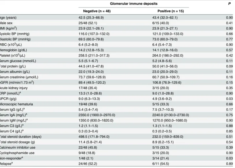
Table 1. Baseline characteristics according to the status of glomerular immune deposits.
Glomerular immune deposits P
Negative (n = 48) Positive (n = 15)
Age (years) 42.5 (25.3–66.9) 43.4 (32.0–62.1) 0.90
Male sex 25/48 (52.1) 6/15 (40.0) 0.41
BMI (kg/m2) 23.9 (22.1–26.1) 23.9 (21.3–27.1) 0.90
Systolic BP (mmHg) 116.0 (107.0–132.0) 121.0 (109.0–133.0) 0.66
Diastolic BP (mmHg) 69.5 (60.0–79.8) 73.0 (60.0–79.0) 0.77
WBC (x103/μL) 6.4 (5.2
–8.0) 6.4 (5.4–7.3) 0.90
Hemoglobin (g/dL) 14.2 (12.8–15.3) 14.1 (12.8–16.0) 0.57
Platelet (x103/μL) 258.5 (211.0
–317.3) 264.0 (186.0–292.0) 0.42
Serum glucose (mmol/L) 5.5 (5.1–6.7) 5.2 (4.8–5.6) 0.11
Total protein (g/L) 44.5 (41.0–47.8) 50.0 (41.0–56.0) 0.09
Serum albumin (g/L) 22.0 (19.3–24.0) 23.0 (20.0–29.0) 0.11
Serum creatinine (μmol/L) 73.7 (59.6–126.9) 60.7 (50.9–109.7) 0.16
eGFR (ml/min/1.73 m2) 89.4 (49.5
–120.2) 106.8 (76.9–129.6) 0.15
Acute kidney injury 17/48 (35.4) 3/15 (20.0) 0.35
CRP (nmol/L)a 13.3 (1.0–28.6) 9.0 (1.0–28.8) 0.90
UPCR (g/g) 9.0 (6.3–13.3) 4.9 (3.6–9.2) 0.03
Microscopic hematuria 19/48 (39.6) 5/15 (33.3) 0.66
Serum IgG (g/L)a 5.4 (3.4
–7.4) 7.5 (3.7–10.3) 0.17
Serum IgA (mg/L)a 2350.0 (1900.0–2970.0) 2240.0 (2130.0–2730.0) 0.75
Serum IgM (mg/L)a 1350.0 (830.0
–1820.0) 1270.0 (950.0–1680.0) 0.90
Serum C3 (g/L)a 1.2 (1.1
–1.5) 1.3 (1.1–1.5) 0.88
Serum C4 (g/L)a 0.3 (0.3
–0.4) 0.3 (0.2–0.5) 0.85
Total steroid duration (days) 498.5 (171.8–794.0) 232.0 (159.0–839.0) 0.51
Total steroid dosage (g) 11.4 (5.8–21.4) 8.9 (6.2–15.1) 0.54
Calcineurin inhibitor use 22/48 (45.8) 5/15 (33.3) 0.39
Cyclophosphamide use 9/48 (18.8) 3/15 (20.0) 0.90
Non-respondera 1/48 (2.1) 3/14 (21.4) 0.03
Relapsea 24/46 (52.2) 6/11 (54.5) 0.89
Values were expressed as median (interquartile range) for continuous variables and n/total (%) for categorical variables. Differences were evaluated by chi square or Fisher`s exact test for categorical variables or the Mann-Whitney U test for continuous variables. BMI, body mass index; BP, blood pressure; WBC, white blood cells; eGFR, estimated glomerularfiltration rate; CRP, c-reactive protein; UPCR, urine protein creatinine ratio.
arepresented incomplete data. The total numbers of negative/positive glomerular immune deposits of CRP, IgG, IgA, IgM, C3 and C4 were 43/14, 40/13,
43/14, 39/13, 45/14, and 45/14, respectively.
kidney injury and microscopic hematuria in the positive glomerular immune deposits group were not different from those in the negative group. The rate of non-responders was signifi-cantly higher in patients with deposits than in those without deposits (P= 0.033).
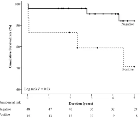
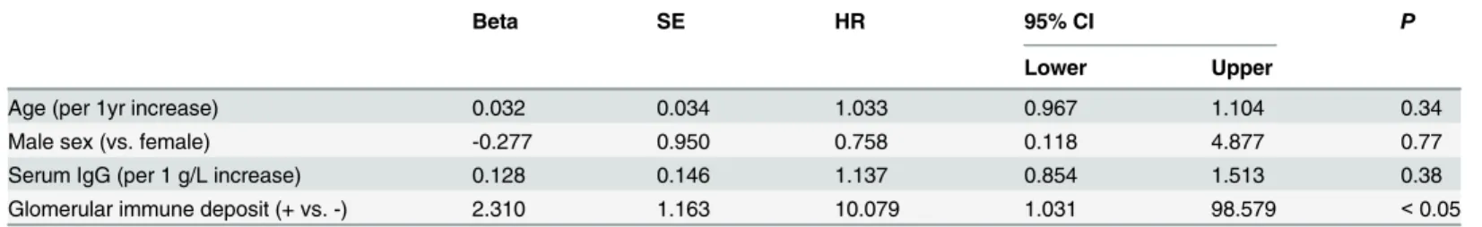
In patients with glomerular immune deposits, 26.7% (4/15) developed the composite out-come, while only 6.3% (3/48) of those without deposits (P= 0.049) developed the composite outcome. In Kaplan-Meier survival curve analysis, the positive glomerular immune deposits group showed significantly shorter survival time than the negative group (Fig 1). The glomeru-lar immune deposits group and serum IgG level were significantly associated with the develop-ment of the composite outcome (Table 2). The confounding effects were adjusted by
multivariate Cox proportional hazards regression analysis (Table 3). The presence of glomeru-lar immune deposits was the only factor associated with the development of the composite out-come (hazard ratio [HR]: 2.310, 95% confidence interval [CI]: 1.031–98.579,P= 0.047).
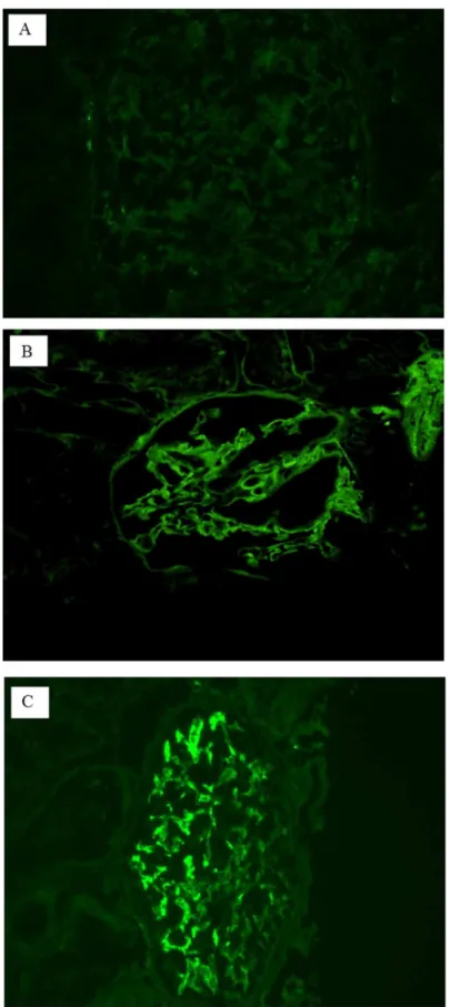
We compared pathologic characteristics according to the status of glomerular immune deposits (Table 4), and there were no differences in lesions of the glomerulus, tubulointersti-tium, and vasculature. Among 15 patients in the positive glomerular immune deposits group, the most common humoral factor deposited was IgM (60%), followed by C1q (26.7%), IgG (26.7%), IgA (20.0%), and C3 (6.7%). Of these 15 patients, 5 and 2 were diagnosed with IgMN and C1qN, respectively. We demonstrated clinical and IF findings in 15 patients in the positive glomerular immune deposits group (Figs2–4). Most IF results showed either a linear
periph-eral (LP) (Fig 2B) or granular mesangial (GM) pattern (Fig 2C). In the positive glomerular immune deposits group, the intensity of deposits was mostly found to be one positive result (Fig 3). Nearly all patients with IgM and C1q deposits showed a GM pattern. All patients with IgA and IgG deposits demonstrated an LP pattern. Of 8 with IgM-GM pattern deposits, 5 were
Fig 1. Kaplan-Meier survival curve according to the status of glomerular immune deposits.Mean (95% CI) survival of negative and positive glomerular immune deposit groups were 4.8 (4.6–5.0) years and 4.1
(3.2–5.0) years, respectively.
diagnosed with IgMN. Of 4 with C1q-GM pattern deposits, 2 were diagnosed with C1qN. Although most of those with a composite outcome and non-responders were identified among patients with either IgM- or C1q-GM pattern deposits, 1 patient with IgG-LP pattern deposits was a non-responder and ultimately died (Fig 4).
Table 2. Factors associated with the composite outcome.
Composite outcome P
Negative (n = 56) Positive (n = 7)
Age (years) 38.7 (26.6–65.6) 50.8 (32.0–80.5) 0.35
Male sex 29/56 (51.8) 2/7 (28.6) 0.43
BMI (kg/m2) 23.9 (22.1
–26.7) 23.2 (21.3–27.7) 0.89
Systolic BP (mmHg) 116.5 (107.5–131.0) 133.0 (105.0–134.0) 0.46
WBC (x103/μL) 6.5 (5.2
–8.0) 6.1 (5.2–6.4) 0.53
Hemoglobin (g/dL) 14.2 (13.0–15.5) 14.1 (12.3–14.8) 0.50
Serum glucose (mmol/L) 5.4 (4.9–6.5) 5.3 (5.1–5.6) 0.49
Serum creatinine (μmol/L) 71.5 (59.6–110.3) 60.7 (21.5–90.1) 0.18
eGFR (ml/min/1.73 m2) 93.2 (51.3
–120.8) 101.9 (68.9–162.8) 0.41
Acute kidney injury 16/56 (28.6) 4/7 (57.1) 0.20
CRP (nmol/L)a 11.4 (1.0
–28.6) 13.3 (1.0–29.5) 0.78
UPCR (g/g) 8.9 (5.8–12.5) 6.2 (3.6–12.1) 0.34
Microscopic hematuria 21/56 (37.5) 3/7 (42.9) 0.90
Serum IgG (g/L)a 5.4 (3.4
–7.5) 10.7 (6.6–12.9) 0.02
Glomerular lesions 32/56 (55.4) 4/7 (57.1) 0.90
Tubulointerstitial lesions 40/56 (71.4) 4/7 (57.1) 0.42
Vascular lesions 19/56 (33.9) 2/7 (28.6) 0.90
Glomerular immune deposits 11/56 (19.6) 4/7 (57.1) <0.05
Total steroid duration (days) 498.5 (161.5–831.0) 292.0 (175.0–338.0) 0.16
Total steroid dosage (g) 10.9 (5.5–21.4) 11.6 (6.9–11.8) 0.74
Calcineurin inhibitor use 25/56 (44.6) 2/7 (28.6) 0.69
Cyclophosphamide use 11/56 (19.6) 1/7 (14.3) 0.90
Non-respondera 2/55 (3.6) 2/7 (28.6) 0.06
Relapsea 28/51 (54.9) 2/6 (33.3) 0.41
Values were expressed as median (interquartile range) for continuous variables and n/total (%) for categorical variables. Differences were evaluated by Fisher`s exact test for categorical variables or the Mann-Whitney U test for continuous variables. BMI, body mass index; BP, blood pressure; WBC, white blood cells; eGFR, estimated glomerularfiltration rate; CRP, c-reactive protein; UPCR, urine protein creatininie ratio
arepresented incomplete data. The total numbers of negative/positive composite outcomes for CRP and IgG were 50/7 and 48/5, respectively.
doi:10.1371/journal.pone.0147387.t002
Table 3. Multivariate Cox proportional hazards regression analysis for the composite outcome.
Beta SE HR 95% CI P
Lower Upper
Age (per 1yr increase) 0.032 0.034 1.033 0.967 1.104 0.34
Male sex (vs. female) -0.277 0.950 0.758 0.118 4.877 0.77
Serum IgG (per 1 g/L increase) 0.128 0.146 1.137 0.854 1.513 0.38
Glomerular immune deposit (+ vs. -) 2.310 1.163 10.079 1.031 98.579 <0.05
Age, sex and variables withP<0.05 in univariate analysis were integrated in the multivariate model. SE, standard error; HR, hazard ratio; CI, confidence interval.
Discussion
MCD is known for excellent long-term outcomes and is considered to be the reference for the clinical features of GN [1–5]. However, poor outcomes such as ESRD and death still occur in
this disease [2,5]. A recent study by Szeto et al. showed relatively poor outcomes in 340 MCD patients: 9.4% had ESRD and 18.2% died [27]. Therefore, identification of risk factors for the development of poor outcomes is important in MCD. To our knowledge, this is the first study suggesting that glomerular immune deposits are associated with increased risk for the develop-ment of composite outcomes in adult MCD patients.
Our study showed the positive rate of glomerular immune deposits in adult MCD patients (23.8%). Consistent with previous studies [1–5], only a few patients progressed to a poor
out-come. However, patients with glomerular immune deposits showed a 10 times higher risk for a poor outcome than those who did not have deposits. The reason why glomerular immune deposits are associated with poor outcome in adult MCD patients remains unclear. One possi-ble explanation is the higher rate of non-responders in the positive deposits group than in the negative group. According to Szeto et al., treatment resistance is an independent risk factor for mortality in MCD patients (HR: 5.87, 95% CI: 1.83–18.85,P<0.001) [27].
We also assume that the poor outcome found in patients positive for glomerular immune deposits is in keeping with the poor outcome shown in those with IgMN and C1qN. Over 30 years ago, several studies identified the disease entities known as IgMN and C1qN, which showed immune complex deposition in morphologically MCD patients [10–12]. According to
subsequent studies, IgMN and C1qN could have a variety of phenotypes ranging from MCD to proliferative glomerulonephritis [13,15], which suggested different disease processes from typ-ical MCD [9]. Moreover, patients with predominantly IgM- or C1q- deposits showed poor treatment results and long-term outcomes [13–15]. To our knowledge, most previous studies
on IgMN and C1qN were case series with small sample size, and only two studies directly
Table 4. Pathologic characteristics according to the status of glomerular immune deposits.
Glomerular immune deposits P
Negative (n = 48) Positive (n = 15)
Glomerular lesions 26/48 (54.2) 9/15 (60.0) 0.70
Tubulointerstitial lesions 35/48 (72.9) 9/15 (60.0) 0.34
Vascular lesions 16/48 (33.3) 5/15 (33.3) 0.90
Electron dense depositsa 3/43 (7.0) 5/13 (38.5) 0.01
IF stain
IgG 0/48 (0.0) 4/15 (26.7) 0.002
IgA 0/48 (0.0) 3/15 (20.0) 0.011
IgM 0/48 (0.0) 9/15 (60.0) <0.001
C3 0/48 (0.0) 1/15 (6.7) 0.24
C1q 0/48 (0.0) 4/15 (26.7) 0.002
Specific diagnosis <0.001
C1qN 1/48 (2.1) 2/15 (13.3) 0.14
IgMN 0/48 (0.0) 5/15 (33.3) <0.001
Typical MCD 47/48 (97.9) 8/15 (53.3) <0.001
Values were expressed as n/total (%). Differences were evaluated by chi square or Fisher`s exact test. IF, immunofluorescence; C1qN, C1q nephropathy; IgMN, IgM nephropathy; MCD, minimal change disease.
aAmong 63 patients,7 (2 in the positive group, 5 in the negative group) had inadequate specimens for electron microscopic evaluation.
Fig 2. Typical results of immunofluorescence (IF) staining.A shows a negative IF result, B shows a positive result for the linear peripheral pattern and C shows a positive result for the granular mesangial pattern.
Fig 3. Intensity of deposits in immunofluorescence stain.GID, glomerular immune deposits.
compared IgMN or C1qN with MCD. Mubarak et al. compared 95 cases of IgMN and 267 cases of MCD in children. In their analysis, more patients with IgMN (15.7%) progressed to renal failure than those with MCD (2.5%,P<0.05) [19]. However, IgMN cases in the study by
Mubarak et al. had various morphologic changes: 65.9% showed glomerular mesangial prolifer-ation, 34.1% had minor changes and 27.4% had focal segmental glomerulosclerosis, which lim-ited direct comparison with our study results. Gunasekara et al. compared 59 MCD children with C1qN (n = 13) and without C1qN (n = 46) [22]. In their analysis, MCD patients with C1qN showed significantly shorter relapse-free periods at final follow-up, compared to MCD patients without C1qN (P= 0.027). However, that study did not report development of ESRD or death, and the results are not comparable with our study results.
Fig 4. Clinical and immunofluorescence findings in patients with glomerular immune deposits.*demonstrates patients with<3.0 g/g of urine protein creatinine ratio. EDD, electron dense deposit.
In our study, the diagnosis of IgMN and C1qN was not consistent. The diagnosis of C1qN was made only when dominant or co-dominant deposits of C1q were accompanied by EDDs. If we used IF criteria alone [14], there were 2 more patients who could be diagnosed with C1qN in our study. The diagnosis of IgMN is a bit more confusing. Of 5 IgMN patients, 2 had evidence of EDD, but the other 3 were diagnosed solely with the results of IF staining in the absence of evidence of EDD (Fig 4: patient 5 due to 2+ intensity; patient 6–7 due to 1+ intensity
with mesangial matrix expansion). We also found 3 more patients who could be diagnosed with IgMN according to IF criteria [16]. However, the inconsistency of diagnosis of IgMN and C1qN was not a problem in our study alone, since many other studies also used different crite-ria for the diagnosis of IgMN and C1qN [14–16,18–21,28]. We assume this reflects the
uncer-tainty of IgMN and C1qN as distinct clinical disease entities [13,15,29].
The current study had several limitations. First, the definition of glomerular immune depos-its was arbitrary, because this was the first study to expand the scope from IgM- or C1q-GM pattern deposits to any types of humoral deposits with any pattern. However, our working defi-nition might be useful because it revealed an exceptional case of a patient who was a non-responder and ultimately died of unidentified causes, even though the diagnosis was clearly not IgMN or C1qN. The usefulness of our definition needs to be validated in a large prospective cohort. Second, the study design was retrospective. Kidney specimens or slides were not always available since many had passed the storage expiration date. Therefore, we only relied on the original pathology reports. Finally, a study from a single center and a single country limited the generalizability.
In conclusion, glomerular immune deposits were associated with increased risk of develop-ment of a composite outcome in adult MCD patients. The higher rate of non-responders among patients with glomerular immune deposits is notable, and is related to the poor out-come. Therefore, adult MCD patients with glomerular immune deposits require careful management.
Supporting Information
S1 Fig. Algorithm for the patients selection.
(PPTX)
Author Contributions
Conceived and designed the experiments: SWL KYN D-WC HJC. Performed the experiments: SWL M-YY SHB. Analyzed the data: SWL S-YA SK. Wrote the paper: SWL HJC.
References
1. Sharpstone P, Ogg CS, Cameron JS. Nephrotic Syndrome Due to Primary Renal Disease in Adults: I. Survey of Incidence in South-east England. Br Med J. 1969; 2(5656):533–5. PMID:5769886; PubMed
Central PMCID: PMC1983477.
2. Lee H, Kim DK, Oh KH, Joo KW, Kim YS, Chae DW, et al. Mortality and Renal Outcome of Primary Glomerulonephritis in Korea: Observation in 1,943 Biopsied Cases. Am J Nephrol. 2013; 37(1):74–83.
doi:10.1159/000345960PMID:23343855.
3. Malafronte P, Mastroianni-Kirsztajn G, Betonico GN, Romao JE Jr., Alves MA, Carvalho MF, et al. Pau-lista registry of glomerulonephritis: 5-year data report. Nephrol Dial Transplant. 2006; 21(11):3098–
105. doi:10.1093/ndt/gfl237PMID:16968733.
5. Chou YH, Lien YC, Hu FC, Lin WC, Kao CC, Lai CF, et al. Clinical Outcomes and Predictors for ESRD and Mortality in Primary GN. Clin J Am Soc Nephrol. 2012; 7(9):1401–8. doi:10.2215/CJN.04500511
PMID:22798538; PubMed Central PMCID: PMC3430959.
6. Lai KW, Wei CL, Tan LK, Tan PH, Chiang GS, Lee CG, et al. Overexpression of interleukin-13 induces minimal-change-like nephropathy in rats. J Am Soc Nephrol. 2007; 18(5):1476–85. doi:10.1681/ASN.
2006070710PMID:17429054.
7. Clement LC, Avila-Casado C, Mace C, Soria E, Bakker WW, Kersten S, et al. Podocyte-secreted Angiopoietin-like-4 mediates proteinuria in glucocorticoid-sensitive nephrotic syndrome. Nat Med. 2011; 17(1):117–22. doi:10.1038/nm.2261PMID:21151138; PubMed Central PMCID: PMC3021185. 8. Garin EH, Diaz LN, Mu W, Wasserfall C, Araya C, Segal M, et al. Urinary CD80 Excretion Increases in
Idiopathic Minimal-Change Disease. J Am Soc Nephrol. 2009; 20(2):260–6. doi:10.1681/ASN.
2007080836PMID:19056875; PubMed Central PMCID: PMC2637046.
9. Shalhoub RJ. Pathogenesis of lipoid nephrosis: a disorder of T-cell function. Lancet. 1974; 2 (7880):556–60. PMID:4140273.
10. Cohen AH, Border WA, Glassock RJ. Nehprotic syndrome with glomerular mesangial IgM deposits. Lab Invest. 1978; 38(5):610–9. PMID:347169.
11. Bhasin HK, Abuelo JG, Nayak R, Esparza AR. Mesangial proliferative glomerulonephritis. Lab Invest. 1978; 39(1):21–9. PMID:355724.
12. Jennette JC, Hipp CG. C1q nephropathy: a distinct pathologic entity usually causing nephrotic syn-drome. Am J Kidney Dis. 1985; 6(2):103–10. PMID:3875286.
13. Wenderfer SE, Swinford RD, Braun MC. C1q nephropathy in the pediatric population: pathology and pathogenesis. Pediatr Nephrol. 2010; 25(8):1385–96. doi:10.1007/s00467-009-1429-xPMID:
20180137.
14. Vizjak A, Ferluga D, Rozic M, Hvala A, Lindic J, Levart TK, et al. Pathology, Clinical Presentations, and Outcomes of C1q Nephropathy. J Am Soc Nephrol. 2008; 19(11):2237–44. doi:10.1681/ASN.
2007080929PMID:18650484; PubMed Central PMCID: PMC2573004.
15. Myllymaki J, Saha H, Mustonen J, Helin H, Pasternack A. IgM Nephropathy: Clinical Picture and Long-Term Prognosis. Am J Kidney Dis. 2003; 41(2):343–50. doi:10.1053/ajkd.2003.50042PMID:
12552495.
16. Arias LF, Prada MC, Velez-Echeverri C, Serna-Higuita LM, Serrano-Gayubo AK, Ochoa CL, et al. IgM nephropathy in children: clinicopathologic analysis. Nefrologia. 2013; 33(4):532–8. doi:10.3265/
Nefrologia.pre2013.Mar.11962PMID:23897185.
17. Vanikar AV, Kanodia KV, Patel RD, Suthar KS, Patel HV, Gumber MR, et al. IgM Nephropathy in India: A Single Centre Experience. Indian J Pediatr. 2012; 79(8):1025–7. doi:10.1007/s12098-012-0693-0
PMID:22290630.
18. Shakeel S, Mubarak M, Kazi JI, Lanewala A. The prevalence and clinicopathological profile of IgM nephropathy in children with steroid-resistant nephrotic syndrome at a single centre in Pakistan. J Clin Pathol. 2012; 65(12):1072–6. doi:10.1136/jclinpath-2012-200933PMID:22930793.
19. Mubarak M, Kazi JI, Shakeel S, Lanewala A, Hashmi S, Akhter F. Clinicopathologic characteristics and steroid response of IgM nephropathy in children presenting with idiopathic nephrotic syndrome. APMIS 2011; 119(3):180–6. doi:10.1111/j.1600-0463.2010.02708.xPMID:21284735.
20. Lau KK, Gaber LW, Delos Santos NM, Wyatt RJ. C1q nephropathy: features at presentation and out-come. Pediatr Nephrol. 2005; 20(6):744–9. doi:10.1007/s00467-004-1810-8PMID:15827744. 21. Fukuma Y, Hisano S, Segawa Y, Niimi K, Tsuru N, Kaku Y, et al. Clinicopathologic correlation of C1q
nephropathy in children. Am J Kidney Dis. 2006; 47(3):412–8. doi:10.1053/j.ajkd.2005.11.013PMID:
16490619.
22. Gunasekara VN, Sebire NJ, Tullus K. C1q nephropathy in children: clinical characteristics and out-come. Pediatr Nephrol. 2014; 29(3):407–13. doi:10.1007/s00467-013-2692-4PMID:24326785. 23. Levey AS, Stevens LA, Schmid CH, Zhang YL, Castro AF 3rd, Feldman HI, et al. A new equation to
esti-mate glomerular filtration rate. Ann Intern Med. 2009; 150(9):604–12. Epub 2009/05/06. 150/9/604 [pii].
PMID:19414839; PubMed Central PMCID: PMC2763564.
24. Kidney Disease: Improving Global Outcomes (KDIGO) Acute Kidney Injury Work Group. KDIGO clini-cal practice guideline for acute kidney injury. Kidney Int Suppl. 2012; 2(1):1–138. Epub 2012/03/01. doi:
10.1038/kisup.2011.32PMID:25018918; PubMed Central PMCID: PMC4089595.
25. Zhou XJ, Rakheja D, Yu X, Saxena R, Vaziri ND, Silva FG. The aging kidney. Kidney Int. 2008; 74 (6):710–20. doi:10.1038/ki.2008.319PMID:18614996.
26. Oh SW, Kim S, Na KY, Chae DW, Kim S, Jin DC, et al. Clinical implications of pathologic diagnosis and classification for diabetic nephropathy. Diabetes Res Clin Pract. 2012; 97(3):418–24. doi:10.1016/j.
27. Szeto CC, Lai FM, Chow KM, Kwan BC, Kwong VW, Leung CB, et al. Long-term outcome of biopsy-proven minimal change nephropathy in Chinese adults. Am J Kidney Dis. 2015; 65(5):710–8. doi:10.
1053/j.ajkd.2014.09.022PMID:25465164.
28. Kersnik Levart T, Kenda RB, Avgustin Cavic M, Ferluga D, Hvala A, Vizjak A. C1Q nephropathy in chil-dren. Pediatr Nephrol. 2005; 20(12):1756–61. doi:10.1007/s00467-005-2040-4PMID:16247648. 29. Vanikar A. IgM nephropathy; can we still ignore it. J Nephropathol. 2013; 2(2):98–103. doi:10.12860/