Do standardization and quantiication
of histopathological criteria improve the
diagnosis of inlammatory bowel disease?
Padronização e quantificação de critérios histopatológicos
melhoram o diagnóstico da doença intestinal inflamatória?
Rosimeri Kuhl Svoboda Baldin1; José Ederaldo Queiroz Telles2; Renato Araújo Bonardi3; Heda Maria Barska dos Santos Amarante4; Antônio Baldin Júnior5
First submission on 25/09/13; last submission on 05/03/14; accepted for publication on 27/03/14; published on 20/06/14
1. Master of Science in Microbiology, Parasitology and Pathology at Universidade Federal do Paraná (UFPR); pathologist at Hospital de Clínicas at UFPR; assistant professor at UFPR-Medical Pathology Department.
2. Doctor in Therapeutic and Internal Medicine at Universidade Federal de São Paulo (UNIFESP); pathologist at Hospital de Clínicas (UFPR); adjunct professor at UFPR-Medical Pathology Department.
3. Doctor in Surgery at UFPR; head of Department of Surgery of UFPR.
4. Master of Medicine by UFPR; assistant professor at UFPR-Internal Medicine Department.
5. Master of Science in Microbiology, Parasitology and Pathology at UFPR; surgeon at Hospital de Clínicas at UFPR.
ABSTRACT
Introduction: Inlammatory bowel disease comprises two major categories: Crohn’s disease and ulcerative rectocolitis, both with different clinical and histological aspects, causing sometimes signiicant morbidity. Objectives: Choose and apply standardized and quantiied histopathological diagnosis method, and compare the results and quality index with the original diagnosis. Materials and methods:
43 histological colonoscopic biopsies of 37 patients were re-evaluated by standardized system. Results and discussion: The original
diagnoses were more inconclusive (23.3%) than those standardized (2.3%). The agreement with gold standard (clinical, colonoscopical, and radiological diagnosis) was higher on standardized diagnoses (95.3%) than in original (74.4%), especially in relation to Crohn’s disease, which percentages were 92.3% and 46.1%, respectively. The quality index was calculated in conclusive diagnosis of each method. For ulcerative rectocolitis, both methods showed sensitivity and negative predictive value of 100%; otherwise the original diagnosis demonstrated speciicity of 85.7%, positive predictive value of 96.3% and accuracy of 97.0%, and the standardized diagnosis 92.3%, 96.7% and 97.6%, respectively. For Crohn’s disease, there is speciicity and positive predictive value of 100% in both methods; the original diagnosis showed sensitivity of 85.7%, negative predictive value of 96.3% and accuracy of 97%, while for the standardized diagnoses 92.3%, 96.7%, and 97.6%, respectively. Conclusion: The standardized diagnosis presented a higher percentage of correct and conclusive diagnoses than those presented in the original diagnosis, especially for Crohn’s disease, as well as equal or slightly higher values in some quality index.
Key words: ulcerative rectocolitis; Crohn’s disease; biopsy; pathology.
INTRODUCTION
Inlammatory bowel disease (IBD) is the generic term for a speciic group of chronic inlammatory disorders of unknown origin affecting the gastrointestinal tract, with outbreaks of acute exacerbation(10). For not presenting speciic pathognomonic signs
the diagnosis is made by the correlation of clinical symptoms
with various signals detected on endoscopic, radiological and histopathological indings, which are add together to a more speciic conclusion. Colonoscopy is essential to make possible the diagnosis and evaluate the extent, severity and distribution of IBD. Information obtained at colonoscopy, associated histopathological criteria, providing data for classify IBD in one of its two major subgroups: ulcerative rectocolitis (UC) and Crohn’s disease (CD).
About 10%-20% of cases are designated as indeterminate IBD, which diagnosis cannot be deined as UC or CD(8). The speciic
diagnosis is important to evaluate surgical treatment as well as long-term follow up since the UC and DC medical management is different as regards the evolution of the disease and the indication for surgery. The relative risk of colorectal cancer is highest in patients with IBD than in the general population(5, 7).
OBJECTIVES
Choose and apply standardized and quantiied histopathological diagnosis method, and compare the results and quality index with
the original diagnosis.
MATERIALS AND METHODS
This study was approved by the Research Ethics Committee at Hospital de Clínicas-Universidade Federal do Paraná (UFPR), under protocol number: 718.137/2003-09, and was recorded in Research Database System at UFPR (BANPESQ) under code number: 2003005726. Retrospective study was performed on IBD of 255 outpatient registers at Hospital de Clínicas at UFPR, in July 2004. There were 130 patients diagnosed with CD and 125 UC. We found 445 histopathological examinations, colonic endoscopic biopsies of these patients in iles of Department of Pathology. Among these, only 43 examinations of 37 patients had all the inclusion criteria (some had made more than one exam that met the inclusion criteria). Each test consisted of a series of 5-8 slides, and each slide assembly (relative to one exam) there was 9-39 fragments of bowel mucosa. All these material totaled 264 slides and 846 fragments. For sample selection we established the following criteria: 1) patients with active colitis at the time of biopsy; 2) clinical diagnosis established; 3) clinical segment of at least 12 months; 4) at least ive different samples of the colon, one of which necessarily rectum. The material was previously set in aqueous 10% formalin and submitted for processing. The capsules were placed in an automatic tissue processor, which held dehydration, diaphanization, impregnation, and embedment in parafin. The specimens were included in parafin for holding samples microtome cuts of 4-5 mm. The material was stained with hematoxylin-eosin (HE) and examined under light microscopy(2, 19, 22). For histopathological evaluation, the method
developed by Tanaka et al.(25) was chosen. It was reproduced
in Table 1 and 2 just like elaborated by the authors. The data
in these tables deined criteria H1 to H9, which were marked as present (1) or absent (0). From these values were made the
calculations to IBD and CD, which scores were used to classify the cases into the categories described in Table 1 and 2. The original histopathological diagnoses of 37 patients included in this study (43 exams) were classiied as UC or CD when these hypotheses were suggested or indicated compatibility with them, and classiied as inconclusive when these possibilities were not expected.
TABLE 1 – Simple criteria for differentiate IBD and other forms of colitis (non IBD)
Category Deinition SCORE IBD (SIBD) = 2H1+ 3H2 + 3H3 + 2H4 - 4
Deinite IBD SIBD ≥ 2 H1: Cryptic atrophy
Probable IBD SIBD = 1 H2: Cryptic distortion
Unknown SIBD = 0 H3: Basal plasmacytosis with intense MMNII
Probable non-IBD SIBD = -1 H4: Paneth cells metaplasia distal to the right
angle of the colon
Deinite non-IBD SIBD ≤ -2
Source: Tanaka et al.(25).
IBD: inlammatory bowel disease; MMNII: monomorfonuclear inlammatory iniltrate.
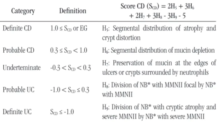
TABLE 2 – Simple criteria for differentiate CD and UC
Category Deinition Score CD (SCD) = 2H5 + 3H6 + 2H7 + 3H8 - 3H9 - 5
Deinite CD 1.0 ≤ SCD or EG H5: Segmental distribution of atrophy and
crypt distortion
Probable CD 0.3 ≤ SCD < 1.0 H6: Segmental distribution of mucin depletion
Underteminate -0.3 < SCD < 0.3 Hulcers or crypts surrounded by neutrophils7: Preservation of mucin at the edges of
Probable UC -1.0 < SCD ≤ 0.3 Hwith MMNII8: Division of NB* with MMNII focal by NB*
Deinite UC SCD ≤ -1.0 Hsevere MMNII by NB* with severe MMNII9: Division of NB* with cryptic atrophy and
Source: Tanaka et al.(25).
CD: Crohn’s disease; UC: ulcerative colitis; EG: epithelioid granuloma; NB*: number of biopsies; MMNII: monomorfonuclear inlammatory iniltrate.
RESULTS
We selected 253 patients − 107 men (42%) and 146 women (58%) − from IBD outpatient, classified into 129 UC (51.0%) and 124 CD (49.0%), which had a total of 445 subjected to histopathological colon biopsies examination, with an average of 1.7 per patient. After surveying the results of 445 biopsies, only 43 (9.6%) met the inclusion criteria for the present study. Table 3 showsclinical data and diagnosis
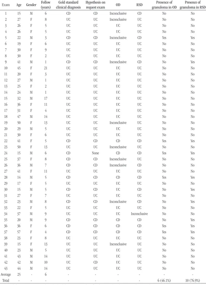
TABLE 3 – Main clinical and histopathological data from analyzed exams (43 exams of 37 patients)
Exam Age Gender (years)Follow Gold standard clinical diagnosis
Hypothesis on
request exam OD RSD
Presence of granuloma in OD
Presence of granuloma in RSD
1 15 M 6 CD CD Inconclusive CD No No
2 27 F 8 UC UC Inconclusive UC No No
3 26 F 5 UC UC UC UC No No
4 26 F 5 UC UC UC UC No No
5 22 M 3 CD CD Inconclusive CD Yes Yes
6 19 F 6 UC UC UC UC No No
7 30 F 9 UC UC UC UC No No
8 30 F 2 UC UC UC UC No No
9 41 M 1 CD CD Inconclusive CD No Yes
10 45 F 21 UC UC UC UC No No
11 20 F 3 UC UC UC UC No No
12 27 M 1 UC UC UC UC No No
13 25 F 2 UC UC UC UC No No
14 24 M 1 UC UC UC UC No No
15 32 M 17 UC UC UC UC No No
16 36 F 11 UC UC UC UC No No
17 31 F 4 UC UC UC UC No No
18 47 M 14 UC UC UC UC No No
19 50 F 13 UC UC Inconclusive UC No No
20 29 M 5 UC UC UC UC No No
21 30 F 6 UC UC UC UC No No
22 41 F 5 CD CD CD CD Yes Yes
23 50 F 13 UC UC Inconclusive UC No No
24 15 M 6 CD None CD CD Yes Yes
25 37 F 8 CD CD Inconclusive UC No No
26 36 M 7 CD CD Inconclusive CD No No
27 41 F 11 UC UC UC UC No No
28 14 M 5 CD CD CD CD Yes Yes
29 17 F 5 UC UC UC UC No No
30 15 M 5 CD CD UC CD No Yes
31 27 F 7 UC UC UC UC No No
32 23 M 8 CD CD Inconclusive CD No Yes
33 22 F 5 UC UC UC UC No No
34 37 M 9 UC UC UC Inconclusive No No
35 20 M 9 CD CD CD CD No Yes
36 36 F 6 CD CD CD CD Yes Yes
37 57 F 4 CD CD CD CD Yes Yes
38 23 F 8 UC UC UC UC No No
39 15 F 13 UC UC Inconclusive UC No No
40 23 M 5 UC UC UC UC No No
41 43 M 14 UC UC UC UC No No
42 42 M 10 UC CD UC UC No No
43 44 M 14 UC UC UC UC No No
Average 25 - 6 - - - - -
-Total - - - 6 (46.1%) 10 (76.9%)
All RSD were classified as definite for one out of the two diseases (CD or UC), except for case 34 that was classified as inconclusive. These 43 exams were discriminated in 30 (69.8%) UC diagnoses, and 13 (30.2%) CD diagnoses. 37 patients presented 43 exams, which were categorized in 25 (67.6%) UC and 12 (32.4%) CD (there was more than one examination for the same patient included in the study). From these 37 patients, 16 (43.2%) were male, and 21 (56.8%) female. From 25 patients with UC, 8 (32.0%) were male, and 17 (68.0%) female. From the 12 individuals with CD, 8 (66.7%) were male, and 4 (33.3%) female. Patients were diagnosed with IBD at 4-57 years of age, mean 25 years ± 10,1. The comparison between OD and RSD regarding the percentage of exams with conclusive and inconclusive diagnoses is presented
in Table 4. To compare OD and RSD results in percentages of assertiveness and conclusiveness of diagnosis, binomial test was applied, since each sample was evaluated by two diagnostic methods (OD and RSD), determining the result classified into two categories. Values of p < 0.05 were considered statistically
significant. The binomial test results indicated rejection of the null hypothesis at a significance level of 5% (p = 0.0117), this allows us to affirm that there is a statistically significant difference between OD and RSD in relation to the percentage of exams with inconclusive diagnoses. Table 4 shows that the percentage of tests considered inconclusive by OD (23.3%) is higher than the well regarded by RSD (2.3%). The assessment of the proportion of cases with correct diagnosis by OD and RSD for each disease (UC and CD) is presented in Table 5. For UC, the test result indicated the non-rejection of the null hypothesis at a signiicance level of 5% (p = 0.1250). Thus, it can be stated
that there is no statistically signiicant difference between OD and RSD in relation to the percentage of samples with correct diagnoses for UC. For CD, the test result indicated rejection of the null hypothesis at a signiicance level of 5% (p = 0.0313). Thus,
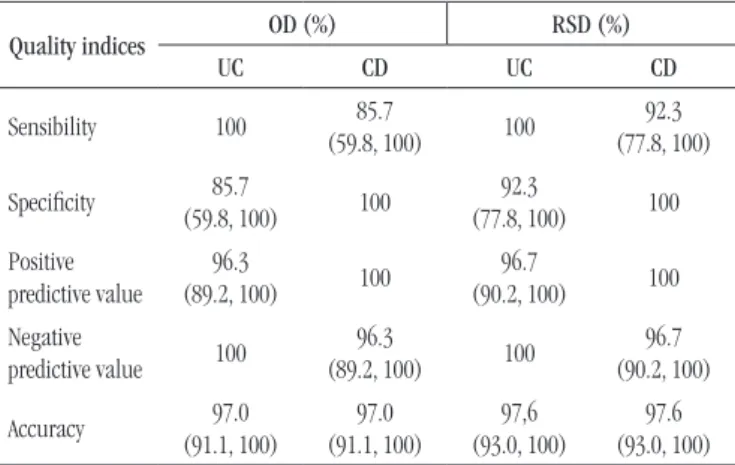
it can be stated that there is a statistically signiicant difference between OD and RSD in relation to the percentage of samples with correct diagnoses for CD. Table 5 shows that the percentage of correct cases by RSD (92.3%) is higher than the percentage for OD (46.1%). The results of the quality rates for the conclusive diagnoses obtained by OD and RSD are shown in Table 6.
TABLE 6 – Quality indices and their estimated 95% interval of conidence obtained from OD and RSD
Quality indices OD (%) RSD (%)
UC CD UC CD
Sensibility 100 (59.8, 100)85.7 100 (77.8, 100)92.3
Speciicity (59.8, 100)85.7 100 (77.8, 100)92.3 100
Positive
predictive value
96.3
(89.2, 100) 100
96.7
(90.2, 100) 100
Negative
predictive value 100
96.3
(89.2, 100) 100
96.7 (90.2, 100)
Accuracy (91.1, 100)97.0 (91.1, 100)97.0 (93.0, 100)97,6 (93.0, 100)97.6
OD: original diagnosis; RSD: reviewed standardized diagnosis; UC: ulcerative colitis; CD: Crohn’s disease.
TABLE 4 – Distribution of diagnosis as conclusive or inconclusive (n = 43 exams)
Diagnosis OD RSD
Conclusive 33 (76.7%) 42 (97.7%)
Inconclusive 10 (23.3%) 1 (2.3%)
Total (exams) 43 43
OD: original diagnosis; RSD: reviewed standardized diagnosis.
TABLE 5 – Comparison of OD and RSD in relation to the percentage of cases with correct diagnosis of UC and CD
Diagnosis
Gold standard
UC CD
OD RSD OD RSD
Correct 26 (86.7%) 29 (96.6%) 6 (46.1%) 12 (92.3%)
Incorrect(*) 4 (13.3%) 1 (3.3%) 7 (53.8%) 1 (7.7%)
Total 30 30 13 13
OD: original diagnosis; RSD: reviewed standardized diagnosis; UC: ulcerative colitis; CD: Crohn’s disease.
Incorrect(*): inconclusive or incorrect in relation with gold standard.
DISCUSSION
Inconclusive diagnoses such as “chronic inlammation” and “nonspeciic inlammation” are of limited value for both the physician and the pathologist(11, 28), and despite the
clinicopathological correlation be the goal of histopathology, it is not always possible because the information is not available, or clinical data provided contradict the histopathological indings. In these cases, the pathologist needs to extract as much information as possible from the biopsy. For an appropriate evaluation of biopsy in IBD, the pathologist should be informed about the duration of symptoms and what kind of treatment was given to the patient because the treatment can produce patchiness or discontinuity of mucosal inlammation in UC making a differential diagnosis more dificult(10). Another study refers that the treatment of UC
can lead to partial cure, which results in focal lesions simulating distribution of CD(29). This fact has already been reported(14)
information about therapeutics should be given to the pathologist
avoid misdiagnosis(17, 18, 27). Other authors state that only the
epithelioid granulomas without ruptured crypts and or chronic active ileitis are features highly suggestive of CD on mucosal biopsy analysis and that some UC exams may show discontinuous disease, no rectal disease, inlammation in ileum, extracolonic involvement, granulomatous inlammation, aphthous ulcers or mural inlammation. In these cases, the differential diagnose with CD can be almost impossible(30). In the appropriate clinical
presentation the presence of granuloma inlammation in gastrointestinal biopsy specimens conirms the diagnosis of CD(16, 32), and when the granuloma is not found, the diagnoses of
CD should be suggested emphasizing the necessity of correlation with others clinical, endoscopic and imaging features(9). In a study
the evaluation of mucosal biopsy in patients with CD demonstrated increased numbers of macrophages and microgranulomas with the help of the technique of immunohistochemistry for CD68+(31). Another dificulty is the presence of granulomas
related with crypts ruptures. To distinguish between epithelioid granuloma and a granulomatous lesion by rupture of crypt, cuts in multiple tissue levels may be necessary to ind the presence of neutrophils, lymphocytes, and foamy macrophages that are
absents in the former(30).
In the present study, the histopathological diagnosis was correct and conclusive in OD at 74.4% of patients, and in 97.7% RSD, demonstrating that the last one increased correlation with the gold standard for diagnosis. This percentage was also higher
than those found by several studies(6, 15, 20, 21, 27, 28) in which the
correlation was 94%, 80%, 75%, 72%, 73%, and 73% respectively. Our hypothesis that the standardized histopathological diagnosis increases the correlation with the gold standard in IBD was supported on two other studies(3, 13). There is several studies on
determining the histopathological criteria more discriminately, and on how better applying them to the diagnosis of IBD. In some studies, the criteria are repeated, consolidating its importance in the diagnosis of IBD, as occurred, for example, with the alteration in cryptic architecture, which is present in seven reviewed
studies(6, 13, 15, 21, 23, 24, 27). The authors of the study that was used
as a model for the standardization review of cases evaluated(24)
observed that basal plasmacytosis (Figure 1) associated with
severe inlammatory iniltrate is more discriminative than plasmocytosis considered individually. They also observed that the diffuse crypt atrophy (Figure 2B) is characteristic of UC, whereas segmental atrophy is related with CD. Figure 2A shows a comparison between a normal mucosa and a mucosa with crypt atrophy. However, they did not consider cryptic abscesses and cryptitis discriminative for the differential diagnosis between UC and CD, contradicting what other authors have stated(13, 15, 21,
FIGURE 1 – Basal plasmacytosis: presence of numerous plasma cells between the crypt base (●) and muscularis mucosa ( ■) (HE, lens 400×)
HE: hematoxylin-eosin.
FIGURE 2A – Normal colonic mucosa with crypts ( ■) tubular, parallel, straight, and juxtaposed to the muscularis mucosa (●) (HE, lens 40×)
HE: hematoxylin-eosin.
27, 28). Paneth metaplasia (Figure 3) was not analyzed in some
studies(13, 20, 21), but was analyzed and considered important in six
other studies(6, 24-28). In the studies reviewed, the histopathological
indings were evaluated taking into account different criteria. The study we have chosen as model for standardized review, assessed 70 criteria (the largest number of criteria among the studies reviewed), which were tested and selected by statistical calculations(24). The
series of three studies of these authors(24-26) consisted of 431, 726,
and 60 cases respectively. Together, it is the largest study that used the same criteria for IBD biopsies histopathological evaluation. In the present study, only one case of CD (number 25 in Table 3) was diagnosed as UC by RSD. In this case, we observed that rectal mucosa was compromised, there was diffuse distribution of atrophy and crypt distortion (Figure 4) (criterion H5), segmental
distribution of mucin depletion (criterion H6) and preservation of
mucin in areas of acute inlammation (criterion H7). A conlict was observed in the histopathological criteria in this case: the diffuse and continuous distribution of cryptic changes from the rectum is characteristic of UC according to some authors
observations(23, 24). Another unusual feature found in this CD case
was the commitment of the rectum, which is usually spared in CD(12). The case inconclusive by RSD (number 34 in Table 3) was
deined by the gold standard as UC, but it presented: segmental distribution of crypt architectural changes (criterion H5), segmental distribution of depletion of mucus (criterion H6), and focal inlammation, which are histopathological characteristics of CD(23, 24). On this exam request there was no information about the
patient being in treatment at the time of biopsies, even though it has endoscopic appearance of UC, but with segmental distribution. In biopsies of children with UC, architectural distortion of crypts occurs in 32.1%, while it happens in 57.9% of adults(29). The
authors correlate this difference with the shorter duration of disease before biopsy in pediatric cases. There is a contradiction between the observations of some studies(23, 29) about the time
for the appearance of architectural changes promoted by IBD. A
study argues(23) that they are precocious and can be detected in
biopsies from patients with seven days of onset of disease activity, and moreover the architectural changes are the most important in the diagnosis of IBD histopathology, once it differ in each type of IBD: diffuse for UC, and focal for CD. These authors also note that these changes are not exclusive to IBD as they can be found in intestinal amebiasis and shigellosis cases, depending on the strain and duration of infection (greater than one week). The quality scores obtained by OD and RSD in the present research were not consistent with one study(4). Here RSD presented speciicity, positive
predictive value, and accuracy for UC slightly larger than OD. RSD also showed sensitivity, negative predictive value, and accuracy for CD slightly larger than OD. In conclusive cases the speciicity for UC and sensitivity to CD (both 92.3%) were higher in RSD than in OD (both 85.7%), agreeing with the results of several
studies(1, 3, 13, 24-27) that consider the standardization of
histopathological criteria a necessary instrument for improving the quality indices of histopathological diagnosis.
CONCLUSION
RSD diagnosis had a higher percentage of correct and conclusive diagnoses than those presented in OD, especially statistically signiicant difference for CD exams. RSD’s conclusive cases obtained slightly higher speciicity rates, positive predictive value, and accuracy for UC; slightly higher sensitivity rates, negative predictive value, and accuracy for CD, when compared to OD.
FIGURE 3 – Paneth cell metaplasia: presence of Paneth cells (●) lined crypts in samples after colon right angle (HE, lens 400×)
HE: hematoxylin-eosin.
FIGURE 4 – Crypt distortion: branching, dilatation, and tortuous crypts ( ■) (HE, lens 100×)
RESUMO
Introdução: Duas são as formas de manifestação da doença intestinal inlamatória: doença de Crohn e retocolite ulcerativa, ambas com evolução clínica, tratamento e aspectos histopatológicos diferentes, causando, por vezes, signiicativa morbidade.
Objetivos: Escolher e aplicar método padronizado e quantiicado de diagnóstico histopatológico e comparar os resultados e os índices de qualidade, com os dos diagnósticos originais. Materiais e métodos: Foram reavaliadas histologicamente 43 biópsias colonoscópicas seriadas de 37 pacientes por sistema padronizado. Resultado e discussão: Os diagnósticos originais foram mais inconclusivos (23,3%) do que os padronizados (2,3%). A concordância com o padrão-ouro (diagnóstico clínico, colonoscópico e radiológico) foi maior nos diagnósticos padronizados (95,3%) do que nos originais (74,4%), principalmente em relação à doença de Crohn, cujos percentuais foram de 92,3% e 46,1%, respectivamente. Para retocolite ulcerativa, ambos os métodos apresentaram sensibilidade e valor preditivo negativo de 100%; já nos diagnósticos originais, foram veriicados especiicidade de 85,7%, valor preditivo positivo de 96,3% e acurácia de 97%, e nos diagnósticos padronizados, 92,3%, 96,7% e 97,6%, respectivamente. Para doença de Crohn, veriicaram-se especiicidade e valor preditivo positivo de 100% nos dois métodos; nos diagnósticos originais, sensibilidade de 85,7%, valor preditivo negativo de 96,3% e acurácia de 97%, e nos diagnósticos padronizados, 92,3%, 96,7% e 97,6%, respectivamente. Conclusão: O diagnóstico padronizado apresentou maior percentual de diagnósticos corretos e conclusivos do que os apresentados no diagnóstico original, principalmente para doença de Crohn, assim como valores iguais ou ligeiramente maiores em alguns índices de qualidade.
Unitermos: retocolite ulcerativa; doença de Crohn; biópsia; patologia.
REFERENCES
1. ALVES, P. R. A. et al. Histological scores on the colonoscopic diagnosis of inlammatory bowel disease. Arq Bras Cir Dig, v. 9, p. 67-70, 1994. 2. BANCROFT, J. D.; STEVENS, A. Theory and practice of histological techniques. New York, NY: Churchill Livingstone, 1977.
3. BENTLEY, E. et al. How could improve the initial diagnosis of colitis? Evidence from an international workshop. J Clin Pathol, v. 55, p. 955- 60, 2002.
4. CROSS, S.S.; HARRISON, R. F. Discriminant histological features in the diagnosis of idiopathic inlammatory bowel disease: analyses of a large dateset by novel data visualization technique. J Clin Pathol, v. 55, p. 51-7, 2002.
5. DANI, R. Gastroenterologia essencial. Rio de Janeiro, RJ: Guanabara
Koogan, 2011.
6. DUNDAS, S. A. C.; DUTTON, J.; SKIPWORTH, P. Reliability of rectal biopsy in distinguishing between chronic inlammatory bowel disease and acute self-limiting colitis. Histopathol, Oxford, v. 31, p. 60-6, 1997. 7. EKBOM, A. et al. Increase risk of large-bowel cancer in Crohn disease with colonic involvement. Lancet, London, v. 336, p. 357-9, 1990. 8. FARMER, M. et al. The importance of diagnostic accuracy in colonic inlammatory bowel disease. Am J Gastroenterol, New York, v. 95, n. 11, p. 3184-8, 2000.
9. GARETH, P. J.; RAVIKUMARA, M. Endoscopic and histologic indings in pediatric inlammatory bowel disease. Gastroenterol Hepatol, v. 6, p.
174-80, 2010.
10. GEBOES, K. Pathology of inlammatory bowel diseases (IBD): variability with time and treatment. Col Dis, v. 3, p. 2-12, 2001.
11. GEBOES, K.; EYKEN, P. V. Inlammatory bowel disease unclassiied and indeterminate colitis: the role of the pathologist. J Clin Pathol, v. 62,
p. 201-5, 2009.
12. HARRISON, T. R. Harrison’s principles of internal medicine. New York, NY: McGraw-Hill, 2011.
13. JENCKINS, B. et al. Guidelines for the initial biopsy diagnosis of suspected chronic idiopathic inlammatory bowel disease: the British Society of Gastroenterology iniciative. J Clin Pathol, v. 50, p. 93-105, 1997. 14. KIM, B. et al. Endoscopic and histological patchiness in treated ulcerative colitis. Am J Gastroenterol, v. 94, p. 3258-62, 1999.
15. LE BERRE, N. et al. Histological discrimination of idiopathic inlammatory bowel disease from others types of colitis. J Clin Pathol, v. 48, p.749-53, 1995.
16. LUDEMAN, L.; SHEPHERD, N. A. Problem areas in the pathology of chronic inlammatory bowel disease. Cur Diag Pathol, v. 12, p. 248-60, 2006. 17. MAGRO, F. et al. European consensus on the histopathology of inlammatory bowel disease. J Crohns Colitis, v. 7, p. 827-51, 2013. 18. MATTEO, C. et al. Chronic idiopathic inlammatory bowel diseases:
the histology report. Digest Liver Dis, v. 43S, p. S293-S303, 2011.
19. MICHALANY, J. Técnica histológica em anatomia patológica: com
instruções para o cirurgião, enfermeira e citotécnico. São Paulo, SP: Editora Pedagógica Universitária, 1980.
disease and ulcerative colitis in adults? Pathol Res Pract,v.183, p. 481-8,
1988.
21. SELDENRIJK, C. A. et al. Histopathological evaluation of colonic
mucosal biopsy specimens in chronic inlammatory bowel disease: diagnostic implications. Gut, v. 32, p. 1514-20, 1991.
22. SPENCER, M. Fundamentals of light microscopy. Cambridge: University Press, 1982.
23. SURAWICZ, C. M. et al. Mucosal biopsy diagnosis of colitis: acute self-limited colitis and idiopathic inlammatory bowel disease. Gastroenterol, v. 107, p. 755-63, 1994.
24. TANAKA, M. et al. Morphologic criteria applicable to biopsy specimens for effective distinction of inlammatory bowel disease from others forms of colitis and of Crohn disease from ulcerative colitis. Scand J Gastroenterol, v. 34, p. 55-67, 1999.
25. TANAKA, M. et al. Simple mucosal biopsy criteria differentiating among Crohn’s disease, ulcerative colitis, and other forms of colitis: measurement of validity. Scand J Gastroenterol, v. 35, p. 281-6, 2000.
26. TANAKA, M. et al. Observer variation of diagnoses based on simple biopsy criteria differentiating among Crohn’s disease, ulcerative colitis, and other forms of colitis. J Gastroenterol Hepatol, v. 16, p. 1368-72,
2001.
27. THEODOSSI, A. et al. Observer variation and discriminatory value of biopsy features in inlammatory bowel disease. Gut, v. 35, p. 961-8, 1994.
28. TSANG, P.; ROTTERDAM, H. Biopsy diagnosis of colitis: possibilities and pitfalls. Am J Surg Pathol, v. 23, p. 423-30, 1999.
29. WASHINGTON, K. et al. Histopathology of ulcerative colitis in initial rectal biopsy in children. Am J Surg Pathol, v. 26, p. 1441-9, 2002. 30. YANTISS, R. K.; ODZE, R. D. Diagnostic dificulties in inlammatory bowel disease pathology. Histopathol, v. 48, p. 116-32, 2006.
31. YANTISS, R. K. et al. Increased CD68 positive macrophages and macrophage microaggregates are prevalent in Crohn’s colitis and aid in its distinction from ulcerative colitis. Mod Pathol,v. 18, p.123A, 2005. 32. TAKASHI, H. et al. Evaluation of diagnostic criteria for Crohn’s disease in Japan. J Gastroenterol, v. 49, p. 93-9, 2014.
MAILING ADDRESS
Rosimeri Kuhl Svoboda Baldin