Article received on June 1, 2014 and accepted for publishing on July 5, 2014.
Imaging in the diagnosis of chronic pancreatitis
Vasile D. Balaban1,2, Andrei M. Lungu1, Dragoș Cuzino2,3, Săndi aBu uri ă1,2, Bogdan Macadon1, Mihăiță Pătrășes u1, Raluca S. Costache1,2, PetruțNuță1, Constantin Ştefani4, Florentina Ioniță-Radu1, Mariana Jinga1,2
Abstract: Chronic pancreatitis is characterised by progressive and irreversible damage of the pancreatic parenchyma and ductal system, which leads to chronic pain, loss of endocrine and exocrine functions. Clinically, pancreatic exocrine insufficiency becomes apparent only after 90% of the parenchima has been lost. Despite the simple definition, diagnosing chronic pancreatitis remains a challenge, especially for early stage disease. Because pancreatic function tests can be normal until late stages and have significant limitations, there is an incresing interest in the role of imaging techniques for the diagnosis of chronic pancreatitis. In this article we review the utility and accuracy of different imaging methods in the diagnosis of chronic pancreatitis, focusing on the role of advanced imaging (magnetic resonance imaging, endoscopic retrograde
cholangiopancreatography and endoscopic ultrasound).
INTRODUCTION
Chronic pancreatitis is represented by a progressive inflammation and fibrosis of the pancreas, resulting in permanent structural damage and loss of function. Regarding its epidemiology, incidence and prevalence worldwide varies depending on the frequency of risk factors (rates mainly paralelling alcohol consumption) and on the diagnostic method used (with higher incidence and prevalance rates where diagnosis was
based on advanced imaging techniques – a study in Japan using advanced diagnostic tools reported a prevalence of 45.4 and incidence of 12.4/100.000, compared to 10-15 and respectively 3.5-4 in western countries). Etiology is represented by heavy alcohol consumption, ductal obstruction (post-traumatic fibrosis, stones, pseudocyst, tumors, pancreas divisium, Oddi sphicter dysfunction), genetic causes (hereditary pancreatitis), tropical pancreatitis,
autoimmune pancreatitis or systemic disease (lupus, hyperparathyroidism).
The natural history of c hronic pancreatitis evolves with a slowly progressing subclinical phase, followed by recurrent episodes of abdominal pain and finally, when over 90% of the pancreatic parenchyma is lost, exocrine insufficiency appears. Although it is not yet known if early diagnosis of chronic pancreatitis can alter its natural history and change the outcomes of this disease, efforts should be made for earlier detection of the disease.
In real-life there are two main diagnostic issues
ega di g this disease: o o e ha d it s o e
-diagnosed by considering a hyperechoic pancreas on
ORIGINAL ARTICLES
1 Carol Davila Central Emergency Military Hospital,
Bucharest
2 Carol Davila University of Medicine and Pharmacy,
Faculty of Medicine, Bucharest
3 Institute of Military Medicine, Bucharest
Vol. CXVII •New Series • No. 3-4/2014 • Romanian Journal of Military Medicine
ultrasound as chronic pancreatitis, and on the other
ha d it s u de-diagnosed in early stages when
patients do t ha e e o i e i suffi ie ut p ese t with recurrent abdominal pain and abdominal dis tention, symptoms which are frequently attributed to irritable bowel syndrome (IBS).
On clinical grounds, early chronic pancreatitis can be asymptomatic or paucisymptomatic; moreover, early symptoms are nonspecific, of low intensity and often attributed to IBS. The classic steathoreea appears late
in the evolution of the disease, but our aim is to detect chronic pancreatitis before overt exocrine insufficiency develops.
Diagnostic tests for chronic pancreatitis are separated in two: those that evaluate pancreatic function and those that detect structural changes2.
Pancreatic function tests (PFT) can be classified as direct (which directly measure pancreatic secretions) and indirect (which measure either the pancreatic enzymes in blood or stool or the effect/lack of effect of the pancreatic enzymes, by administering a substrate which requires pancreatic enzymes for digestion and measuring the metabolites of the substrate in stool, urine, blood or breath). Direct PFT is done by collecting and measuring intraduodenal bicarbonate and lipase levels after stimulation with a secretagogue (secretin-cholecystokinin); this method has a sensitivity of 96% (which means a normal result can accurately rule out chronic pancreatitis) and
specificity of 37%, but it has major limitations – it s not standardized, not widely available, it depends on the patient to tolerate the oroduodenal tube/endoscope for an hour (here we add the costs and risks of prolonged sedation) and it has many false positive results (in Billroth II anastomosis, diabetes mellitus, celiac disease, cirrhosis and recent acute pancreatitis)2. Other PFT such as fecal elastase, fecal chymotrypsin or serum trypsinogen have proven useful only for advanced disease2.
Histopathological examination of the pancreas could be diagnostic in the early stages, however tissue sampling in chronic pancreatitis is rarely used because of many reasons: histopathological changes are non-specific (chronic inflammation and fibrosis),
sampling can be nonrepresentative in focal disease, normal aging can produce changes similar to those seen in chronic pancreatitis and pancreatic biopsy has a high risk of complications (acute pancreatitis, fistula, pseudocyst).
In this setting, imagery has emerged as a very valuable tool for evaluation of chronic pancreatitis.
Imaging methods used for the diagnosis and staging (with regard to severity and complications) consist of: plain abdominal x-ray, transabdominal ultrasound,
computed tomography (CT), magnetic resonance imaging (MRI) with magnetic resonance cholangiopancreatography (MRCP) and secretin stimulation (S-MRCP), endoscopic retrograde cholangiopancreatography (ERCP) and endoscopic ultrasound (EUS).
Plain abdominal X-Ray
Plain abdominal radiography can reveal diffuse calcifications on the projection area of the pancreas, which are considered pathognomonic of chronic pancreatitis3.
Abdominal ultrasound (US)
Abdominal ultrasound is a simple, non-invasive, widely available imaging tool. Its limitations are
ep ese ted the fa t that it s highl e a i e -dependent and patient--dependent (excessive adipose tissue, history of upper GI tract surgery or overlying gas can lead to a poor view of the pancreas).
Examination is done using a convex probe, with a
frequency of 3.5-5 Mhz, using epigastric and left subcostal approaches. To optimise visualisation of the pancreas, some recommend drinking 500 ml of water before the examination, to make the stomach an acoustic window.
position of intraductal or pseudocystogastric stent and it can evaluate the efficacy of extracorporeal lithotripsy by showing the disappearance of stones and reduction in pancreatic duct diameter.
Computed tomography (CT)
From its first use in chronic pancreatitis back in 1976, CT is now widely used for the evaluation of pancreatic pathology. Computed tomography has a reported sensitivity of 75-90% and specificity of 85% in the diagnosis of chronic pancreatitis, but it can be normal
in patients with early stage disease3,6.
CT scan can reveal chages in size (enlargement, atrophy) and contour (irregularity of the anterior border) of the pancreas, calcifications (with higher detection rates than ultrasound, being the imaging method of choice for calcifications), dilatation of the main pancreatic duct or common bile duct, alterations in peripancreatic fat – see figures 1, 2. It can also detect complications such as pseudocysts, arterial pseudoaneuyrisms or splenic vein thrombosis6.
Contrast-enhanced computed tomography
(CECT) is also useful in differentiating pseudotumoral chronic pancreatitis from pancreatic cancer6.
The disadvantages of the CT scan consist of irradiation, risk of contrast-induced nephropathy and its contraindication in patients with iodine-allergy6.
Endoscopic retrograde cholangiopancreatography (ERCP)
ERCP is a very useful tool for detecting morphological changes in the pancreatic ductal system. It is done using a lateral-view endoscope by cannulating the main pancreatic duct from the duodenum and injecting contrast to create a pancreatogram. The pancreatogram can show structural changes (dilatation, narrowing, irregularity, stones, leaks) of the pancreatic duct, abnormal side branches, communicating pseudocysts, which are reported in a standardized manner, using the Cambridge criteria, (Table 1). In interpreting the pancreatogram, one should keep in mind that focal ductal changes can follow an episode of severe acute pancreatitis,. Also a
normal pancreatogram does not exclude early chronic pancreatitis.
Table 1 Cambridge Classification for ERCP (Banks PA. J Gastroenterol 2007)
Grade Main duct Abnormal side
branches
Normal Normal None
Equivocal Normal < 3
Mild Normal > 3
Moderate Abnormal > 3
Marked
Abnormal with at least one additional feature: large cavity (>10 mm),
obstruction, filling defects, severe dilatation or irregularity
> 3
The main disavantages of ERCP are represented by
the fa t that it s st o gl ope ato depe de t,it s a
invasive technique with potential serious complications (pancreatitis, hemorrhage, infection) and it only reveals ductal changes6. Over the last years, ERCP has become a primarily therapeutic tool, so that diagnostic studies have been abandoned in favor of other imaging methods such as MRI/MRCP or EUS. However, ERCP remains of great importance in pancreatic endotherapy (pancreatic sphincterotomy, stenting and cyst drainage).
Magnetic resonance imaging (MRI). Magnetic resonance cholangiopancreatography (MRCP)
Due to its noninvasivity and lack of irradiation, MRI with MRCP, gadolinium enhancement and secretin stimulation is being increasingly seen as the imaging method of choice in the evaluation of pancreatic disease. MRI can be used in renal impairment and in
patients with iodine allergy, in whom CECT cannot be performed7. One of the drawbacks of MRI is represented by the artefacts caused by respiratory movements, but this has improved with the latest technology.
Vol. CXVII •New Series • No. 3-4/2014 • Romanian Journal of Military Medicine
function) and there is a delayed enhancement after gadolinium administration (due to fibrosis, blood flow inside the pancreas is altered and the gland peaks in the venous phase instead of the arterial one)6. MRI is inferior to CT in detecting calcifications, because they are seen as signal void.
Pancreatic ductal system is best evaluated by magnetic resonance cholangiopancreatography.
MRCP images are obtained without injecting contrast medium, by using heavily T2-weighted sequences.
On T2-weighted images, fluid from the bilio-pancreatic system has a bright signal and by adding fat-supression, the signal of fluid within the ducts becomes more pronounced6. MRCP can show dilatation, strictures, irregularities in the Wirsung duct or similar changes in the side-branch ducts – see figures 3,4.
The advantages of MRCP over ERCP are lack of instrumentation of the Wirsung duct and lack of need for sedation during the procedure; however, MRCP is only diagnostic, whereas ERCP is therapeutic. Another
difference between the two is that while during ERCP the collapsed secondary branches are distended by the contrast agent, MRCP visualizes the ductal system in a physiologic state, thus being less acurate for small duct disease. To overcome this difference a combined functional-structural test has been developped, called MRCP-S (magnetic resonance cholangiopancreatography with secretin stimulation): secretin stimulates secretion in the ductal system and it also increases the tonus of the Oddi sphincter
(preventing the release of secretion in the duodenum), thus filling in the collapsed branches and better delineating minimal duct changes in patients in whom MRCP showed no abnormalities under physiological conditions; the study of Manfredi et al. showed that visualization of the secondary branches improved significantly in both patients with severe disease (from 71% to 100%) and those with a mild-moderate disease (from 4% to 63%). MRCP-S also allows for calculation of the change in pancreatic duct size before and after secretin stimulation (index called pancreatic duct compliance, PDC), which is reduced in chronic pancreatitis6.
Table 2. Rosemont criteria for chronic pancreatitis
Rank Criteria Feature Definition
Pa
re
n
ch
ymal
fe
atu
re
s
1 Major A Hyperechoic foci with shadowing
Echogenic structures i le gth a d width that shadow
2 Major B Lobularity with honeycombing
Well-i u s i ed, st u tu es ith enhancing rim and relatively echo-poor e te , o tiguous lo ules
Minor Lobularity without honeycombing
Same as above, noncontiguous lobules
3 Minor Hyperechoic foci without shadowing
E hoge i st u tu es fo i i oth length and width with no shadowing
4 Minor Cysts Anechoic, rounded/elliptical structures with or without septations
5 Minor Stranding H pe e hoi li es of i le gth i at least 2 different directions with respect to the imaged plane
Du
ctal
feat
u
re
s
1 Major A MPD calculi Echogenic structure(s) within MPD with acoustic shadowing
2 Minor Irregular MPD contour
Uneven or irregularoutline and ectatic course
3 Minor Dilated side branches
3 or more tubular anechoic structure seach easu i g mm in width,budding from the MPD
4 Minor MPD dilation . -mm body or >1.5-mm tail
Endoscopic ultrasound (EUS)
Endoscopic ultrasound is done using a linear/radial transducer integrated into the tip of the scope, using 7.5-12 Mhz frequencies. It is a great method of evaluating the pancreas, considering that the transducer gets in very close range of the gland, from which is separated only by the digestive tract wall.
EUS is especially useful for early chronic pancreatitis, being able to reveal small structural changes. Its advantage over ERCP is that it detects both
parenchymal a d du tal ha ges a d does t use ionizing radiation. One of the disadvantages of EUS is that it s ope ato-dependent.
Add-on modules such as elastography and contrast-enhanced EUS (CEEUS) provide further details in evaluating pancreatic disease. Moreover, linear EUS can also provide tissue sampling (to differentiate an inflammatory mass from adenocarcinoma) and can guide therapy in chronic pancreatitis (celiac plexus neurolysis for unresponsive pain, pseudocyst drainage, main pancreatic duct drainage).
EUS characteristics of chronic pancreatitis have been grouped into ductal and parenchymal, focusing on 9 criteria which correspond to histopathological changes: 5 of them are parenchymal (hyperechoic foci, hyperechoic strands, lobularity, cysts/ pseudocysts, calcifications) and 4 are ductal (dilated pancreatic duct, irregular pancreatic duct,
hyper-echoic pancreatic duct walls, visible side branches) – see figures 5,6,7,8. Because not all of these criteria have the same importance in the diagnosis of chronic pancreatitis, these sonographic features have been redefined as major and minor features in a consensus classification called Rosemont.
According to the Rosemont criteria, EUS result can be23: consistent with chronic pancreatitis (1 major A
featu e + i o featu es, o ajo A feature + major B feature, or 2 major A features), suggestive of
chronic pancreatitis (1 major A feature + <3 minor features, o ajo ţ featu e + i o features, or
i o featu es , i dete i ate for chronic
pancreatitis (3 to 4 minor features, no major features or major B feature alone or with <3 minor features)
o o al o ajo featu es, minor features –
excluding cysts, dilated MPD, hyperechoic non-shadowing foci, dilated side branch).
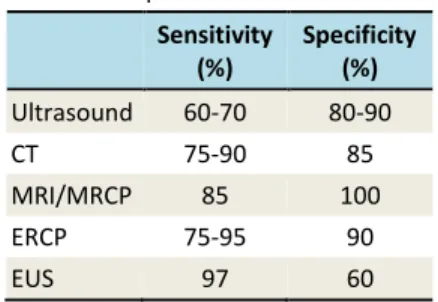
Table 3. Sensitivity and specificity of imaging studies for the diagnosis of chronic
pancreatitis10
Sensitivity (%)
Specificity (%)
Ultrasound 60-70 80-90
CT 75-90 85
MRI/MRCP 85 100
ERCP 75-95 90
EUS 97 60
Figures 1,2: CT scan of a 40 years-old male, smoker, with chronic alcohol intake and history of multiple episodes of acute pancreatitis: Enlarged head of the pancreas with multiple cystic structures (up to
Vol. CXVII •New Series • No. 3-4/2014 • Romanian Journal of Military Medicine
Figures 3,4: MRI and MRCP, same patient: Pseudotumoral chronic pancreatitis, 9/7 cm pseudocyst in the bodytail with splenic vein occlusion
Figures 5-8: EUS examination, same patient: Inhomogeneous head of the pancreas with multiple calcifications (up to 6 mm) and cystic structures; large cystic lesion in the body of the pancreas with hyperechoic debris
CONCLUSIONS
Imaging is becoming more and more important in the evaluation of chronic pancreatitis.
Among the imaging methods, plain abdominal radiograph is useful when calcifications are present and abdominal ultrasound is valuable for follow-up.
CT is best for identifying pancreatic calcifications and for evaluating complications of chronic pancreatitis.
ERCP can accurately detect changes in the ductal
s ste , ut it s o ada s a epted o l fo
References:
1 Tandon RK, Sato N, Garg PK. Chronic pancreatitis. Asia-Pacific consensus report. J Gastroenterol Hepatol 2002; 17: 508-518.
2 Feldman M. Friedman LS, Brandt LJ. Chronic pancreatitis.
I : “leise ge a d Fo dt a s Gast oi testi al a d Li e
Disease. 9th Edition. Elsevier 2010.
3 Steer ML, Waxman I, Freedman S. Chronic pancreatitis. N Engl J Med 1995; 332:1482-1490.
4 Forsmark C, Adams PC. Pancreatic function testing – valuable but underused. Can J Gastroenterol. 2009; 23: 529–530.
5 Parsi MA, Conwell DL, Zuccaro G, et al. Findings on endoscopic retrograde cholangiopancreatography and pancreatic function test in suspected chronic pancreatitis and negative cross-sectional imaging. Clin Gastroenterol Hepatol. 2008; 6:1432-1436.
6 Choueiri NE, Balci NC, Alkaade S, Burton FR. Advanced imaging of chronic pancreatitis. Curr Gastroenterol Rep. 2010; 12:114-120.
7 Munoz JED: Chronic pancreatitis. In Clinical Pancreatology for Practicing Gastroenterologists and Surgeons. Malden: Blackwell; 2006: 180–253.
8 Badea R, Diaconu B. Contributions of US to the diagnosis of chronic pancreatitis and to evaluating its main complications. Rom J Gastroenterol 2005; 14:183-189.
9 Haaga JR, Alfidi RJ, Zelch MG, et al. Computed tomography of the pancreas. Radiology 1976; 120:589–595.
10Nair JR, Lawler L, Miller MR. Chronic pancreatitis. Am Fam Physician. 2007; 76:1679-1688.
11Malfertheiner P, Büchler M. Correlation of imaging and function in chronic pancreatitis. Radiol Clin North Am. 1989; 27:51-64.
12Axon ATR, Classen M, Cotton PB, Cremer M, Freeny PC, Lees WR. Pancreatography in chronic pancreatitis: International definitions. Gut 1984; 25:1107-1112.
13Banks PA. Classification and diagnosis of chronic pancreatitis. J Gastroenterol 2007; 42 (Suppl XVII): 148– 151.
14Angelini G, Cavallini G, Pederzoli P, et al. Long term outcome of acute pancreatitis: A prospective study with 118 patients. Digestion 1993; 54: 143–147.
15Tandon RK, Sato N, Garg PK and The Consensus Study Group. Chronic pancreatitis: Asia–Pacific consensus report. Journal of Gastroenterology and Hepatology 2002; 17: 508– 518.
16Remer EM, Baker ME. Imaging of chronic pancreatitis. Radiol Clin North Am 2002; 40:1229–1242.
17Sai JK, Suyama M, Kubokawa Y, Watanabe S. Diagnosis of mild chronic pancreatitis (Cambridge classification): comparative study using secretin injection-magnetic resonance cholangiopancreatography and endoscopic retrograde pancreatography. World J Gastroenterol 2008; 14:1218–1221.
18Balci NC, Alkaade S, Magas L, Momtahen AJ, Burton FR. Suspected chronic pancreatitis with normal MRCP: findings on MRI in correlation with secretin MRCP. J Magn Reson Imaging 2008; 27:125–131.
19Manfredi R, Costamagna G, Brizi MG, et al. Severe chronic pancreatitis versus suspected pancreatic disease: dynamic MR cholangio-pancreatography after secretin stimulation. Radiology 2000; 214:849–855.
20Cappeliez O, Delhaye M, Devière J. Chronic pancreatitis: evaluation of pancreatic exocrine function with MR pancreatography after secretin stimulation. Radiology 2000; 215:358-364.
21Seicean A. Endoscopic ultrasound in chronic pancreatitis: Where are we now? World J Gastroenterol. 2010; 16:4253– 4263.
22The International Working Group for Minimum Standard Terminology for Gastrointestinal Endosonography. Reproduction of minimal standard terminology in gastrointestinal endosonography. Dig Endosc 1998; 10:158-188.