Open Access
Research
Animal models for clinical and gestational diabetes: maternal and
fetal outcomes
Ana CI Kiss, Paula HO Lima, Yuri K Sinzato, Mariana Takaku,
Marisa A Takeno, Marilza VC Rudge and Débora C Damasceno*
Address: Laboratory of Experimental Research of Gynecology and Obstetrics, Department of Gynecology and Obstetrics, Botucatu Medical School - São Paulo State University (Unesp), Botucatu, São Paulo, Brazil
Email: Ana CI Kiss - ana.inhasz@gmail.com; Paula HO Lima - paulalima@fmb.unesp.br; Yuri K Sinzato - yuri_sinzato@yahoo.com.br; Mariana Takaku - xokimxl@yahoo.com; Marisa A Takeno - matakeno@yahoo.com.br; Marilza VC Rudge - marilzarudge@ig.com.br; Débora C Damasceno* - damasceno@fmb.unesp.br
* Corresponding author
Abstract
Background: Diabetes in pregnant women is associated with an increased risk of maternal and neonatal morbidity and remains a significant medical challenge. Diabetes during pregnancy may be divided into clinical diabetes and gestational diabetes. Experimental models are developed with the purpose of enhancing understanding of the pathophysiological mechanisms of diseases that affect humans. With regard to diabetes in pregnancy, experimental findings from models will lead to the development of treatment strategies to maintain a normal metabolic intrauterine milieu, improving perinatal development by preventing fetal growth restriction or macrosomia. Based on animal models of diabetes during pregnancy previously reported in the medical literature, the present study aimed to compare the impact of streptozotocin-induced severe (glycemia >300 mg/dl) and mild diabetes (glycemia between 120 and 300 mg/dl) on glycemia and maternal reproductive and fetal outcomes of Wistar rats to evaluate whether the animal model reproduces the maternal and perinatal results of clinical and gestational diabetes in humans.
Methods: On day 5 of life, 96 female Wistar rats were assigned to three experimental groups: control (n = 16), severe (n = 50) and mild diabetes (n = 30). At day 90 of life, rats were mated. On day 21 of pregnancy, rats were killed and their uterine horns were exposed to count implantation and fetus numbers to determine pre- and post-implantation loss rates. The fetuses were classified according to their birth weight.
Results: Severe and mild diabetic dams showed different glycemic responses during pregnancy, impairing fetal glycemia and weight, confirming that maternal glycemia is directly associated with fetal development. Newborns from severe diabetic mothers presented growth restriction, but mild diabetic mothers were not associated with an increased rate of macrosomic fetuses.
Conclusion: Experimental models of severe diabetes during pregnancy reproduced maternal and fetal outcomes of pregnant women presenting uncontrolled clinical diabetes. On the other hand, the mild diabetes model caused mild hyperglycemia during pregnancy, although it was not enough to reproduce the increased rate of macrosomic fetuses seen in women with gestational diabetes. Published: 19 October 2009
Diabetology & Metabolic Syndrome 2009, 1:21 doi:10.1186/1758-5996-1-21
Received: 4 March 2009 Accepted: 19 October 2009
This article is available from: http://www.dmsjournal.com/content/1/1/21
© 2009 Kiss et al; licensee BioMed Central Ltd.
Background
Diabetes mellitus (DM) is a disease characterized by dis-arrangements in carbohydrate, protein and lipid metabo-lism caused by the complete or relative insufficiency of insulin secretion and/or insulin action [1]. Diabetes in pregnant women is associated with an increased risk of maternal and neonatal morbidity and remains a signifi-cant medical challenge. Diabetes during pregnancy may be divided into clinical diabetes (women previously diag-nosed with type 1 or type 2 diabetes) and gestational dia-betes, defined as any glucose intolerance detected during pregnancy that has evolved from a diagnosis associated with the metabolic risk of type 2 diabetes to a clinical con-dition associated with higher risks for maternal and peri-natal morbidity [2]. Fortunately, the prognosis has changed dramatically due to an increased clinical aware-ness of the potential risks for the mother and the infant.
Experimental models are developed with the purpose of enhancing understanding of the pathophysiological mechanisms of diseases that affect humans. With regard to diabetes in pregnancy, experimental findings from models will lead to the development of treatment strate-gies to maintain the closest to normal metabolic intrauter-ine milieu, improving perinatal development by preventing fetal growth restriction or macrosomia. The rat (and the rabbit) is often used in reproductive toxicity studies [3]. In general, the uncontrolled human type 1 DM clinical status during pregnancy is reproduced by streptozotocin (STZ) administration (40 mg/kg) to rats during adult life using the venous route [4-6]. In this experimental model, rats present with severe diabetes, with glycemia above 300 mg/dl, and the fetuses of dams are classified as small fetuses for gestational age, character-izing intrauterine growth restriction. Human type 2 DM and gestational DM conditions are reproduced in animals by administration of different doses of STZ in the neona-tal period [7-16], before mating [17-20] or during preg-nancy [21-30]. Adult animals present with glycemia between 120 and 300 mg/dl, characterizing moderate or mild diabetes [31-33]. Merzouk and colleagues [23-25] and Soulimane-Mokhtari and colleagues [30] verified that mildly hyperglycemic dams have fetuses that are large for gestational age, classified as macrosomic.
Evidence in the literature indicates that neonatal rats treated with STZ at birth exhibit altered insulin and glu-cose tolerance tests [8,9,13] and plasmatic insulin [11,15]. Based on the insulin action response and glucose tolerance test, Triadou and colleagues [15] established an experimental design that reproduces the development of gestational diabetes in women. Several reports in the liter-ature describe the effects of severe and mild diabetes on pregnancy, fetal glycemia and development, but these studies did not investigate correlations between maternal
and fetal repercussions in these two different glycemic ranges. Therefore, the present study aimed to compare the impact of STZ-induced severe and mild diabetes on glyc-emia and maternal reproductive and fetal outcomes of
Wistar rats to evaluate whether the animal model
repro-duces the maternal and perinatal results of clinical and gestational diabetes in humans.
Methods
Subjects
Wistar rats were obtained from São Paulo State University
(Unesp) Botucatu, São Paulo State, Brazil. They were maintained in an experimental room under controlled conditions of temperature (22 ± 2°C), humidity (50 ± 10%), and a 12-hour light/dark cycle. All experimental procedures presented in this study were approved by the local Committee of Ethics in Animal Experimentation, which assures adherence to the standards established by the Guide for the Care and Use of Laboratory Animals.
Experimental procedures
On day 5 of life, 64 female Wistar rats were randomly
dam pups [34]. Blood pool glycemia levels were deter-mined from three newborns from each litter.
Statistical analysis
Results are presented as mean ± standard error of mean. The proportion test (Chi-square) was used for fetal weight classification. Two-way analysis of variance (ANOVA) fol-lowed by the Student-Newman-Keuls test was employed to compare the data for maternal glycemia, food intake, body weight during pregnancy, number of implantation sites and number of live fetuses. Pre- and post-implanta-tion loss rates were analyzed by Mann Whitney non-para-metric test. Maternal and fetal glycemia correlation was determined using Pearson correlation. The statistical sig-nificance interval is considered as P < 0.05 for all data. All
statistical analyses were performed with Statistica software (Statsoft, Tulsa, OK, USA).
Results
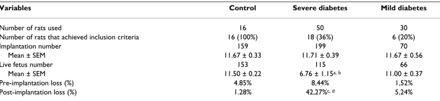
All 16 rats assigned to the control group were mated, had a positive pregnancy diagnosis and were included in this study. Only 18 of 50 rats administered STZ as adults (severe diabetic rats) had a positive pregnancy diagnosis and were included in this study following the inclusion criteria for their experimental group. All 30 rats adminis-tered streptozotocin as neonates were also mated, but only 16 presented with a positive pregnancy diagnosis and only 6 achieved the inclusion criteria. The rats that did not reach inclusion criteria were used in another study. There were no significant differences in the number of implantation sites in the severe and mild diabetic groups compared to the control group nor between the severe and mild diabetic groups. A lower mean number of live fetuses and a higher post-implantation loss rate were observed in severe diabetic rats compared to the control and mild diabetes groups (Table 1).
Rats with severe diabetes had a higher food intake com-pared to mild diabetic rats on days 14 to 21 of pregnancy,
and compared to control rats on all days of pregnancy. Mild diabetic rats had a higher food intake compared to the control group only on day 0 of pregnancy (Figure 1A). Both severe and mild diabetic rats had lower body weight compared to the control group (Figure 1B).
During their entire pregnancy, control rats had normal glycemic values (around 80 mg/dl). Glycemia remained above 300 mg/dl in the severe diabetic rats and between 120 and 300 mg/dl in the mild diabetic rats. Both severe and mild diabetic rats had higher glycemia levels through-out pregnancy compared to the control group. When compared to the mild diabetes group, severe diabetic dams had higher glycemia levels prior to mating and dur-ing pregnancy. Newborns from severe diabetic dams had higher glycemia levels compared to newborns from both the control and mild diabetic groups (Table 2). There was a positive correlation (P < 0.05) between maternal and
fetal glycemia in all experimental groups.
In both the severe and mild diabetes groups, there was a higher proportion of SPA fetuses and a reduced percent-age of APA and LPA fetuses compared to the control group. Severe diabetic rats also had higher SPA and lower APA rates compared to mild diabetic rats. The proportions of LPA fetuses from the severe and mild diabetes groups were similar (Table 3).
Discussion
STZ is often used to induce DM in experimental animals due to its toxic effects on pancreatic beta-cells [35,36]. It is a potent alkylating agent able to methylate DNA [37-39] and although it is generally accepted that the cytotoxicity produced by STZ depends on DNA alkylation [37,39], several lines of evidence indicate that free radicals play an essential role in its mechanism of DNA damage and cyto-toxicity. The nitrosurea moiety of STZ is responsible for its cellular toxicity, which is probably mediated through a decrease in NAD levels and the production of intracellular
Table 1: Maternal reproductive outcomes of control, severe diabetic and mild diabetic rats
Variables Control Severe diabetes Mild diabetes
Number of rats used 16 50 30
Number of rats that achieved inclusion criteria 16 (100%) 18 (36%) 6 (20%)
Implantation number 159 199 70
Mean ± SEM 11.67 ± 0.33 11.71 ± 0.39 11.67 ± 0.56
Live fetus number 153 115 66
Mean ± SEM 11.50 ± 0.22 6.76 ± 1.15a, b 11.00 ± 0.37
Pre-implantation loss (%) 4.85% 8,44% 1,52%
Post-implantation loss (%) 1.28% 42,27%c, d 5,24%
Effect of streptozotocin induced diabetes on food intake and body weight of rats during pregnancy Figure 1
Effect of streptozotocin induced diabetes on food intake and body weight of rats during pregnancy. (A) Food intake and (B) body weight on days 0, 7, 14 and 21 of pregnancy of rats injected with citrate buffer solution (control), strepto-zotocin as adults (severe diabetes) and streptostrepto-zotocin during the neonatal period (mild diabetes). Values are presented as mean ± standard error of mean. aP < 0.05 - statistically significant difference compared to control group (Student Newman Keuls); bP < 0.05 - statistically significant difference compared to the mild diabetes group (Student Newman Keuls).
A
0
5
10
15
20
25
30
35
40
45
50
D0
D7
D14
D21
Day of Pregnancy
F
o
od I
n
ta
ke
(
g
)
Control
Severe diabetes Mild diabetes
a,b
a,b
a a
a
B
0
50
100
150
200
250
300
350
400
450
D0
D7
D14
D21
Pregnancy Day
B
ody
W
e
ight
(
g)
Control
Severe diabetes Mild diabetes
a a
a a
free radicals. The deoxyglucose moiety of STZ facilitates its transport across the cell membrane, in which the GLUT-2 glucose-transporter appears to play an essential role. The insulin-producing beta-cells of the islets of Langerhans not only express high levels of GLUT-2 transporters but also have a relatively low NAD content, making them par-ticularly vulnerable to STZ toxicity [40].
In the mild diabetes group, STZ treatment created a range of damage to beta cells, leading to a variable range of insu-lin insufficiency. Only 6 (20%) of the initial 30 rats had a positive pregnancy diagnosis and presented with mild diabetes on pregnancy day 0 according to the inclusion criteria previously established (glycemia between 120 and 300 mg/dl). Although the success rate of this model may appear low, models in which high doses of STZ are administered in the neonatal period to achieve mild dia-betes are well established [7-16]. However, these studies do not mention how many animals achieved hyperglyc-emia in adult life. STZ has a beta-cell specific toxicity that produces severe and permanent diabetes when given to adult rats. When given during the neonatal period, there is a spontaneous recovery from the damage caused to the beta-cells in the first 2 weeks of life. However, beta-cell regeneration is incomplete and this reduced beta-cell mass results in the appearance of a form of diabetes in adult life that resembles DM type 2 in humans [9]. Indi-vidual differences in STZ metabolism [41] and beta-cell
regeneration capacity [9] may explain why so many rats that receive STZ do not present mild diabetes in adult life.
In the present study, rats with glycemia above 300 mg/dl (severe diabetes) had higher food intake but reduced body weight during pregnancy, both common features of the severe diabetic state. The reduced body weight is a consequence of metabolic alterations caused by hypergly-cemia/hypoinsulinemia, such as asthenia, as described by Damasceno and colleagues [4]. Rats injected neonatally with STZ had mild diabetes (glycemia from 120 to 300 mg/dl) without a significant increase in food intake, but reduced body weight, which can also be explained by met-abolic alterations despite the lower glycemia compared to the severely diabetic rats. Although maternal hypoin-sulinemia/hyperglycemia has a major impact on fetal weight, the reduced maternal body weight of mild dia-betic rats, resulting from low weight gain during preg-nancy, could be a cause of the low number of LPA fetuses in this group.
Severe diabetic rats had glycemia levels above 300 mg/dl throughout pregnancy. This result was expected and is in agreement with other studies previously performed in our laboratory [4-6], reproducing the hyperglycemia that some women with uncontrolled clinical diabetes present during pregnancy. The mild diabetic rats maintained their glycemia between 120 and 300 mg/dl during pregnancy. STZ administration in the neonatal period caused mild
Table 2: Glycemia of control, severe diabetic and mild diabetic rats throughout pregnancy and of newborns
Control (n = 16) Severe diabetes (n = 18) Mild diabetes (n = 6)
Prior mating 84.33 ± 0.76 343.56 ± 14,36a, b 177.12 ± 45.53
Day 0 78.17 ± 3.89 351.78 ± 12.79a, b 186.67 ± 26.04a
Day 7 77.67 ± 3.68 294.11 ± 11.01a, b 177.67 ± 32.84a
Day 14 77.17 ± 5.85 327.44 ± 12.70a, b 179.69 ± 39.85a
Day 21 79.83 ± 6.65 322.61 ± 17.95a, b 170.67 ± 30.21a
Newborns 68.25 ± 7.94 464.33 ± 28.95a, b 115.9 ± 37.57
Glycemia (mean ± standard error of mean) were taken prior to mating and on days 0, 7, 14 and 21 of pregnancy from rats injected with citrate buffer solution (control), streptozotocin as adults (severe diabetes) and streptozotocin during the neonatal period (mild diabetes). Blood pool glycemia was determined from three newborns from each litter. aP < 0.05 - statistically significant difference compared to control group (Student Newman Keuls);
bP < 0.05 - statistically significant difference compared to mild diabetes group (Student Newman Keuls).
Table 3: Fetal weight classification of offspring born to control, severe diabetic and mild diabetic rats
Variable/groups Control Severe diabetes Mild diabetes
SPA 46/192 (24%) 112/129 (87%)a, b 39/65 (60%)a
APA 99/192 (52%) 14/129 (11%)a, b 22/65 (34%)a
LPA 47/192 (24%) 3/129 (2%)a 4/65 (6%)a
hyperglycemia during pregnancy, which has also been reported by Triadou and colleagues [15], Capobianco and colleagues [10] and Kiss [42], reproducing the hyperglyc-emia that some women with gestational diabetes present during pregnancy.
In our study, the lower number of live fetuses and the high post-implantation loss rate in the severe diabetes group are characteristic of a hyperglycemic (glycemia above 300 mg/dl) intrauterine milieu, and are in agreement with other studies [6,43]. In the present study, the high glyc-emic levels did not prevent embryo implantation but did impair development, leading to fetal death, as confirmed by the low number of live fetuses. Our results also show that rats with severe diabetes had newborns with intrau-terine growth restriction. This can be explained by fetal beta-cell collapse, which eventually leads to fetal hypoin-sulinemia that causes the growth restriction [19,44,45].
There is evidence that the hyperglycemic intrauterine milieu of a mildly diabetic mother stimulates the fetal endocrine pancreas to hyperinsulinemia and accelerated anabolism, resulting in fetal and neonatal macrosomia. Many reports in the literature indicate that animal models in which STZ is injected during the neonatal period are compatible with human gestational diabetes conditions, with the presence of macrosomic fetuses [23-25,30] that are intolerant to glucose [44,46]. In contrast, our results show that the mild diabetic dams did not have an increased percentage of newborns classified as LPA. Simi-larly, Kervran and colleagues [19] also did not obtain macrosomic fetuses when studying the offspring of rats with mild hyperglycemia during pregnancy, and suggest that the differences between the clinical findings in humans and the experimental results using rats are due to the short pregnancy time in the rat and differences in the percentages of adipose tissue in rat fetuses (1%) and human offspring (16%) and the greater weight gain in the human species.
The offspring of the mild diabetic dams did not have impaired glycemia compared to the control group. How-ever, the offspring of the severe diabetic dams showed higher glycemia levels compared to both the control and mild diabetes groups. Many clinical and experimental studies have shown that offspring that developed in an intrauterine milieu that has been modified by hyperglyc-emia show intolerance to glucose [44,46]. In the present study, offspring were not submitted to the glucose toler-ance test, so there is no evidence that they are intolerant to glucose, but their glycemia levels correlate positively with those of their mothers. Kervran and colleagues [19] also observed a positive correlation between maternal and fetal glycemia levels in both severe and mild diabetic dams.
Conclusion
STZ-induced severe and mild diabetic dams showed dif-ferent glycemic responses during pregnancy, although both adversely affected fetal glycemia and weight, con-firming that maternal glycemia is directly associated with fetal development. Newborn from severe diabetic moth-ers presented intrauterine growth restriction, but mild dia-betic mothers did not have an increased percentage of LPA fetuses. The experimental model of severe diabetes during pregnancy reproduced maternal and fetal outcomes of women with uncontrolled clinical diabetes. On the other hand, the mild diabetes model caused mild hyperglyc-emia during pregnancy, although it was not enough to reproduce the increased rate of macrosomic fetuses seen in women with gestational diabetes.
Abbreviations
APA: appropriate for pregnancy age; DM: diabetes melli-tus; LPA: large for pregnancy age; SPA: small for pregnancy age; STZ: streptozotocin.
Competing interests
The authors declare that they have no competing interests.
Authors' contributions
ACIK participated in the acquisition, analysis and inter-pretation of data and helped to draft the manuscript. PHOL participated in the acquisition of data and helped to draft the manuscript. YKS participated in the acquisi-tion of data and helped to draft the manuscript. MT par-ticipated in the acquisition of data and helped to draft the manuscript. MAT participated in the acquisition of data and helped to draft the manuscript. MVCR helped to draft the manuscript. DCD conceived the study, participated in its design, coordination, analysis and interpretation of data and helped to draft the manuscript. All authors read and approved the final manuscript.
Acknowledgements
The authors are grateful to CAPES (Brazil) for financial support and to the Research Support Center (RSC) of the Botucatu Medical School, São Paulo State University (Unesp), for their invaluable contribution to the study design and statistical analysis.
References
1. American Diabetes Association: Diagnosis and classification of diabetes mellitus. Diabetes Care 2009, 32(Suppl 1):S62-67. 2. Forsbach-Sanchez G, Tamez-Perez HE, Vazquez-Lara J: Diabetes
and pregnancy. Arch Med Res 2005, 36:291-299.
3. de Rijk EP, van Esch E, Flik G: Pregnancy dating in the rat:
pla-cental morphology and maternal blood parameters. Toxicol
Pathol 2002, 30:271-282.
4. Damasceno DC, Volpato GT, Calderon Ide M, Aguilar R, Rudge MV:
Effect of Bauhinia forficata extract in diabetic pregnant rats:
maternal repercussions. Phytomedicine 2004, 11:196-201. 5. Rudge MV, Damasceno DC, Volpato GT, Almeida FC, Calderon IM,
6. Volpato G, Damasceno D, Campos K, Rocha R, Rudge M, Calderon I:
Avaliação do efeito do exercício físico no metabolismo de ratas diabéticas prenhes. Revista Brasileira de Medicina do Esporte
2006, 12:229-233.
7. Blondel O, Bailbe D, Portha B: Relation of insulin deficiency to impaired insulin action in NIDDM adult rats given streptozo-cin as neonates. Diabetes 1989, 38:610-617.
8. Blondel O, Bailbe D, Portha B: Insulin resistance in rats with non-insulin-dependent diabetes induced by neonatal (5 days) streptozotocin: evidence for reversal following phlorizin treatment. Metabolism 1990, 39:787-793.
9. Bonner-Weir S, Trent DF, Honey RN, Weir GC: Responses of neo-natal rat islets to streptozotocin: limited B-cell regeneration and hyperglycemia. Diabetes 1981, 30:64-69.
10. Capobianco E, Jawerbaum A, White V, Pustovrh C, Sinner D, Gonzalez ET: Elevated levels of endothelin-1 and prostaglan-din E2 and their effect on nitric oxide generation in placental tissue from neonatal streptozotocin-induced diabetic rats.
Prostaglandins Leukot Essent Fatty Acids 2003, 68:225-231.
11. Movassat J, Saulnier C, Portha B: Insulin administration enhances growth of the beta-cell mass in streptozotocin-treated new-born rats. Diabetes 1997, 46:1445-1452.
12. Murali B, Goyal RK: Improvement in insulin sensitivity by
losa-rtan in non-insulin-dependent diabetic (NIDDM) rats.
Phar-macol Res 2001, 44:385-389.
13. Portha B, Kergoat M: Dynamics of glucose-induced insulin release during the spontaneous remission of streptozocin
diabetes induced in the newborn rat. Diabetes 1985,
34:574-579.
14. Portha B, Levacher C, Picon L, Rosselin G: Diabetogenic effect of streptozotocin in the rat during the perinatal period. Diabetes
1974, 23:889-895.
15. Triadou N, Portha B, Picon L, Rosselin G: Experimental chemical diabetes and pregnancy in the rat. Evolution of glucose tol-erance and insulin response. Diabetes 1982, 31:75-79.
16. Tsuji K, Taminato T, Usami M, Ishida H, Kitano N, Fukumoto H, Koh G, Kurose T, Yamada Y, Yano H, et al.: Characteristic features of insulin secretion in the streptozotocin-induced NIDDM rat model. Metabolism 1988, 37:1040-1044.
17. Caluwaerts S, Holemans K, van Bree R, Verhaeghe J, Van Assche FA:
Is low-dose streptozotocin in rats an adequate model for
gestational diabetes mellitus? J Soc Gynecol Investig 2003,
10:216-221.
18. Eriksson U, Dahlstrom E, Larsson KS, Hellerstrom C: Increased incidence of congenital malformations in the offspring of dia-betic rats and their prevention by maternal insulin therapy.
Diabetes 1982, 31:1-6.
19. Kervran A, Guillaume M, Jost A: The endocrine pancreas of the fetus from diabetic pregnant rat. Diabetologia 1978, 15:387-393. 20. Kinney BA, Rabe MB, Jensen RA, Steger RW: Maternal hyperglyc-emia leads to gender-dependent deficits in learning and memory in offspring. Exp Biol Med (Maywood) 2003, 228:152-159. 21. Heinze E, Vetter U: Skeletal growth of fetuses from streptozo-tocin diabetic rat mothers: in vivo and in vitro studies. Diabe-tologia 1987, 30:100-103.
22. Lopez-Soldado I, Herrera E: Different diabetogenic response to moderate doses of streptozotocin in pregnant rats, and its
long-term consequences in the offspring. Exp Diabesity Res
2003, 4:107-118.
23. Merzouk H, Madani S, Boualga A, Prost J, Bouchenak M, Belleville J:
Age-related changes in cholesterol metabolism in macro-somic offspring of rats with streptozotocin-induced diabetes.
J Lipid Res 2001, 42:1152-1159.
24. Merzouk H, Madani S, Chabane Sari D, Prost J, Bouchenak M, Bel-leville J: Time course of changes in serum glucose, insulin, lip-ids and tissue lipase activities in macrosomic offspring of rats
with streptozotocin-induced diabetes. Clin Sci (Lond) 2000,
98:21-30.
25. Merzouk H, Madani S, Hichami A, Prost J, Belleville J, Khan NA: Age-related changes in fatty acids in obese offspring of streptozo-tocin-induced diabetic rats. Obes Res 2002, 10:703-714. 26. Mulay S, Philip A, Solomon S: Influence of maternal diabetes on
fetal rat development: alteration of insulin receptors in fetal liver and lung. J Endocrinol 1983, 98:401-410.
27. Oh W, Gelardi NL, Cha CJ: Maternal hyperglycemia in pregnant rats: its effect on growth and carbohydrate metabolism in the offspring. Metabolism 1988, 37:1146-1151.
28. Oh W, Gelardi NL, Cha CJ: The cross-generation effect of neo-natal macrosomia in rat pups of streptozotocin-induced dia-betes. Pediatr Res 1991, 29:606-610.
29. Plagemann A, Harder T, Janert U, Rake A, Rittel F, Rohde W, Dorner
G: Malformations of hypothalamic nuclei in hyperinsulinemic
offspring of rats with gestational diabetes. Dev Neurosci 1999,
21:58-67.
30. Soulimane-Mokhtari NA, Guermouche B, Yessoufou A, Saker M, Moutairou K, Hichami A, Merzouk H, Khan NA: Modulation of lipid metabolism by n-3 polyunsaturated fatty acids in
gesta-tional diabetic rats and their macrosomic offspring. Clin Sci
(Lond) 2005, 109:287-295.
31. Gelardi NL, Cha CJ, Oh W: Glucose metabolism in adipocytes of obese offspring of mild hyperglycemic rats. Pediatr Res 1990,
28:641-645.
32. Guermouche B, Yessoufou A, Soulimane N, Merzouk H, Moutairou K, Hichami A, Khan NA: n-3 fatty acids modulate T-cell calcium
signaling in obese macrosomic rats. Obes Res 2004,
12:1744-1753.
33. Vercheval M, De Hertogh R, Pampfer S, Vanderheyden I, Michiels B, De Bernardi P, De Meyer R: Experimental diabetes impairs rat embryo development during the preimplantation period.
Diabetologia 1990, 33:187-191.
34. Calderon I, Rudge M, Ramos M, Peraçoli J: Estudo longitudinal, bioquímico e histoquímico de placentas de ratas diabéticas: relação com a macrossomia e o retardo de crescimento intra-uterino. Revista Brasileira de Ginecologia e Obstetrícia 1999,
21:91-99.
35. Junod A, Lambert AE, Orci L, Pictet R, Gonet AE, Renold AE: Studies of the diabetogenic action of streptozotocin. Proc Soc Exp Biol Med 1967, 126:201-205.
36. Rakieten N, Rakieten ML, Nadkarni MV: Studies on the
diabe-togenic action of streptozotocin (NSC-37917). Cancer
Chem-other Rep 1963, 29:91-98.
37. Bennett RA, Pegg AE: Alkylation of DNA in rat tissues following
administration of streptozotocin. Cancer Res 1981,
41:2786-2790.
38. Randerath K, Reddy MV, Gupta RC: 32P-labeling test for DNA damage. Proc Natl Acad Sci USA 1981, 78:6126-6129.
39. Tjälve H: Streptozotocin: distribution, metabolism and mech-anisms of action. Uppsala J Med Sci 1983:145-147.
40. Schnedl WJ, Ferber S, Johnson JH, Newgard CB: STZ transport and cytotoxicity. Specific enhancement in GLUT2-express-ing cells. Diabetes 1994, 43:1326-1333.
41. Kim E, Sohn S, Lee M, Jung J, Kineman RD, Park S: Differential responses of the growth hormone axis in two rat models of
streptozotocin-induced insulinopenic diabetes. J Endocrinol
2006, 188:263-270.
42. Kiss A: Análise do desenvolvimento, atividade geral, compor-tamento sexual e prenhez de ratas com diabete induzido por
streptozotocin no período neonatal. UNESP, Faculdade de
Medicina de Botucatu; 2008.
43. Volpato GT, Damasceno DC, Rudge MV, Padovani CR, Calderon IM:
Effect of Bauhinia forficata aqueous extract on the
maternal-fetal outcome and oxidative stress biomarkers of streptozo-tocin-induced diabetic rats. J Ethnopharmacol 2008, 116:131-137. 44. Aerts L, Van Assche FA: Animal evidence for the transgenera-tional development of diabetes mellitus. Int J Biochem Cell Biol
2006, 38:894-903.
45. Holemans K, Aerts L, Van Assche FA: Fetal growth restriction
and consequences for the offspring in animal models. J Soc
Gynecol Investig 2003, 10:392-399.