Original article doi: 10.12980/jclm.3.2015j5-154 ©2015 by the Journal of Coastal Life Medicine. All rights reserved.
Soybean and
tempeh
total isolavones improved antioxidant activities in normal and scopolamine-induced
rat brain
Aliya Ahmad1,2, Vasudevan Mani1,2*, Kalavathy Ramasamy2,3, Atish Prakash1,2, Abu Bakar Abdul Majeed1,2
1
Faculty of Pharmacy, Campus Puncak Alam, Universiti Teknologi MARA, 42300 Bandar Puncak Alam, Selangor Darul Ehsan, Malaysia
2
Brain Degeneration and Therapeutics Group, Pharmaceutical and Life Sciences Communities of Research, Universiti Teknologi MARA, 40450 Shah Alam, Selangor Darul Ehsan, Malaysia
3
Collaborative Drug Discovery Research Group, Pharmaceutical and Life Sciences Communities of Research, Universiti Teknologi MARA, 40450 Shah Alam, Selangor Darul Ehsan, Malaysia
Journal of Coastal Life Medicine
*Corresponding author: Dr. Vasudevan Mani, Faculty of Pharmacy, Campus Puncak Alam, Universiti Teknologi MARA, 42300 Bandar Puncak Alam, Selangor Darul Ehsan, Malaysia.
E-mail: vasudevan@puncakalam.uitm.edu.my
Foundation Project: supported by the Research Excellence Funds (600-RMI/ DANA 5/3/REI (3/2013) and 600-RMI/ST/DANA 5/3/DST (465/2011), Research Management Centure, Universiti Teknologi MARA, Malaysia and Institute of Graduate Studies, Universiti Teknologi MARA, Malaysia.
1. Introduction
Recent studies have gained much interest in free radicals or reactive oxygen species due to their role in the progression of cancer, diabetes, arteriosclerosis, arthritis and various neurodegenerative disorders[1,2]. Even though the brain comprises only 2% of the total body weight, it receives 15% of cardiac output and uses 20% of total body oxygen consumption. Therefore, the brain is easily susceptible to oxidative stress due to the high usage of oxygen, high level of polyunsaturated fatty acids and relatively low level of antioxidants[3]. Apart from this, the generation of action potential is also crucial in the brain. Hence, in order to maintain the ion gradients across the plasma membrane, neuronal cells consume large amount of energy. This intense energy requirement is continuous; even brief periods of oxygen or glucose deprivation can result in neuronal changes[4]. These suggested that excessive oxidation processes lead to the release of free radicals in the biological system.
Free radicals are released during normal metabolic and oxidation processes. The increased concentration in body can lead to oxidative damage to proteins, lipids, DNA and RNA[5]. The perpetual oxidative damages in turn may interfere with the activities in biological systems and weaken the defence mechanisms against any abnormal substances in animal tissues thus promoting neuronal death. In
age-related neurodegenerative Alzheimer’s disease (AD), the
high level of free radicals escalates the risk of AD by increasing the production of short fragment of β-amyloid (Aβ). It has been reviewed that oxidizing agents increase the expression of ammonium polyphosphate, which results in high amount of potential-to-be-Aβ ammonium polyphosphate protein. Increased β-secretase 1, which is the key enzyme in the production of Aβ is also linked to oxidizing agents[6]. Over time, the aggregation of Aβ plaques in the extracellular environment also further intensifies the oxidative stress to the brain.
Dietary interventions have been postulated to play a role in the prevention of oxidative stress and cognitive decline among the elderly. Soybean (Glycine max L.) is a legume that is rich in indigenous isoflavones. Intake of soybean and its fermented products has been linked to many health benefits mainly in lowering the incidences of cardiovascular disease, risk of ischemic stroke and cholesterol levels that in turn reduce the incidence of atherosclerosis[7,8]. Efficacy of soybean phytochemicals has also A RT I C L E I N F O A B S T R AC T
Objective: To highlight the comparative studies between total isoflavone extracts from soybean and tempeh on the neuronal oxidative stress and antioxidant activities.
Methods: The total isoflavones were administered orally for 15 days with 3 selected doses (10, 20 and 40 mg/kg). Piracetam (400 mg/kg, p.o.) was used as a standard drug while scopolamine (1 mg/kg, i.p.) was used as a drug that promoted amnesia in selected groups. The oxidative markers (thiobarbituric acid reactive substances and nitric oxide) were measured in brain homogenate. The antioxidant activities evaluated were catalase, superoxide dismutase, glutathione reductase and glutathione.
Results: Our results showed that soybean and tempeh isoflavones significantly improved the levels of catalase, superoxide dismutase, glutathione reductase and glutathione while decreased levels of thiobarbituric acid reactive substances and nitric oxide in both the brain of normal as well as scopolamine-induced animals.
Conclusions: Our findings suggested that soybean and tempeh isoflavones could be useful in the management and prevention of age-related neurodegenerative changes including
Alzheimer’s disease through its antioxidant activities.
Article history:
Received 19 Aug 2015
Received in revised form 31 Aug 2015 Accepted 10 Sep 2015
Available online 6 Nov 2015
Keywords:
Soybean Tempeh Isoflavones Antioxidant Neuroprotective
been demonstrated for its anticancer properties against breast, prostate and also endometrial cancers[9,10]. Besides that, soybean has always been associated with lowering menopausal symptoms in women and reducing the chances of diabetic type 2[11,12]. Although soybean is well known for its antioxidant activity, the effect of soybean and its fermented products on neuro-oxidative stress is not well documented. In recent years, interest on the health benefit of soybean as a neuroprotective nutrient in the management of AD has increased. Tempeh is a fermented soybean (with Rhizopus oligosporus) food that is commonly found in Indonesia and Malaysia. Chang et al., highlighted the antioxidant properties of tempeh through α,α -diphenyl-β-pricryl-hydrazyl and superoxide-scavenging assays
in vitro[13]. The present study is therefore aimed to evaluate and compare the neuronal antioxidant activities of total isoflavones from soybean and tempeh in rat brain.
2. Materials and methods
2.1. Soybean and
tempeh
isoflavones preparation
Soybeans (1 kg) were soaked overnight in tap water and the beans were dehulled by soaking for another 24 h. The dehulled beans were boiled for 30 min then cooled and divided into two parts. Part of the soybeans (500 g) was directly dried by placing at -80 °C (soybean).
The other part (500 g) was fermented with Rhizopus sp. and incubated in air tight plastic at 28 °C for 3 days (tempeh). The tempeh
was then cut into small pieces and stored at -80 °C. After 2 days, both
soybean and tempeh were lyophilized. The samples were grinded before being stored in air tight condition. The extraction method and standardization of both isoflavones were similar to that reported in our previous study[14].
2.2. Vehicles
Piracetam (400 mg/kg) and scopolamine (1 mg/kg) were diluted in normal saline. The freeze-dried soybean and tempeh total isoflavones (SI and TI) were suspended separately in 0.5% w/v carboxy methyl cellulose sodium to obtain concentrations of 10, 20 and 40 mg/kg. The isoflavones (SI and TI) and piracetam were administered orally. Scopolamine was injected intraperitoneally.
2.3. Animals
All the experiments were carried out using male Sprague-Dawley rats which were purchased from Institute of Medical Research,
Kuala Lumpur, Malaysia. Young (3–4 months) rats weighing about (180 ± 20) g were used in the present study. The animals were kept
at the Laboratory and Facility of Animal Management, Faculty of Pharmacy, Universiti Teknologi MARA (UiTM), Puncak Alam, Malaysia, of which the temperature and light were controlled at 26
°C and a 12 h light cycle starting from 07:00 to 19:00. The animals
had free access to standard laboratory food and water ad libitum. The Research Committee on the Ethical Use in Research (UiTMCare) UiTM, Malaysia, approved the experimental protocol [600-FF(PT.5/2)] and the care of laboratory animals was taken as per the guidelines of the Guide for the Care and Use of Laboratory Animals (National Research Council, 2011).
2.4. Acute toxicity studies
Acute toxicity studies were performed according to the Organization for Economic Co-operation and Development (No. 423) guidelines[15]. The animals were employed by random sampling technique. The animals were fasted for 4 h with access to water only.
SI and TI were administered orally at a dose of 5 mg/kg initially and any signs of mortality were observed for 3 days. If mortality was observed in two out of three animals, then the dose administered was considered as toxic dose. However, if the mortality was observed in only one out of three animals, then the same dose was repeated again to confirm the toxic effect. If no mortality was observed, then only higher (50, 300 and 2 000 mg/kg) doses were employed for further toxicity studies.
2.5. Drug administration
The rats were totally divided into 16 groups (6 animals per group). Different sets of animals were used for normal and scopolamine-induced groups. Control group was administered continuously for 15 days with normal saline while piracetam (400 mg/kg) acted as the reference drug group. For normal groups, SI10, SI20 and SI40, and TI10, TI20 and TI40 were administered orally for 15 days at three concentrations (10, 20 and 40 mg/kg). Additionally, for scopolamine-induced groups, after 90 min of oral administration, the animals were injected with scopolamine (1 mg/kg, i.p.) in order to induce amnesia. On Day 15, after 60 min dose, the animals were sacrificed.
2.6. Collection of brain samples
At the end of the treatment, animals were sacrificed by cervical decapitation under light ether anaesthesia. Immediately after decapitation, the whole brain was carefully removed from the skull. For preparation of brain homogenate, the fresh whole brain was weighed and transferred into a glass homogenizer and homogenized using ice bath after adding 10 volumes of cold phosphate buffer saline (PBS) solution. The homogenate was centrifuged at 3 000 r/min for 10 min and the resultant cloudy supernatant liquid was used for estimation of following parameters.
2.7. Catalase assay
The catalase assay was performed according to the manual using the catalase assay kit from Cayman Chemical Company, USA. In brief, brain tissue homogenate in cold PBS was centrifuged for 5 min at 8 500 r/min at 4 °C. The clear supernatant obtained was diluted to a
concentration of 100 mg/mL for the assay. The sample was analyzed using spectrophotometer at 540 nm. Catalase activity was calculated by using equation as mentioned below and was expressed in µmol/L/ min in 100 mg/mL of brain homogenate.
Formaldehyde (µmol/L) = Sample absorbance – (y – intercept) 0.17 mL
Slope × 0.02 mL
[
]
Catalase activity (µmol/L/min) =Formaldehyde produced (µmol/L) 20 min
2.8. Superoxide dismutase (
SOD) assay
SOD (IU/mL) =
Sample linearized
rate - (y - intercept) 0.23 mL
Slope × 0.01 mL
× Sample dilution
[(
)
]
2.9. Glutathione (
GSH) assay
The GSH assay was performed according to the manual using the GSH assay kit from Cayman Chemical Company, USA. In brief, brain tissue homogenate in cold PBS was centrifuged for 5 min at 14 000 r/min at 4 °C. The clear supernatant obtained was diluted to a
concentration of 100 mg/mL for the assay. The sample was analyzed using spectrophotometer (Infinite M200, Tecan) at 405 nm. The GSH concentration of the sample was calculated in the following equation. GSH (μmol/L) = Absorbance - (y - intercept)
Slope × 2
2.10. Glutathione reductase (GR) assay
The GR assay was performed according to the manual using the assay kit from Cayman Chemical Company, Michigan, USA. The supernatant of brain homogenates was standardized to 100 mg/ mL each. For the non-enzymatic well, 120 μL of assay buffer (50 mmol/L potassium phosphate, pH 7.5 containing 1 mmol/L ethylene diamine tetraacetic acid) was added to 20 μL oxidized glutathione. In the positive control well, 100 μL assay buffer was added to 20 μL of oxidized glutathione, and 20 μL of GR, while in the sample wells, 20
μL of sample was added instead of GR. The reaction was initiated by adding 50 μL of nicotinamide adenine dinucleotide phosphate. The optical density was read once every minute to obtain at least 5 time points. Two points on the linear portion of the curve were selected and the GR activity was determined by using the equation mentioned below. The ∆OD/min at 340 nm was determined by the difference of the optical densities at two time points divided by the time difference in minutes.
GR activity (nmol/mim/mL) = 吤OD/min 0.19 mL
0.00373 µmol×0.02 mL × Sample dilution
2.11. Thiobarbituric acid reactive substances (
TBARS) assay
Malondialdehyde (MDA) was measured according to the manual using the TBARS assay kit from Cayman Chemical Company, USA. Brain tissue homogenate in cold PBS was centrifuged for 5 min at 8 500 r/min. The clear supernatant obtained was diluted to a concentration of 100 mg/mL for the assay. Each sample was assayed in triplicates. Using the assay kit, 100 µL of sodium dodecyl sulphate solution was added to 100 µL of MDA standards (ranging from 0 to 50 µmol/L) or sample. To these vials, 4 mL of colour reagent was added forcefully, and then the vials were capped and boiled for 1 h. The vials were removed and immediately placed on ice for 10 min. The vials were then centrifuged for 10 min. A volume of 150 µL from each vial was placed in a plate, and the absorbance at 540 nm was read using spectrophotometer. The MDA value of samples and standards were calculated as described in the following equation.
MDA (µmol/L) = (Corrected absorbance) - (y - Intercept) Slope
2.12. Nitric oxide (NO) assay
The NO was measured according to the manual using the nitrate/ nitrite assay kit from Cayman Chemical Company, USA. In brief, brain tissue homogenate in cold PBS was centrifuged at 8 500 r/ min for 5 min at 4 °C. The clear supernatant obtained was diluted
to a concentration of 100 mg/mL for the assay. The samples were analyzed using spectrophotometer (Infinite M200, Tecan) at 540 nm.
The NO concentration of the sample was expressed in μmol/L. The NO level was calculated according to the equation.
NO = Absorbance540 - y - Intercept 200 µL
Slope 40 µL
(
)
(
)
2.13. Statistical analysis
All the results were expressed as mean ± SEM. Data was analyzed
using One-way ANOVA followed Dunnett’s t-test. P < 0.05 was considered as statistically significant.
3. Results
3.1.
Tempeh
isoflavone increased catalase level
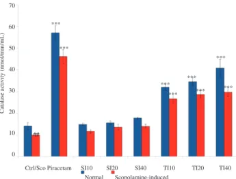
Figure 1 shows that brain catalase activity was significantly increased (P < 0.001) in normal and animals administered with piracetam (57.02 nmol/min/mL), while control was 14.10 nmol/ min/mL. The same significant increase was also observed in all the tempeh isoflavone treated groups (TI10, TI20 and TI40) as compared to the control group. The catalase levels were recorded as 31.92, 34.49 and 40.82 nmol/min/mL at TI10, TI20 and TI40 respectively. In contrast, no significant changes were observed in groups treated with soybean isoflavone in all three administered doses (SI10, SI20 and SI40) when compared to the control group. It is evident that tempeh isoflavone improved the catalase activity within the brain better than soybean. However, tempeh isoflavone was not able to improve the level of catalase as much as piracetam.
Ctrl/Sco Piracetam SI10 SI20 SI40 TI10 TI20 TI40 70
60
50
40
30
20
10
0
Catalase acti
vity (nmol/min/mL)
##
Normal Scopolamine-induced ***
***
*** ***
***
*** ***
***
Figure 1. Effect of soybean (10, 20 and 40 mg/kg) and tempeh (10, 20
and 40 mg/kg) administered orally for 15 days on brain catalase activity in normal and scopolamine-induced groups.
Values were in mean ± SEM (n = 6). ***: P < 0.001 as compared to control and scopolamine-induced groups respectively; ##
: P < 0.01 was the significant value of scopolamine-induced group when compared to control. Ctrl: Control; Sco: Scopolamine.
3.2. Soybean and tempeh isoflavones increased
SODlevel
The effect on SOD activities by soybean and tempeh isoflavones was shown in Figure 2. In normal animal model, piracetam treated group (11.05 IU/mL) showed significant increase (P < 0.01) in the SOD activity as compared to control group (8.99 IU/mL). Low and medium doses of soybean (SI10 and SI20) did not show any significant changes in the SOD activity. However, at a high dose of soybean (SI40 at 10.98 IU/mL) and low dose of tempeh isoflavone (TI10 at 11.30 IU/mL), the SOD activity was significantly increased (P < 0.01) when compared to control. Higher doses of tempeh (TI20, 11.96 IU/mL and TI40, 13.03 IU/mL) showed higher and significant (P < 0.001) increase of SOD activities as compared to control. The results also indicated that TI20 and TI40 significantly improved (P < 0.001) the catalase activity within the brain better than the piracetam group.
Figure 2 also shows the effect of soybean and tempeh isoflavones on SOD activity in scopolamine-induced animal model. Scopolamine significantly reduced (P < 0.001) SOD activity as compared to control group. In contrast, standard drug piracetam (10.66 IU/mL) showed significant (P < 0.001) elevation in SOD as compared to the scopolamine-induced group (6.68 IU/mL). It was observed that
all doses of soybean (8.83–9.24 IU/mL) and tempeh isoflavones (9.51–9.96 IU/mL) showed significant increase in the SOD activities at P < 0.001. Tempeh isoflavone showed higher SOD activities as compared to soybean group. This further supported the ability of tempeh to reverse the memory impairment by scopolamine better than soybean[16].
Scopolamine-induced
Figure 2. Effect of soybean (10, 20 and 40 mg/kg) and tempeh (10, 20 and
40 mg/kg) administered orally for 15 days on the activity of brain SOD in normal and scopolamine induced group.
Values were in mean ± SEM (n = 6). **
: P < 0.01 and ***
: P < 0.001 as compared to control group and scopolamine-induced group respectively; ##
: P < 0.01 was the significant value of scopolamine-induced group when compared to control. Ctrl: Control; Sco: Scopolamine.
Ctrl/Sco Piracetam SI10 SI20 SI40 TI10 TI20 TI40 Normal
##
** ** **
***
*** *** *** *** ***
*** ***
*** 14
12
10
8
6
4
2
0
SOD acti
vity (IU/mL)
3.3. Soybean and
tempeh
isoflavones increased
GSHactivity
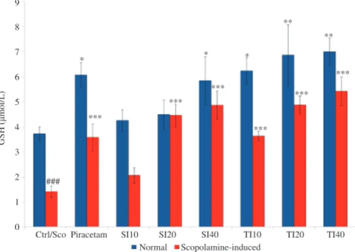
The effect of soybean and tempeh isoflavones on the level of brain GSH was shown in Figure 3. In normal animal model, low doses of soybean isoflavone (SI10 and SI20) did not affect the brain GSH level. However, at a high dose of soybean isoflavone (SI40), significant increase (P < 0.05) in brain GSH level (5.84 μmol/L) was observed as compared to control group (3.73 μmol/L). In tempeh treated groups, all doses displayed a significant increase in the level of GSH (6.23, 6.86 and 6.99 μmol/L in TI10, TI20 and TI40 respectively). In TI10 group, the increase was at the level of P < 0.05 whilst, TI20 and TI40 showed further significant increases at the level of P < 0.01 as compared to control. Piracetam (400 mg/kg) also showed a significant (P < 0.05)
elevation in brain GSH level (6.07 μmol/L). From the results, it can be concluded that TI20 and TI40 possessed better ability to improve the level of GSH within the brain as compared to the piracetam group. Tempeh isoflavone treated groups also had the ability to enhance GSH better than soybean treated groups and it showed that the low dose of tempeh isoflavone (TI10) increased the GSH level similar to that of the higher dose of soybean (SI40).
Scopolamine-induced
Figure 3. Effect of soybean (10, 20 and 40 mg/kg) and tempeh (10, 20 and 40
mg/kg) administered orally for 15 days on level of brain GSH in normal group and scopolamine induced group.
Values were in mean ± SEM (n = 6). *
: P < 0.05, **
: P < 0.01 and *** : P < 0.001 as compared to control and scopolamine-induced group for each model respectively; ###
: P < 0.001 was the significant value of scopolamine-induced group when compared to control. Ctrl: Control; Sco: Scopolamine.
Ctrl/Sco Piracetam SI10 SI20 SI40 TI10 TI20 TI40 Normal
9
8
7
6
5
4
3
2
1
0
GSH (
µ
mol/L)
### ***
*** ***
*** ***
***
* * *
** **
Scopolamine-induced group showed a significant decrease (P <
0.001) in the level of GSH (1.41 μmol/L) as compared to control group (Figure 3). The low dose (SI10) of soybean isoflavone did not show any effect in brain GSH but at higher doses of soybean isoflavone (4.45 and 4.86 μmol/L in SI20 and SI40 respectively) significant increase (P < 0.001) was observed. For tempeh isoflavone, all doses showed significant increase (P < 0.001) of brain GSH (3.64, 4.87 and 5.41 μmol/L in TI10, TI20 and TI40 respectively) as compared to scopolamine-induced group. Piracetam also showed a significant (P < 0.001) elevation in the level of brain GSH (3.58 μmol/L) when compared to scopolamine-induced group. Based on these results, the study suggested that tempeh isoflavone showed better significant improvement in the level of GSH as compared to soybean isoflavone. This suggested tempeh being more health beneficial than soybean.
3.4. Soybean and
tempeh
isoflavones increased GR level
Figure 4 shows the effect of soybean and tempeh isoflavones on the level of brain GR in normal animal model. In normal animal model, low and medium doses (SI10 and SI20) of soybean isoflavone did not affect the brain GR activity. At a high dose of 40 mg/kg of soybean isoflavone (32.74 nmol/min/mL in SI40), a significant increase (P <
0.001) in GR was documented when compared with control (24.03 nmol/min/mL). For tempeh isoflavone, at doses TI20 and TI40 (29.65 and 32.54 nmol/min/mL correspondingly) showed a significant increase in brain GR activities with values of P < 0.05 and P < 0.001 respectively. This suggested that tempeh treated groups showed better ability to improve the GR activity than soybean did. Piracetam also significantly increased (P < 0.001) the brain GR activity as compared to control.
(26.43 nmol/min/mL) while, SI10 and SI20 did not produce any significant changes. High concentration of tempeh isoflavone (TI40) showed significant increase (P < 0.001) in GR activity (29.72 nmol/ min/mL). TI20 increased GR activity (26.39 nmol/min/mL) at the level of P < 0.05 while TI10 did not show any significant differences. Piracetam treated scopolamine induced rats showed improvement in the GR activity as compared to the control scopolamine-induced group (P < 0.001).
Scopolamine-induced
Figure 4. Effect of soybean (10, 20 and 40 mg/kg) and tempeh (10, 20 and 40
mg/kg) administered orally for 15 days on level of brain GR in normal group and scopolamine induced groups.
Values are in mean ± SEM (n = 6). *
: P < 0.05 and ***
: P < 0.001 as compared to control and scopolamine-induced group for each model respectively; ##
: P < 0.01 was the significant value of scopolamine-induced group when compared to control. Ctrl: Control; Sco: Scopolamine.
Ctrl/Sco Piracetam SI10 SI20 SI40 TI10 TI20 TI40 Normal
## ***
***
***
***
*** *** *
* 45
40
35
30
25
20
15
10
5
0
GR acti
vity (nmol/min/mL)
3.5. Soybean and tempeh isoflavones reduced
TBARSlevel
In normal group, all doses (10, 20 and 40 mg/kg) of soybean and tempeh isoflavones exerted a significant reduction of MDA (P < 0.001) as compared to the control group (Figure 5). Soybean isoflavone reduced the MDA to 24.40 μmol/L, 24.12 μmol/L and 23.82 μmol/L (SI10, SI20 and SI40 respectively) as compared to control, 28.05 μmol/ L. Tempeh isoflavone (16.83 μmol/L–20.50 μmol/L) reduced the MDA even greater than soybean did. Piracetam (13.91 µmol/L) being the standard drug used also showed a significant decrease (P < 0.001) compared to control group.
Scopolamine-induced
Figure 5. Effect of soybean (10, 20 and 40 mg/kg) and tempeh (10, 20 and
40 mg/kg) administered orally for 15 days on brain MDA levels in normal groups and scopolamine induced groups.
Values are in mean ± SEM (n = 6). ***: P < 0.001 as compared to control and scopolamine-induced group for each model respectively; ###
: P < 0.001 was the significant value of scopolamine-induced group when compared to control. Ctrl: Control; Sco: Scopolamine.
Ctrl/Sco Piracetam SI10 SI20 SI40 TI10 TI20 TI40 Normal
###
*** ***
*** *** ***
*** ***
*** ******
****** *** *** 45
40
35
30
25
20
15
10
5
0
MD
A (
µ
mol/L
)
A similar trend (Figure 5) was observed in the scopolamine-induced
group, which exerted a significant reduction in the production of MDA (P < 0.001) at all doses of both soybean and tempeh isoflavones (10, 20 and 40 mg/kg). The level of MDA in scopolamine-induced group was higher (38.09 µmol/L) than control (28.05 µmol/L). Soybean isoflavone reduced the level of MDA to 22.73 µmol/L, 21.66
µmol/L and 20.95 µmol/L (SI10, SI20 and SI40 respectively) while tempeh isoflavone showed even greater reduction with the values of 20.59 µmol/L, 16.61 µmol/L and 14.60 µmol/L (TI10, TI20 and TI40 respectively) as compared to scopolamine-induced group. The scopolamine-induced animals treated with piracetam also showed a significant decrease (P < 0.001) in the level of MDA with value of 15.51 µmol/L as compared to scopolamine-induced group.
3.6. Soybean and tempeh isoflavones reduced NO
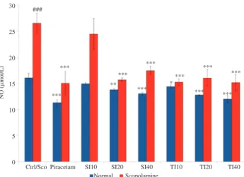
The effect of soybean and tempeh isoflavones on NO level in brain was shown in Figure 6. In normal animal model, the level of NO was observed at 16.06 µmol/L. With treatment of piracetam at 400 mg/kg, significant decrease of NO level (11.29 µmol/L) was observed at the level of P < 0.001. The same significant level was also observed in SI40, TI20 and TI40 groups with values 13.03 µmol/L, 12.77 µmol/L and 12.00 µmol/L respectively. SI20 significantly reduced (P < 0.01) NO to 13.76 µmol/L. Low dose of tempeh isoflavone group, TI10 also showed significant reduction at the level of P < 0.05 with value of 14.35 µmol/L. Meanwhile, SI10 did not show significant reduction. All groups were statistically compared to control group.
Figure 6. Effect of soybean (10, 20 and 40 mg/kg) and tempeh (10, 20 and 40
mg/kg) administered orally for 15 days on brain NO levels in normal groups and scopolamine induced groups.
Values are in mean ± SEM (n = 6). *
: P < 0.05, **
: P < 0.01 and *** : P < 0.001 as compared to control and scopolamine-induced group for each model respectively; ###
: P < 0.001 was the significant value of scopolamine-induced group when compared to control. Ctrl: Control; Sco: Scopolamine.
Ctrl/Sco Piracetam SI10 SI20 SI40 TI10 TI20 TI40 Normal
###
*** ***
***
*** ***
***
*** ***
*** ***
** *
Scopolamine 30
25
20
15
10
5
0
NO (
µ
mol/L)
4. Discussion
Oxidative stress can be very deleterious in the biological system especially in brain as it will lead to cell damage and death. Antioxidants, free radical scavengers and chelators are three agents that have been studied to directly suppress the formation of free radicals within the biological system. In contrast, free radical inhibitors that can be classified into antioxidant enzymes and other compounds may indirectly improve oxidative stress. The indirect reaction may be through the inhibition of the pro-oxidant enzymes or oxidative activity[17]. The free radical scavengers may react with free radical which produces inactive, stable radicals. The chelators act by chelating the active transition metals to form inactive complexes hence delay in oxidation.
Our in vivo experiments in the present study further confirm our previous in vitro study that tempeh isoflavone was a better free radical scavenger and ferrous ion chelator as compared to soybean isoflavone[14]. The tempeh isoflavone showed an elevated level of GSH and other antioxidant enzymes such as SOD, catalase and GR. Wang et al.[18] found that douchi, soybean fermented with Aspergillus oryzae increased the level of glutathione peroxidase, catalase and SOD in rats. Yet another study by Wu and Chu showed fermentation of soybean with Bacillus subtilis exerted an increment of SODin vitro[19]. These suggest that the fermentation process of soybean increased the antioxidant enzymes both in vitro and in vivo.
SOD is an antioxidant enzyme that plays a vital role in regulation of free radical processes in biological systems. In general, SOD stabilises free radicals by donating its electron producing H2O2. Theoretically,
the dismutation of superoxide occurs at copper, manganese or iron centres of SOD isoenzymes CuZnSOD, MnSOD or FeSOD but only CuZnSOD and MnSOD are used by eukaryotes[17]. According to Deng et al.[20], superoxide activates cytokines, by which CuZnSOD may exhibit neuroprotective effect by suppressing the microglial activation by the cytokines. The MnSOD has a role in preventing the mitochondrial production of oxygen radicals. It is also believed that the overexpression of MnSOD in the brain prevents apoptosis induced by tumor necrosis factor-α[21]. Previous studies indicated that soybean is able to increase the SOD level within the brain[5,18,22,23]. In the present study, the results showed that in normal and scopolamine-induced animal models, tempeh isoflavones at all doses exhibited significant increase of SOD in the brain and better than soybean isoflavones due to the higher amount of aglycones after fermentation process[14]. This suggests that tempeh is a better antioxidant when compared to soybean. Tempeh was able to reverse the scopolamine-induced amnesia by increasing the level of SOD in scopolamine-induced animals. Thus, the risk of getting AD might be reduced by consuming soybean and its fermented product tempeh.
Catalase is known as a heme containing enzyme that decomposes H2O2 to oxygen and water. This reduces the deleterious effect of H2O2
such as cell damage and induction of apoptosis. Many recent studies supported that the increase of catalase in animals treated with soybean food products[5,18,22,23]. The H2O2 within the biological system should
be balanced between the oxidation and repair mechanisms. The present study proved that soybean isoflavones significantly increased catalase within the brain. However, tempeh isoflavone treated animals showed a more significant increase of catalase in the brain as compared to soybean isoflavone treated animals. This suggested that tempeh possessed better antioxidant ability as compared to soybean. The consumption of tempeh may be more beneficial to health. GSH is a tripeptide (γ-L-glutamyl-L-cysteinylglycine) which is the most abundant thiol antioxidant in mammalian biological systems. GSH is involved in metabolism, catalysis and the most important as an antioxidant. GSH is capable to act as free radical scavenger and electron donor in enzymatic redox cycle of glutathione peroxidase and GR catalyzing the reduction of peroxides[17]. As a water-soluble
antioxidant, GSH is very important to reduce the level of free radicals that prevent the cells from oxidative stress and cell damage in the brain. When the cells and neurons within the brain are depleted, brain losses its functions especially in memory, emotion, motor cognitive ability etc. These can be supported by a study which found that the depletion of GSH in the brain led to cytotoxicity, which correlated with the formation of excessive NO in the brain[24]. The NO has been well debated as one of the causes to oxidative stress whilst antioxidants have been reported to inhibit NO. Therefore, high levels of GSH as observed in this study may lead to the low NO level. The results showed that the tempeh isoflavone groups exhibited higher amount of GSH within the brain than soybean isoflavone groups did.
GR is important in GSH redox cycle. The GR is important in
production of GSH asa result from reaction with glutathione disulfide, nicotinamide adenine dinucleotide phosphate and H+[25]. In this study, the results showed that tempeh isoflavone treated animals exhibited higher level of GR as compared to soybean isoflavone treated animals. This would suggest that the consumption of tempeh is more beneficial in preventing oxidative series within the brain that causes neurodegeneration and AD.
In this study, the measurement of MDA was performed to measure the effects of soybean and tempeh isoflavones on the magnitude of lipid peroxidation within the brain. MDA is a biomarker for oxidative stress as it is one of the end products of lipid peroxidation process[26]. Measurement of the MDA within the brain tissue represents the magnitude of the deleterious effect of lipid peroxidation in
neurodegenerative diseases such as AD, Parkinson’s disease and Huntington’s disease[27]. Therefore, to overcome this process, it is crucial to inhibit the peroxidation that occurs within the brain. Much has been documented recently on food based products with health benefits especially in preventing oxidation. But to date, very few studies reported on the ability of tempeh as an anti-oxidative food. Based on the results obtained in this study, it showed that treatment with 40 mg/kg of soybean isoflavone reduced the MDA level significantly as compared to control group. In fact, even a low dose of 10 mg/kg of tempeh isoflavone showed more significant results as compared to 40 mg/kg of soybean isoflavone treatment. These results reflected that tempeh is rich with aglycones which are easily absorbed as compared to glycoside. Besides that, the results obtained in antioxidant studies also correlated with the result of MDA levels. Tempeh isoflavones facilitated the increasing level of GSH within the brain better than soybean isoflavone did. It suggested that antioxidant GSH would reduce the free radicals so that the peroxidation of lipids is weakened. A previous study showed that soybean attenuated oxidative stress by reducing TBARS formation and increasing GSH level within the rat brain[18]. While, another study found that soybean supplementation reversed the scopolamine-induced amnesia and significantly increased the GSH levels and lowered the levels of TBARS within the brain[22]. Both studies supported the results obtained from the present study, where soybean isoflavone decreased the level of lipid peroxidation. Tempeh however, has excellent ability to reduce lipid peroxidation as compared to soybean suggesting that tempeh has better health beneficial effects.
observed in scopolamine-induced model, by which both soybean and tempeh isoflavones treated groups showed significant reduction in the level of NO as compared to scopolamine-induced group. Interestingly, tempeh isoflavone treated groups showed more significant reductions as compared to soybean isoflavone treated groups. Previous study also suggested that soybean genistein has the ability to alleviate the excessive NO level within biological system[29]. Besides that, high level of NO also can induce inflammation and tissue damage. Therefore, this study also correlated the significant reduction of NO by soybean and tempeh isoflavones.
From the SOD and catalase activities results, this study concluded that the increase in SOD activity was followed by the increase in catalase activity. The tempeh isoflavone showed an elevated level of GSH and other antioxidant enzymes such as SOD, catalase and GR. Also, both soybean and tempeh isoflavones showed significant reduction in the oxidative biomarkers, which are MDA and NO. Both isoflavones also reversed the scopolamine-induced effects, which also significantly reduced the NO production within the brain. However, tempeh isoflavone showed more significant results as compared to soybean isoflavone. The low level in these lipid peroxidation biomarkers would suggest that soybean and tempeh are beneficial to health especially in promoting anti-AD, with regards that tempeh is much more beneficial to health.
Conflict of interest statement
We declare that we have no conflict of interest.
Acknowledgments
This study was supported by the Research Excellence Funds (600-RMI/DANA5/3/REI (3/2013) and 600-RMI/ST/DANA5/3/DST (465/2011), Research Management Centure, Universiti Teknologi MARA, Malaysia and Institute of Graduate Studies, Universiti Teknologi MARA, Malaysia.
References
[1] Yang Y, Karakhanova S, Werner J, Bazhin AV. Reactive oxygen species in cancer biology and anticancer therapy. Curr Med Chem 2013; 20: 3677-92.
[2] Uttara B, Singh AV, Zamboni P, Mahajan RT. Oxidative stress and neurodegenerative diseases: a review of upstream and downstream antioxidant therapeutic options. Curr Neuropharmacol 2009; 7: 65-74. [3] Floyd RA, Hensley K. Oxidative stress in brain aging. Implications for
therapeutics of neurodegenerative diseases. Neurobiol Aging 2002; 23: 795-807.
[4] Moreira PI, Zhu X, Wang X, Lee HG, Nunomura A, Petersen RB, et al. Mitochondria: a therapeutic target in neurodegeneration. Biochim Biophys Acta 2010; 1802: 212-20.
[5] Bagheri M, Joghataei MT, Mohseni S, Roghani M. Genistein ameliorates learning and memory deficits in amyloid β(1-40) rat model of
Alzheimer’s disease. Neurobiol Learn Mem 2011; 95: 270-6.
[6] Guglielmotto M, Giliberto L, Tamagno E, Tabaton M. Oxidative stress
mediates the pathogenic effect of different Alzheimer’s disease risk
factors. Front Aging Neurosci 2010; 2: 3.
[7] Liang W, Lee AH, Binns CW, Huang R, Hu D, Shao H. Soy consumption reduces risk of ischemic stroke: a case-control study in southern china. Neuroepidemiology 2009; 33: 111-6.
[8] Nagarajan S. Mechanisms of anti-atherosclerotic functions of soy-based diets. J Nutr Biochem 2010; 21: 255-60.
[9] Korde LA, Wu AH, Fears T, Nomura AM, West DW, Kolonel LN, et al. Childhood soy intake and breast cancer risk in Asian American women. Cancer Epidemiol Biomarkers Prev 2009; 18: 1050-9.
[10] Hwang YW, Kim SY, Jee SH, Kim YN, Nam CM. Soy food consumption
and risk of prostate cancer: a meta-analysis of observational studies. Nutr Cancer 2009; 61: 598-606.
[11] Villa P, Costantini B, Suriano R, Perri C, Macrì F, Ricciardi L, et al. The differential effect of the phytoestrogen genistein on cardiovascular risk factors in postmenopausal women: relationship with the metabolic status. J Clin Endocrinol Metab 2009; 94: 552-8.
[12] Pipe EA, Gobert CP, Capes SE, Darlington GA, Lampe JW, Duncan AM. Soy protein reduces serum LDL cholesterol and the LDL cholesterol: HDL cholesterol and apolipoprotein B: apolipoprotein A-I ratios in adults with type 2 diabetes. J Nutr 2009; 139: 1700-6.
[13] Chang CT, Hsu CK, Chou ST, Chen YC, Huang FS, Chung YC. Effect of fermentation time on the antioxidant activities of tempeh prepared from fermented soybean using Rhizopus oligosporus. Int J Food Sci Technol 2009; 44: 799-806.
[14] Ahmad A, Ramasamy K, Majeed AB, Mani V. Enhancement of
β-secretase inhibition and antioxidant activities of tempeh, a fermented soybean cake through enrichment of bioactive aglycones. Pharm Biol 2015; 53: 758-66.
[15] Ecobichon DJ. The basis of toxicity testing. 2nd ed. Boca Raton: CRC Press; 1997.
[16] Ahmad A, Ramasamy K, Jaafar SM, Majeed AB, Mani V. Total isoflavones from soybean and tempeh reversed scopolamine-induced amnesia, improved cholinergic activities and reduced neuroinflammation in brain. Food Chem Toxicol 2014; 65: 120-8.
[17] Denisov ET, Afanas’ev IB. Oxidation and antioxidant in organic chemistry and biology. Boca Raton: CRC Press; 2005.
[18] Wang D, Wang L, Zhu F, Zhu J, Chen X, Zou L, et al. In vitro and in vivo studies on the antioxidant activities of the aqueous extracts of Douchi (a traditional Chinese salt-fermented soybean food). Food Chem 2008; 107: 1421-8.
[19] Wu CH, Chou CC. Enhancement of aglycone, vitamin K2 and superoxide dismutase activity of black soybean through fermentation with Bacillus subtilis BCRC 14715 at different temperatures. J Agric Food Chem 2009; 57: 10695-700.
[20] Deng YY, Lu J, Ling EA, Kaur C. Role of microglia in the process of inflammation in the hypoxic developing brain. Front Biosci (Schol Ed) 2011; 3: 884-900.
[21] Keller JN, Kindy MS, Holtsberg FW, St Clair DK, Yen HC, Germeyer A, et al. Mitochondrial manganese superoxide dismutase prevents neural apoptosis and reduces ischemic brain injury: suppression of peroxynitrite production, lipid peroxidation, and mitochondrial dysfunction. J Neurosci 1998; 18: 687-97.
[22] Bansal N, Parle M. Soybean supplementation helps reverse age- and scopolamine-induced memory deficits in mice. J Med Food 2010; 13: 1293-300.
[23] Yang H, Jin G, Ren D, Luo S, Zhou T. Mechanism of isoflavone
aglycone’s effect on cognitive performance of senescence-accelerated
mice. Brain Cogn 2011; 76: 206-10.
[24] Aquilano K, Baldelli S, Cardaci S, Rotilio G, Ciriolo MR. Nitric oxide is the primary mediator of cytotoxicity induced by GSH depletion in neuronal cells. J Cell Sci 2011; 124: 1043-54.
[25] Wheeler CR, Salzman JA, Elsayed NM, Omaye ST, Korte DW Jr. Automated assays for superoxide dismutase, catalase, glutathione peroxidase, and glutathione reductase activity. Anal Biochem 1990; 184: 193-9.
[26] Markesbery WR, Kryscio RJ, Lovell MA, Morrow JD. Lipid peroxidation is an early event in the brain in amnestic mild cognitive impairment. Ann Neurol 2005; 58: 730-5.
[27] Reed TT. Lipid peroxidation and neurodegenerative disease. Free Radic Biol Med 2011; 51: 1302-19.
[28] C h u n g K K , D av i d K K . E m e rg i n g r o l e s o f n i t r i c o x i d e i n neurodegeneration. Nitric Oxide 2010; 22: 290-5.