*Corresponding author: Eduardo Alves de Almeida; e-mail: ealmeida@ibilce.unesp.br
Biochemical and genotoxic effects of a commercial formulation of the
herbicide tebuthiuron in Oreochromis niloticus of different sizes
M.F. F
ranco-B
ernardes; L.r. M
aschio; M.T.V.
dea
zeredo-o
LiVeira& e.a.
dea
LMeidaUniversidade Estadual Paulista Júlio de Mesquita Filho. Rua Cristóvão Colombo, 2265. 15054-000. São José do Rio Preto, SP. Brazil.
(Received April 16, 2014; Accept June 17, 2014)
Abstract
Pesticides are serious contaminants because they are designed to eliminate pests, but they also affect non-target species. The present study aimed to evaluate the biochemical and genetic effects of the herbicide tebuthiuron in Oreochromis niloticus of different sizes. Thus, we analyzed biomarkers in small and large O. niloticus specimens exposed to 62.5, 125 and 250 mg L-1 of
tebuthiuron for 72 hours. Fish exposed to 250 mg L-1 had high mortality rates; therefore, the data could not be used. The results
showed an increase in EROD activity in ish exposed to 125 mg L-1, but no GST alteration. Antioxidant enzymes GPx and CAT
were altered only in the liver of treated ish compared to the control group: CAT decreased in large ish, and GPx increased in small ish. The MDA analysis did not evidence lipid peroxidation. High DNA damage in exposed small ish (not in large ish)
was observed using comet assay, but a micronucleus test did not show mutagenicity. Moreover, a comparison between control
groups with specimens of different sizes revealed that small ish are more susceptible than large ish to the tebuthiuron effects, since increased comet scores was observed only for smaller ish.
Keywords: aquatic toxicology; biomarkers; ecotoxicology; ish; pesticide.
INTRODUCTION
According to estimates from the Brazilian agency known
as UNICA (Sugarcane Industry Union) (Sugarcane Industry
Union, 2013), the Center-South region of Brazil will produce 589 million tons of sugarcane during the 2013/2014 harvest. This impressive productivity is supported by the extensive use of several agrochemicals for pest control that, as a
consequence, can result in signiicant negative impacts on the environment. In the case of herbicides alone, more than
30 compounds present in different formulations are used in sugarcane cultivation in Brazil (Martini & Durigan, 2004), such as 2,4-D, acetochlor, ametryne, clomazone, atrazine, diuron, hexazinone, glyphosate and tebuthiuron, though the toxic effects of some of these compounds on non-target organisms are not known (Oruç & Üner, 2000; Cattaneo et al., 2008; Xiao et al., 2006; Paiva and Takahashi, 1996; Sikkema
& Shropshire, 2007; Nwani et al., 2011; Akcha et al., 2012;
Mei et al., 2012; Lancetõt et al., 2013; Bicalho & Langenbach,
2013). Tebuthiuron (1-(5-tert-Butyl-1,3,4-thiadiazol-2-yl)-1,3-dimethylurea) is a pre- and post-emergent substituted urea herbicide used for the control of broadleaf and woody weeds,
grasses and brush through the inhibition of photosystem II.
In Brazil, it is widely used on sugarcane crops (Tofoli et al.,
2009), and it is present in different formulations. In some
cases, it is present in combination with other herbicides. Tebuthiuron has a long half-life of about one year in soils
(Helling, 2005), especially in high-carbon or low-rainfall
environments (Chang & Strizke, 1977), but may have a
half-life as low as 20 days under speciic conditions (Cerdeira et
al., 2007). Due to its high water solubility, relatively strong persistence, and low absorption in soil particles, it is expected that tebuthiuron can reach aquatic environments, thus causing negative effects on non-target organisms.
According to data from EPA (2004) and the U.S. National
Library of Medicine (2013), tebuthiuron is slightly to
practically non-toxic to ish and other aquatic species, with
trout, and 87 to 112 mg L-1 in bluegill sunish; however, the
sublethal effects of tebuthiuron on ish metabolism are not
yet completely known. Due to its intense use in agriculture, especially in sugarcane cultivation in Brazil, any additional
information on the sublethal effects of tebuthiuron to ish
are important for a better understanding of the risks that
this herbicide poses to ish communities that inhabit aquatic environments near agricultural zones. In this context, an
assessment of biochemical biomarkers can be useful, since it
generally represents the irst line of metabolic responses of
cells to the contaminants.
The activities of biotransformation enzymes such as cytochrome P450 isoforms or glutathione S-transferases are
commonly evaluated as biomarkers in ish, since they can be signiicantly induced by several contaminants and therefore
indicate a response from the organism to the pollutant (Van Der Oost et al., 2003). Oxidative stress markers, such as the activities of the antioxidant enzymes superoxide dismutase (SOD), glutathione peroxidase (GPx) and catalase (CAT), or the levels of oxidative damage to macromolecules, are also useful biomarkers. Several environmental contaminants can lead cells to increase their rates of oxygen reactive species (ROS) generation due to increases in biotransformation reactions, cyclic redox reactions and/or increases in oxygen uptake by mitochondria for energy production.
If not counteracted by antioxidant defense systems, ROS
can oxidize macromolecules, thus leading to oxidative damages to proteins, lipids and nucleic acids and generating oxidative stress (Oruç & Üner 2000; Van Der Oost et al., 2003). Furthermore, the genotoxic effects of environmental pollutants can be monitored using a broad range of assays, such as the micronucleus test and the comet assay (Çavas & Könen, 2007).
Although numerous studies exist in the literature that report oxidative stress and genotoxic effects caused by different photosystem inhibitor herbicides (i.e. diuron, atrazine and
paraquat) in ish, there is no such data available regarding
the effects of tebuthiuron. Due to both the intense use of tebuthiuron formulations in sugarcane cultivation in Brazil and the lack of studies regarding the sublethal effects of this
herbicide in ish, this work considered whether tebuthiuron affects some classic biochemical biomarkers in Nile Tilapia
(Oreochromis niloticus): the phase I enzyme
ethoxyresoruin-O-deetilase (EROD, indicative of CYP1A), the phase II
enzyme glutathione S-transferase (GST), the antioxidant enzymes SOD, CAT and GPx, the levels of oxidative damage to lipids (lipid peroxidation, as malondialdehyde levels, MDA), and levels of genotoxic markers (micronucleous tests, comet assay and nuclear abnormality levels).
Because biomarker responses can vary signiicantly according to the size of the ish (Peixoto & Santos, 2009),
a factor that can indicate differences in the organism’s susceptibility to the pollutant, we were also interested in evaluating the discrepancies in biomarker responses among
ish of the same species (O. niloticus) but with signiicant differences in body length and weight. The sizes of aquatic
animals have been shown to play an important role in tissue pollutant loads. Recently Kanak et al. (2014) showed that younger tilapias were affected from metal exposure much
more than large ones (two fold in length and ive fold in weight), as their antioxidant parameters signiicantly
decreased even in controls. Therefore, although EPA
reported low toxicity of tebuthiuron to ish, we hypothesize
that this herbicide can poses important metabolic alterations in tilapias, which can be helpful to understand its effects
in ish, and that smaller ish are more susceptible to the
tebuthiuron effects than larger ones.
MATERIAL AND METHODS
Chemicals
All reagents were purchased from Sigma Chemical. In our
study, the herbicide was tested as the complex commercial mixture because this is the form in which it is routinely applied in agriculture and introduced into the environment. The commercial preparations of 1-(5-tert-butyl-1,3,4-thiadiazol-2-yl)-1,3-dimethylurea (tebuthiuron) (known as Combine*
500SC) was used. It is composed of the herbicide (50% w / v, or 500 g / L) and inert ingredients (63.3% w / v, or 633g / L).
Animals and experimental design
Adult nile tilapia (O. niloticus) of both sexes were obtained from the Aquaculture Center of Universidade Estadual Paulista, Jaboticabal Campus, and transferred to the Laboratory of Animal Ecology, at Universidade Estadual Paulista, Campus of São José do Rio Preto. The specimens were kept in tanks with clean tap water for at least one week before the experiment. A total of 48 animals were
used; 24 were small ish (mean length 11.68 ± 1.23 cm, mean weight 47.19 ± 15.5 g) and 24 were large ish (mean length 20.74 ± 1.79 cm, mean weight 284.16 ± 77.95 g).
Fish were placed individually into 48 aquariums of 17 L in
eight groups of six ish each (N=6, real replicas). Male and female was not separated. For each ish size, the animals were
divided into four groups of six animals: the control group and three groups with different concentrations of the herbicide. The nominal concentrations of tebuthiuron that were used were 62.5, 125 and 250 mg L-1 (corresponding to 0.125, 0.25
and 0.500 mL L-1 of the commercial Combine *500SC).
Due to the lack of studies regarding subcellular effects of
tebuthiuron on ish, and due to the reported low toxicity of this herbicide in ish according to the EPA pesticide database, we
chose to test concentrations near the LC50 values reported for rainbow trout (143 mg L-1) and bluegill sunish (106 mg L-1).
Because we used a formulation of tebuthiuron instead of the pure standard, we also considered the LC50 values reported
by EPA for formulated tebuthiuron products (20 to 80%)
for fathead minnow (>180 mg L-1). We also considered the
fact that the ish in our study were exposed to the formulated
database, chronic exposure (45 days) of rainbow trout to 52 mg L-1 of tebuthiuron caused no mortality, but only effects on
growth. This further justiies the elevated concentrations used
in our study.
The aquariums were kept under constant aeration and
temperature. No food was supplied to the ish during the
experiment, and the animals were exposed to the herbicide for 72h. After this period, they were anesthetized with benzocaine for removal of the liver, gills and blood. The liver and gills were frozen at -80° C for subsequent biochemical analysis. The blood was used for micronucleus tests and comet assays on the same day.
Preparation of samples for Biochemical analysis
For the analysis of enzymes EROD, GST, SOD, GPx and CAT, tissues (liver and gills) were homogenized (1:4, w:v)
in Tris-HCl 20 mM, pH 7.5 containing 0.5M of sucrose,
1mM of etylenediamine tetraacetic acid (EDTA), 1mM of dithiothreitol (DTT), and 0.15M of KCl containing 1 mM
of the protease inhibitor phenylmethylsulfonyl luoride
(PMSF). The homogenate was centrifuged at 10,000g for 30 min at 4ºC, and the resulting supernatants were centrifuged at 50,000g for 60 min at 4ºC. The supernatant obtained after this second centrifugation was used for the analyses of GST, SOD, GPx and CAT, and the pellet of the liver samples was
re-suspended in 100 ml of 0.1-M Tris-HCl, pH 7.5, containing
1mM of EDTA, 1mM of DTT, and 0.1 M of KCl and used to analyze EROD activity. The prepared samples were aliquoted for later analysis. For the lipid peroxidation analysis, tissues (liver and gills) were homogenized (1:3 weight: volume) in
0.1M of Tris HCl, pH8.0. Prepared samples were analyzed on
the same day.
BIOCHEMICAL ANALYSIS
EROD, GST, SOD, GPx and CAT
The EROD assay measures the 0-dealkylation, mediated
by CYP1A, of non-luorescent substrate 7-ethoxy-resoruin in resoruin, a luorescent product detected by luorimeter (λexcit = 537 nm λemiss = 583 nm) (Sarasquete and Segner, 2000).
The assay mixture contained 80 mM of potassium phosphate
buffer (pH 7.4), 335 lM 7-ethoxyresoruin, 20 mM of NADPH,
and microsomal liver extract (prepared sample). The reaction was observed for 3 min at 30 ºC. EROD activity (pmol min-1
mg-1 of protein) was calculated based on a previously prepared
resoruin standard curve.
GST activity was evaluated according to Keen et al. (1976). The assay mixture contained 0.2M of potassium phosphate
buffer, pH 6.5, the substrate 1-chloro-2,4-dinitrobenzene (CDNB), GSH, and the sample containing GST. The activity was
determined by measuring the increase in absorbance at 340 nm.
The method for analyzing SOD uses a system of superoxide (xanthine / xanthine oxidase) generation coupled
with cytochrome c reduction by the superoxide anion radical, which causes an increase in absorbance at 550 nm at 25 ° C. The addition of the sample containing SOD promotes an inhibition of cytochrome c reduction, because the enzyme competes with cytochrome c by superoxide (Mccord & Fridovich, 1969).
GPx activity was evaluated according to Sies et al. (1979).
In the analysis, the consumption of NADPH was monitored at 340 nm. NADPH is used by glutathione reductase (GR)
for the reduction of glutathione, which had been previously oxidized by GPx in the conversion of t-butyl hydroperoxide in its corresponding alcohol.
CAT activity was measured following the method reported by Beutler (1975), in which the rate of decomposition of
hydrogen peroxide by the enzyme is quantiied through
the decrease in absorbance at 240 nm at 30 °C.
Protein levels were measured following the method of
Bradford (1976). In a microtube, the Bradford reagent and
the sample were combined. After 40 min in the dark, the samples were measured in a spectrophotometer at 595 nm. The protein concentration was calculated using the calibration curve prepared from bovine serum albumin (BSA) and the Bradford reagent.
Lipid peroxidation
Lipid peroxidation levels were determined by measuring the product formed from the combination of malondialdehyde,
and thiobarbituric acid (TBA) through High Performance Liquid Chromatography (HPLC) and UV/Vis detection.
After homogenizing the sample, the TBA was dissolved in
0.2 M of HCl and the solution was added to the sample. The
reaction mixture was then heated at 90
º
C for 60 min. Thecolored derivative was extracted with butanol and quantiied using HPLC at 532 nm, in terms of a malondialdehyde (MDA)
standard calibration curve that had been previously prepared
using the same procedure used for the samples. The HPLC
system consisted of an ESA584 pump and an ESA526 UV/ Vis detector. The column used was an ACE 5 C18 (250_x 4.6 mm, 5 µm). Chromatogram monitoring and peak identiication
and quantiication were performed using the EZChrom Elite
software (Agilent Technologies). The mobile phase was 0.05
M of KH2PO4, pH 7.0, with 40% methanol, and was pumped
at an isocratic low of 1 mL min-1.
GENOTOXIC EFFECTS
Micronucleus test
In the micronucleus test (Al Sabti, 1986; Al Sabti and
Metcalfe, 1995), a cardiac puncture was performed to remove 3 cc of blood from each animal. The smear technique was used
to prepare three slides for each ish. The material was ixed in
3000 erythrocytes per ish. This analysis was performed with an optical microscope under 100 x magniication, and the
frequency of micronuclei in treated animals was compared to the negative control. The frequencies of nuclear abnormalities (notched, lobed, broken eggs and bebbled, as described by
Carrasco 1990), was also determined. No positive control was used. Since all cells in the ield of vision are counted, the
total number of cells analyzed per animal can vary. Thus, the total number of cells counted was transformed into percentage values. Therefore, the frequency is measured as a percentage.
Comet assay
The comet assay was performed followed the method described by Singh et al. (1988). Blood samples were each diluted in 1000 µl of saline solution. Slides were made with 10 uL of this cell suspension and 120 µl of low melting
point agarose (0.5%) at 37 °C. The slides remained in a lysis
solution (1 mL of Triton X-100, 10 mL of DMSO and 89 mL
of lysing solution stock, pH 10.0 - stock solution: 2.5 M of NaCl, 100 mM of EDTA100, 10 mM to 1 L of Tris) in the
refrigerator for one hour. After their time in the lysis solution, the slides were placed inside a horizontal electrophoresis system for 20 minutes at 25 V, 300 mA. The slides were
neutralized with 0.4M of Tris (pH 7.5), for 15 minutes and ixed in ethanol for 10 minutes. Cells with no DNA damage migrate homogeneously, while cells with damaged DNA form
fragments of different sizes, and the smaller ones migrate faster during electrophoresis, thus forming the tail of a comet.
From each ish, two slides were prepared. On each
slide, 50 nucleoides were analyzed. The slides were stained with ethidium bromide (0.002 mg mL-1). The analysis was
performed with a luorescence microscope, ilter B - 34 (excitation: λ = 420-490 nm, barrier λ = 520 nm) under 40 x magniication. The comets were classiied according to
the comet length using the visual parameters proposed by Kobayashi et al. (1995): class 0 (no damage), class 1 (slight damage); class 2 (moderate damage), class 3 (major damage). The injury score was calculated by multiplying the total
number of cells in each class by class value (0 -3). No positive
control was used.
Statistical analysis
To verify the effect of the herbicide on the animals, the treatments were compared to the control. The small ish
group was analyzed separately from the large ish group. Tests
for normality (Shapiro–Wilk) and homogeneity of variances (Levene) were applied. For the parametric data, the
one-way ANOVA was used, followed by the Tukey test. For
non-parametric data, the Kruskal–Wallis test was used, followed by multiple comparisons of mean ranks.
To see whether the size of the ish inluences the effect
of the biomarkers studied, the differences between small and
large ish and their respective control groups were calculated.
For the parametric data, the Student T-test was used, and
for non-parametric data, the Mann-Whitney test was used.
Signiicant differences were accepted only when p < 0.05.
Analyses were performed with the Statistica 7.0 software.
RESULTS
Mortality
During the exposure period, a high mortality rate of the animals was observed in aquariums containing 250 mg L-1 of
tebuthiuron. In aquariums containing small ish, four of them died, leaving only two animals to be analyzed. In aquariums containing large ish, two of them died, leaving four for analysis.
Due to this mortality rate, statistical analyses were infeasible
with the group of ish exposed to the high concentration of the herbicide, and for this reason, data for the ish exposed to 250
mg L-1 of the herbicide were not considered any further. None
of the ish died in the other treatments.
Biotransformation enzymes
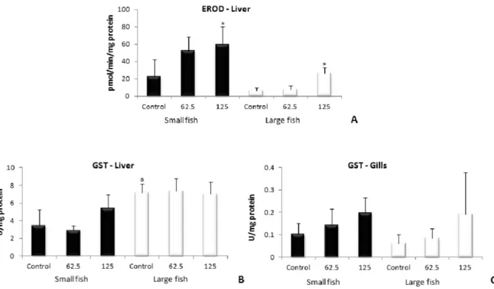
The results regarding EROD and GST activities are shown in Figure 1. EROD activity was measured only in the liver, due to a lack of gill samples available for measurement. The
activity of the enzyme increased in small and large ish exposed
to 125 mg L-1 when compared to their respective controls. The
analysis did not show signiicant difference between animals
of different sizes. With respect to GST activity, no differences were observed between the treated groups and the respective
control groups in neither the liver nor the gills from ish of either size. When the GST activity of ish of different sizes
was compared, it was noted that GST activity in the liver was
higher in large ish than in small ish, but no such difference
was observed in the gills.
Oxidative stress parameters
Oxidative stress parameters (SOD, CAT, and GPx
activities, and MDA levels) measured in ish from the
exposure experiment are shown in Figure 2. The hepatic SOD
activity showed no signiicant differences in small and large ish treated with tebuthiuron in comparison to their respective control groups. The analysis also showed no signiicant
differences between O. niloticus of different sizes. The SOD
activity in the gills did not differ signiicantly in exposed
groups when compared to their controls; however, the enzyme
activity was higher in large ish than in small ish.
GPx activity was signiicantly higher in the liver of small
ish exposed to 125 mg L-1 than in the control group, though
no signiicant difference was found in large ish treated with the herbicide when compared to their control group. No
difference was observed in GPx activity among animals with
In the liver, the CAT activity did not differ signiicantly
in small O. niloticus exposed to herbicide when compared
to the control. However, large ish treated with 62.5 mg L-1
had lower CAT activity than the control group. There was no
signiicant difference between the control groups of large or
small animals. The CAT activity of the gills did not differ
signiicantly in exposed groups compared with their controls, nor was there any signiicant difference between ish of
different sizes.
No signiicant difference was observed in MDA levels in livers of ish exposed to the herbicide compared to the
control groups. However, large O. niloticus showed a higher
level of lipid peroxidation than small specimens. The MDA levels in the gills remained unchanged when treated and
control animals were compared. There was also no signiicant
difference between the lipid peroxidation levels in animals of different sizes.
Genotoxic markers
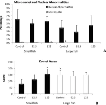
The micronucleous frequency in erythrocytes from O. niloticus specimens of both sizes exposed to tebuthiuron did not differ from their respective controls (Fig. 3). On the other
hand, large ish had lower micronucleous frequency than small ish. Some nuclear abnormalities were also observed, being
the notched the most predominant, but no differences were observed between treated and control groups. The nuclear
abnormalities did not differ between small and large ish, either. The comet assay showed that small ish exposed to 125
mg L-1 of the herbicide presented a higher level of damaged
erythrocytes than the control group. Furthermore, the analysis
revealed that large ish had a higher comet score than small ish.
DISCUSSION
Although studies regarding the biological effects of pesticides have increased substantially over the last few years, there are no studies on the effects of tebuthiuron on biochemical
biomarker in ish. For this reason, the present work brings
important information about the susceptibility and responses
of ish to a commercial formulation of this herbicide, which
has been extensively used in sugarcane agriculture in Brazil.
EROD activity was higher in the group of large ish exposed
to 125 mg L-1 of the tebuthiuron formulation when compared
to the controls. Numerous ield studies have demonstrated a signiicant increase in hepatic CYP1A activity in many species of ish from polluted environments, though no studies
exist regarding the effects of tebuthiuron on this enzyme.
Nevertheless, Schoket & Vincze (1990) observed a signiicant
increase in EROD activity in rats intragastrically treated with diuron (0.73, 1.62, 2.43 and 3.65 mmol/kg) and chlorotoluron (2.35, 5.22, 7.83 and 11.74 mmol/Kg), both phenylurea herbicides, for 3 days. As demonstrated in this study, the herbicide tebuthiuron was also able to induce EROD activity
in tilapia, an increase that was not followed by the phase II
enzyme GST. This result suggests that GST is not involved
in the metabolism of tebuthiuron. Morton & Hoffman (1976)
reported that the main tebuthiuron metabolite excreted by Figure 1 - Activity of biotransformation enzymes EROD in liver (A) and GST in liver (B) and gills (C) of small and large O. niloticus exposed to tebuthiuron
for 72 h at concentrations of 62.5 and 125 mg L-1. *: Signiicant difference compared to the respective control group; a: Signiicant difference compared to
ish is formed by N-demethylation of the substituted urea
side chain; but in our work, no conjugated compounds were found, which is consistent with EROD induction and the lack
of GST effects on tilapias. Interestingly, neither EROD nor GST activities varied according to the size of the ish, and both large and small ish presented similar responses to the
pesticide exposure in terms of these enzymes.
SOD activity did not vary in treated ish of either size
compared to their controls, in the liver nor in the gills. This result indicates that elevated levels of anion radical are not being
formed because of tebuthiuron exposure, which is consistent
with the lack of increase in MDA levels. Nevertheless, the GPx
activity was higher in the livers of small O. niloticus specimens exposed to 125 mg L-1 of tebuthiuron, which may indicate the
production of reactive intermediates as a result of tebuthiuron
exposure. Even though a small but signiicant decrease in CAT activity in the liver of large ish exposed to the lower
concentration of the herbicide formulation could increase
the susceptibility of ish to oxidative stress, the increase
in GPx activity can also be considered to be a protective Figure 2 - Oxidative stress parameters: SOD in liver (A) and gills (B), CAT in liver (C) and gills (D), GPx in liver (E) and gills (F) and MDA in liver (A) and
gills (B) of small and large O. niloticus exposed to tebuthiuronfor 72 h at concentrations of 62.5 and 125 mg L-1. *: Signiicant difference compared to the
response of hepatocytes against ROS. This response would
explain the lack of increases in lipid peroxidation levels. No studies were found regarding MDA levels in ish exposed to
photosynthesis inhibitor herbicides, but an investigation with male Wistar rats exposed to atrazine (300 mg/kg) through
gavage for 7, 14 and 21 days showed a signiicant increase
in hepatic lipid peroxidation (Singh et al., 2011). It is likely that the concentrations of herbicide used in the present study
were not enough to generate oxidative stress. Nevertheless,
it should be mentioned that the concentrations used herein were very high, and were close to both the concentrations that are recommended for agricultural use and to the LC50
values reported for other ish species. In any case, these
concentrations are not expected to be found in natural aquatic environments. According to data from the EPA , tebuthiuron was found in several surface water samples from the U.S. in the ’80s and ’90s at levels of 1 to 200 ng L-1 (Stavola, 2013) –
much lower than the concentrations used in the present study. Therefore, considering the fact that there was no evidence of
oxidative stress in ish in the present study, we can suppose that tebuthiuron is not able to generate oxidative stress in ish
at environmentally relevant concentrations, after the duration of exposure used in the present work.
With respect to DNA damage, our results also showed
no difference in the frequency of micronuclei and nuclear abnormalities in treated animals compared to the control, which indicates that tebuthiuron poses no potential genotoxic
effects on ish, as previously reported by the EPA. However,
small ish exposed to 125 mg L-1 had a higher comet score
than the control. This data shows that, in the concentrations
tested, tebuthiuron may cause DNA damage, though likely not
at realistic environmental concentrations. Again, more studies
are necessary to verify the genotoxic effects after longer
exposure periods. Indeed, the presence of increased comet
scores without increases in micronucleous levels suggests that the herbicide caused genotoxicity, but not mutagenicity.
In a study with atrazine in erythrocytes of Channa punctatus,
Nwani et al. (2011) observed an increase in micronuclei and
an increase in DNA damage in treated animals through comet assay, results which are consistent with our indings.
Comparisons were also made between biomarker
responses in ish of different sizes. In general, larger tilapias
presented higher GST activity in the liver and higher SOD activity in the gills, results which are consistent with higher
levels of MDA. Similarly, DNA damage levels measured by the comet assay were higher in larger ish. Smaller ish,
on the other hand, presented higher micronuclei frequencies
than larger ish. This data suggests that larger ish may be more susceptible to lipid peroxidation and DNA damage,
and they therefore possess higher activities of protective
enzymes, such as SOD and GST. Higher GST activity and EROD induction may also indicate that larger ish present a
better biotransformation response to tebuthiuron compared to
smaller ish. In addition, the higher micronucleous frequency in small ish compared to large ish can indicates that larger ish also present better DNA repair mechanisms than smaller ish. All of this data indicates that small ish could be more susceptible to tebuthiuron exposure than large ish, as hypothesized. Small ish were also more responsive than large ish in terms of the number of biomarkers that responded to the
herbicide exposure. EROD increased in both small and large
ish exposed to the 125 mg L-1 concentration. In fact, in the
case of large ish, this was the only alteration observed among the biomarkers tested. In small ish, in addition to EROD
induction, a decrease in CAT activity, an increase in GPx activity, and an increase in comet score were also observed.
Larger ish, with larger body mass and larger organs, such as
the liver, have more bioaccumulation and biotransformation
capacity compared to smaller ish, which can explains these differences. Nevertheless, it is also known that the metabolic
activity of a young individual is normally higher than the older individual (Kanak et al., 2014), so the most frequent
variations observed in biomarkers from smaller ish compared to large ish could be also due to a better response capacity in smaller ish, an hypothesis that remains to be clariied. Indeed, it should be considered that both experiments with small and large ish were done in a aquariums with the same water volume, so both small and large ish received the same amount of the herbicides that, proportionally to the ish size,
implies that there was more thebutiuron available in water to
small ish than to larger ones, which can also account for the increased biomarker responses in smaller ish.
CONCLUSIONS
In conclusion, this study shows that, at the concentrations
and exposure duration tested, tebuthiuron can increase the
fase I biotransformation enzymes in ish of both sizes, and
Figure 3 - Genotoxic markers: Micronuclei, Nuclear Abnormalities (A)
and Comet Assay (B) in erythrocytes of and large O. niloticus exposed to tebuthiuron for 72 h at concentrations of 62.5 and 125 mg L-1. *: Signiicant
can increase the production of reactive intermediates and
generate genotoxicity in small ish. Larger ish seem to be less
susceptible to the effects of tebuthiuron compared to smaller
ish, which were more responsive, a result that agrees with a
study recently published in this same journal by Kanak et al. (2014), in which small tilapias were more susceptible to metal effects compared to larger ones.
ACKNOWLEDGMENTS
The authors would like to thank the Brazilian foundation “Fundação de Amparo à Pesquisa do Estado de São Paulo –
FAPESP” (2010/04028-9) for their inancial support for this
project.
REFERENCES
AKCHA, F., SPAGNOL, C. & ROUXEL, J. 2012. Genotoxicity
of diuron and glyphosate in oyster spermatozoa and embryos. Aquat Toxicol, 106-107: 104– 113. http://dx.doi.org/10.1016/j. aquatox.2011.10.018
A1-SABTI, K. 1986. Comparative micronucleated erythrocyte cell induction in three cyprinids by ive carcinogenic-mutagenic
chemicals, Cytobios, 47: 147-154.
AL-SABTI, K. & METCALFE, C.D. 1995. Fish micronuclei for
assessing genotoxicity in water. Mutat Res, 343: 121–135. http:// dx.doi.org/10.1016/0165-1218(95)90078-0
BEUTLER, E. 1975. Red Cell Metabolism: A Manual of Biochemical
Methods. New York.
BICALHO, S. T. T. & LANGENBACH, T. 2013. The fate of
tebuthiuron in microcosm with riparian forest seedlings. Geoderma, 207–208: 66–70. http://dx.doi.org/10.1016/j. geoderma.2013.04.032
BRADFORD, M. M. 1976. A rapid and sensitive method for the quantitation of microgram quantities of protein utilising the principle of proteindye binding assay. Anal Biochem, 72: 248-254. http://dx.doi.org/10.1016/0003-2697(76)90527-3
CARRASCO, K. R., TILBURY, K. L. & MYERS, M. S. 1990. An
assessment
of the piscine micronuclei test as an in situ biological indicator of chemical contaminant effects, Can J Fish Aquat Sci, 47: 2123-2136. http://dx.doi.org/10.1139/f90-237
CATTANEO, R., LORO, V. L., SPANEVELLO, R., SILVEIRA, F. A., LUZ, L., MIRON, D. S. & FONSECA, M. B. 2008. Metabolic and histological parameters of silver catish (Rhamdia quelen) exposed to commercial formulation of 2,4-dichlorophenoxiacetic acid (2,4-D) herbicide. Pest Biochem Physiol, 92: 133–137. http://dx.doi.org/10.1016/j.pestbp.2008.07.004
ÇAVAS, T. & KÖNEN, S. 2007. Detection of cytogenetic and DNA damage in peripheral erythrocytes of goldish (Carassius auratus)
exposed to a glyphosate formulation using the micronucleus test and the comet assay. Mutagenesis, 22: 263–268. http://dx.doi. org/10.1093/mutage/gem012
CERDEIRA, A. L., DESOUZA, M. D., QUEIROZ, S. C., FERRACINI, V. L., BOLONHEZI, D., GOMES, M. A, ROSA, M. A., BALDERRAMA, O., RAMPAZZO, P., QUEIROZ, R. H., NETO, F. C. & MATALLO, M. B. 2007. Leaching and
half-life of the herbicide tebuthiuron on a recharge area of Guarany
aquifer in sugarcane ields in Brazil. J Environ Sci Health, 42:
635-9. http://dx.doi.org/10.1080/03601230701465593
CHANG, S. S. & STRIZKE, J. F. 1977. Sorption, movement, and
dissipation of tebuthiuron in soils. Weed Sci, 25: 184-187. EPA (Environmental Protection Agency). 2004. Tebuthiuron
analysis of risks to endangered and threatened paciic salmon and
steelhead. http://www.epa.gov/espp/litstatus/effects/tebuthiuron/ tebuthiuron_analysis.pdf. Accessed 25 september 2013.
HELLING, C. S. 2005. “The science of soil residual herbicides,” in: Soil Residual Herbicides: Science and Management, ed. R. C.
Van Acker (Topics in Canadian Weed Science,
Saint-Anne-de-Bellevue, Quebec: Canadian Weed Science Society), 3 - 22. KANAK, E. G., DOGAN, Z., EROGLU, A., ATLI, G. & CANLI, M.
2014. in press. Effects of ish size on the response of antioxidant
systems of Oreochromis niloticus following metal exposures. Fish Physiol Biochem. http://dx.doi.org/10.1007/s10695-014-9907-x
KEEN, J. H., HABIG, W. H. & JAKOBY, W. B. 1976. Mechanism
for the several activities of the glutathione S-transferases. J Biol Chem, 251: 6183–8. http://www.jbc.org/content/251/20/6183
KOBAYASHI, H., SUGIYAMA, C., MORIKAMA, Y., HAYASHI, M. & SOFUNI, T. 1995. A comparison between manual
microscopic analysis and computerized image analysis in the cell gel electrophoresis. MMS Commun, 3: 103-115.
LANCETÕT, C., ROBERTSON, C., NAVARRO-MARTÍN, L., EDGE, C., MELVIN, S. D., HOULAHAN, J. & TRUDEAU,
V. L. 2013. Effects of the glyphosate-based herbicide RoundupWeatherMax® on metamorphosis of wood frogs (Lithobates sylvaticus) in natural wetlands. Aquat Toxicol, 140– 141: 48–57. http://dx.doi.org/10.1016/j.aquatox.2013.05.012
MARTINI, G. & DURIGAN, J. C. 2004. Inluência do teor de água na superfície do solo sobre a eicácia e seletividade do lazasulfuron, na cultura de cana-de-açúcar. Planta Daninha, 22:
259-267. http://dx.doi.org/10.1590/S0100-83582004000200013
MCCORD, J. M. & FRIDOVICH, I. 1969. Superoxide dismutase.
An enzymic function for erythrocuprein (hemocuprein), J Biol Chem, 224: 6049–55. http://www.jbc.org/content/244/22/6049
MEI, M., DU, Z., XU, R., CHEN, Y., ZHANG, H. & QU, S. 2012.
Photocatalytic degradation of hexazinone and its determination
in water via UPLC–MS/MS. J Hazard Mater, 221–222: 100–108.
http://dx.doi.org/10.1016/j.jhazmat.2012.04.018
MORTON, D. M. & HOFFMAN, D. G. 1976. Metabolism of a
new herbicide, tebuthiuron {1-[5-(1,1-dimethylethyl)-1,3,4-thiadiazol-2-yl]-1, 3-dimethylurea}, in mouse, rat, rabbit, dog,
duck, and ish. J Toxicol Environ Health, 1: 757-768. http://
dx.doi.org/10.1080/15287397609529374
NWANI, C. D., NAGPURE, N. S., KUMAR, R., KUSHWAHA, B.,
KUMAR, P. & LKRA, W. S. 2011. Mutagenic and genotoxic
assessment of atrazine-based herbicide to freshwater ish Channa
punctatus (Bloch) using micronucleus test and single cell gel electrophoresis. Environ Toxicol Pharmacol, 31: 314–322. http:// dx.doi.org/10.1016/j.etap.2010.12.001
ORUÇ, E. Ö. & ÜNER, N. 2000. Combined effects of 2,4-D and
azinphosmethyl on antioxidant enzymes and lipid peroxidation in liver of Oreochromis niloticus. Comp Biochem Physiol C.
127: 291–296. http://dx.doi.org/10.1016/S0742-8413(00)00159-6
PAIVA, W. J. M. & TAKAHASHI, C. S. 1996. Evaluation of
mutagenic potential of the active principle of the herbicide Ametrine in in vivo and in vitro systems. Braz J Genet, 19: 65– 71. http://www.gmb.org.br/Revistas/V19/v19a009.pdf
PEIXOTO, F. P., SANTOS, D. L., VILELA, S. & FONTAÍNHAS-FERNANDES, A. 2009. Caracterização da mitocôndria isolada
de fígado de tilápia-do-nilo (Oreochromis niloticus) e alterações da bioenergética mitocondrial causadas pela exposição ao
herbicida oxiluorfena. Arq Bras Med Vet Zoot, 61: 386-392.
SARASQUETE, C. & SEGNER, H. 2000. Cytochrome P4501A (CYP1A) in teleostean ishes. A review of immunohistochemical
studies. Sci of Total Environ, 247: 313–32. http://dx.doi. org/10.1016/S0048-9697(99)00500-8
SCHOKET, B. & VINCZE, I. 1990. Dose-related induction of rat
hepatic drug metabolizing enzymes by diuron and chlorotoluron, two substituted phenylurea herbicides. Toxicol Lett, 50: 1-7.
http://dx.doi.org/10.1016/0378-4274(90)90246-I
SIES, H. et al 1979. Increased biliary glutathione disulide release in
chronically ethanol-treated rats. FEBS Lett, 103: 287–90. http:// dx.doi.org/10.1016/0014-5793(79)81346-0
SIKKEMA, P. H., SHROPSHIRE, C. & SOLTANI, N. 2007. Effect
of clomazone on various market classes of dry beans. Crop Prot, 26: 943–947. http://dx.doi.org/10.1016/j.cropro.2006.08.014
SINGH, N. P., MCCOY, M. T., TICE, R. R. & SCHNEIDER, L.L.
1988. A simple
technique for quantitation of low levels of DNA damage in
individual cells. Exp Cell Res, 175: 184–191. http://dx.doi. org/10.1016/0014-4827(88)90265-0
SINGH, M., SANDHIR, R. & KIRAN, R. 2011. Effects on
antioxidant status of liver following atrazine exposure and its attenuation by vitamin E. Exp Toxicol Pathol, 63: 269–276.
http://dx.doi.org/10.1016/j.etp.2010.01.005
STAVOLA A. Tebuthiuron Analysis of Risks to Endangered and
Threatened Paciic Salmon and Steelhead. http://www.epa.
gov/espp/litstatus/effects/tebuthiuron/tebuthiuron_analysis.pdf. Accessed 30 jul 2013.
Sugarcane Industry Union – UNICA. http://www.unicadata.com.br/.
Accessed 30 jul 2013.
TOFOLI, G. R., VELINI, E. D., NEGRISOLI, E., CAVENAGHI, A. L. & MARTINS, D. 2009. Dinâmica do tebuthiuron em palha
de cana-de-açúcar. Planta Daninha, 27: 815-821. http://dx.doi. org/10.1590/S0100-83582009000400020
U.S. National Library of Medicine, Hazardous Substances Databank,
Bethesda (MD). http://toxnet.nlm.nih.gov/cgi-bin/sis/search/f?./
temp/~1xsHYZ:1. Accessed 30 jul 2013.
VAN DER OOST, R., BEYER, J. & VERMEULEN, N. P. E. 2003.
Fish bioaccumulation and biomarkers in environmental risk assessment: a review. Environ Toxicol Pharmacol, 13: 57-149. http://dx.doi.org/10.1016/S1382-6689(02)00126-6
XIAO, N., JING, B., GE, F. & LIU, X. 2006. The fate of herbicide