Diagnosis of Enteropathogenic and Enterohemorrhagic
Escherichia coli
Infection in Developing World: Defining
the Biomarker, Antibody and Method
Letı´cia B. Rocha., Anna R. R. Santos., Danielle D. Munhoz, Lucas T. A. Cardoso, Daniela E. Luz, Fernanda B. Andrade, Denise S. P. Q. Horton, Waldir P. Elias, Roxane M. F. Piazza*
Laborato´rio de Bacteriologia, Instituto Butantan, Sa˜o Paulo, Sa˜o Paulo, Brazil
Abstract
Background: Enteropathogenic and enterohemorrhagic Escherichia coli (EPEC/EHEC) are human intestinal pathogens responsible for diarrhea in both developing and industrialized countries. In research laboratories, EPEC and EHEC are defined on the basis of their pathogenic features; nevertheless, their identification in routine laboratories is expensive and laborious. Therefore, the aim of the present work was to develop a rapid and simple assay for EPEC/EHEC detection. Accordingly, the EPEC/EHEC-secreted proteins EspA and EspB were chosen as target antigens.
Methodology:First, we investigated the ideal conditions for EspA/EspB production/secretion by ELISA in a collection of EPEC/EHEC strains after cultivating bacterial isolates in Dulbecco’s modified Eagle’s medium (DMEM) or DMEM containing 1% tryptone or HEp-2 cells-preconditioned DMEM, employing either anti-EspA/anti-EspB polyclonal or monoclonal antibodies developed and characterized herein. Subsequently, a rapid agglutination latex test (RALT) was developed and tested with the same collection of bacterial isolates.
Principal findings:EspB was defined as a biomarker and its corresponding monoclonal antibody as the tool for EPEC/EHEC diagnosis; the production of EspB was better in DMEM medium. RALT assay has the sensitivity and specificity required for high-impact diagnosis of neglected diseases in the developing world.
Conclusion: RALT assay described herein can be considered an alternative assay for diarrhea diagnosis in low-income countries since it achieved 97% sensitivity, 98% specificity and 97% efficiency.
Citation:Rocha LB, Santos ARR, Munhoz DD, Cardoso LTA, Luz DE, et al. (2014) Development of a Rapid Agglutination Latex Test for Diagnosis of Enteropathogenic and EnterohemorrhagicEscherichia coliInfection in Developing World: Defining the Biomarker, Antibody and Method. PLoS Negl Trop Dis 8(9): e3150. doi:10.1371/journal.pntd.0003150
Editor:Mathieu Picardeau, Institut Pasteur, France
ReceivedMarch 7, 2014;AcceptedJuly 29, 2014;PublishedSeptember 25, 2014
Copyright:ß2014 Rocha et al. This is an open-access article distributed under the terms of the Creative Commons Attribution License, which permits unrestricted use, distribution, and reproduction in any medium, provided the original author and source are credited.
Data Availability:The authors confirm that all data underlying the findings are fully available without restriction. All data are included within the manuscript.
Funding:This work was supported by grants 09/14845-7 from Sa˜o Paulo Research Foundation (FAPESP) and from Conselho Nacional de Desenvolvimento Cientı´fico e Tecnolo´gico (CNPq – 304033/2010-3) to RMFP. LBR (FAPESP - 07/58610-8), ARRS (FAPESP - 12/02618-9) and DEL were recipients of FAPESP fellowships and DDM and LTAC were recipients of CNPq fellowships. The funders had no role in study design, data collection and analysis, decision to publish, or preparation of the manuscript.
Competing Interests:The authors have declared that no competing interests exist. * Email: [email protected]
.These authors contributed equally to this work.
Introduction
Annually, nearly five million cases of diarrhea are reported around the world leading to 800 thousand deaths per year in under-fives [1,2], andEscherichia coliis the etiological agent responsible for most of them [3]. TheE. coliisolates associated with diarrhea are classified into pathotypes on the basis of specific virulence factors, pathogenesis or clinical manifestation [4]. Among them, enteropathogenicE. coli
(EPEC) and enterohemorrhagicE. coli(EHEC) continue to represent a threat to human health worldwide [5].
Both pathotypes can induce the attaching and effacing (A/E) lesion on the intestinal mucosa, characterized by intimate bacterial
intimate bacterial attachment via binding of Tir to the bacterial adhesin intimin [9,10].
EHEC but not EPEC produces the Shiga toxins, which are associated with the development of severe complications of infection, namely hemorrhagic colitis (HC) and the hemolytic uremic syndrome (HUS) [11]. Moreover, some EPEC strains may carry a large plasmid known as the EPEC adherence factor plasmid (pEAF) [12,13], which encodes the bundle-forming pilus (BFP) [14,15]. Since pEAF is not present and BFP is not produced by all isolates, this pathotype has been divided in the subgroups typical EPEC (tEPEC) and atypical EPEC (aEPEC), where BFP is produced only by tEPEC [14,16–18].
Epidemiologically, EHEC is more common as a food or water-borne pathogen in industrialized countries, and EPEC remains a significant cause of diarrhea in low-income countries, responsible for high rates of infant morbidity and mortality [15,19,20], but it is worth to mention that aEPEC has been now considered an emerging pathogen in both industrialized and developing coun-tries [21–27].
EPEC and EHEC have been defined on the basis of their pathogenic properties; however, this detection in routine laboratories is expensive and laborious for developing coun-tries. Therefore, in these settings they are defined only with a serogroup agglutination-based test [28]. As LEE-encoded virulence factors are common to EPEC and EHEC strains, intimin has been considered the first target for diagnosis [29], mainly its conserved region (Int388–667) [30,31]. Essentially,
intimin detection in EPEC and EHEC isolates is appropriate by immunofluorescence and/or immunoblotting, i.e., after bacte-rial permeabilization, allowing anti-intimin antibody accessi-bility [32–34].
Alternative targets for EPEC/EHEC diagnosis are the LEE-secreted proteins, including EspA and EspB. For production and delivery of EspA and EspB, special culture conditions are required [7,9,35–38]. Only a few developed antibodies against EspA or EspB have been used either in the characterization of EPEC or EHEC [7,39,40], and only anti-EspB polyclonal antibodies have been evaluated for diagnosis [41,42]. Therefore the goal of the present work was to develop a rapid and simple assay for EPEC/ EHEC detection, especially for EPEC, a pathotype that lacks an internationally recognized standard diagnostic test [43]. Accord-ingly, we first investigated the ideal conditions for EspA/EspB production/secretion in a collection of EPEC/EHEC isolates, employing either anti-EspA/anti-EspB polyclonal or monoclonal antibodies developed and characterized herein. Subsequently, we defined EspB as a target antigen and EspB monoclonal antibodies as a tool for the rapid agglutination latex test (RALT) to be
considered an alternative assay for diarrhea diagnosis in develop-ing countries.
Methods
Bacterial isolates
We analyzed in this study a collection of 71 aEPEC [17], 31 tEPEC [18,32] and 23 EHEC [44], belonging to different serotypes characterized as LEE-positive isolates. We also included for ELISA cut-off definition and specificity of the RALT, 20
LEE-negative diarrheagenic E. coli (DEC/LEE2), 20 fecal E. coli
negative for DEC virulence factors (NVFE. coli) isolates and 20 Enterobacteriaceae isolates (Aeromonas hydrophila, Edwardsiella tarda, Enterobacter cloacae, Enterococcus faecalis, Klebsiella pneumoniae, Morganella morganii, Pseudomonas aeruginosa,
Proteus mirabilis, Providencia spp., Salmonella spp., Serratia marcescens, Shigella boydii and Shigella flexneri) from our laboratory collection. The prototype tEPEC E2348/69 [45] was included in the assays as a positive control for EspA/EspB-producing strain.
Ethics statement
These experiments were conducted in agreement with the Ethical Principles in Animal Research, adopted by the Brazilian College of Animal Experimentation, and they were approved by the Ethical Committee for Animal Research of Butantan Institute (Protocol 469/08).
EspA and EspB antibodies: development and characterization
EspA and EspB recombinant proteins were obtained from E.
coliBL21 clones containing the pET28a-EspA or pET28a-EspB
plasmid. Protein induction, production and purification were done as described elsewhere [7]. These proteins were employed for raising the rabbit polyclonal (PAb) [31] and the monoclonal (MAb) antibodies [44,46].
Detection of secreted proteins EspA and EspB
Bacterial isolates were cultivated in Luria Bertani (LB) broth at 37uC for 18 h. Each culture was then inoculated at a 1:50 dilution
at 37uC for 6 h (250 rpm) into Dulbecco’s modified Eagle’s
medium (DMEM), DMEM containing 1% tryptone (DMEM-T) or preconditioned DMEM (DMEM-PC), which was prepared by incubation of DMEM without antibiotics or fetal bovine serum with monolayers of HEp-2 for 24–48 h. The supernatant referred to as ‘‘preconditioned medium’’ was collected, adjusted to pH 7.4, and filtered through a 0.2 mm membrane [47].
After growth of the bacteria, the cultures were centrifuged at 13,0006gfor 10 min and the supernatants were stored at 4uC for 16–18 h. A 100-mL aliquot of supernatants was used to coat the microplates in indirect ELISA assays. The microplates (MaxiSorp microplates, Nunc, Rochester, NY, USA) were then kept at 37uC for 2 h. After blocking with 1% bovine serum albumin (BSA) at 37uC for 30 min, the microplates were incubated with anti-EspA
MAb (5mg/mL) or MAb anti-EspB (10mg/mL) or with 30mg/
mL anti-EspA PAb or anti-EspB PAb at 37uC for 1 h.
Antigen-antibody binding was detected by the addition of either peroxidase-conjugated goat anti-mouse IgG (1:5,000) or peroxi-dase-conjugated goat anti-rabbit IgG (1:5,000) and OPD (0.5 mg/ mL) and H2O2as enzyme substrates. The peroxidase reaction was
stopped by the addition of 1 N HCl. The absorbance was measured at 492 nm in a Multiskan EX ELISA reader (Labsystems, Milford, MA, USA). The absorbance values from duplicates of three independent experiments from LEE-positive and LEE-negative isolates after reaction with EspA or anti-EspB antibodies were analyzed by GraphPrism 5.01, using Author Summary
Figure 1. EspA and EspB production in different culture media.Atypical EPEC (aEPEC), typical EPEC (tEPEC) and EHEC isolates were cultivated in DMEM or DMEM-T or DMEM-PC. The supernatants were tested by indirect ELISA for EspA detection using anti-EspA IgG-enriched fraction (30mg/ mL) (A) and anti-EspA MAb (5mg/mL) (B) and for EspB detection using anti-EspB IgG-enriched fraction (30mg/mL) (C) and anti-EspB MAb (10mg/mL) (D). The optical densities obtained for the isolates reacted with anti-EspA or anti-EspB polyclonal or monoclonal antibodies were analyzed by GraphPrism 5.01, using Student’sttest and two-away ANOVA. The differences were considered statistically significant when p#0.05.
Student’s t-test and two-away ANOVA. The differences were considered statistically significant when p#0.05. The receiver operating characteristic (ROC) curve was employed for determin-ing the cut-off value usdetermin-ing the ELISA data, considerdetermin-ing the highest sensitivity and specificity.
Rapid agglutination latex test (RALT)
Prior to testing the isolates by rapid agglutination latex test (RALT), the beads were coupled with anti-EspB MAb. Briefly,
beads in a 2.5% aqueous suspension (1mm diameter –
Polyscience, Warrington, PA, USA) were washed three times with PBS and incubated with 8% glutaraldehyde in PBS at room
temperature for 4 h. Next, 200mg anti-EspB MAb were added
and the mixture incubated at room temperature for 16–18 h for coupling, followed by further incubation in the presence of 0.2 M ethanolamine and BSA. Both incubations were with gentle mixing at room temperature for 30 min. Between incubations,
the coated beads were washed and centrifuged (7,2006g) for
6 min. After the last washing procedure, the pellet was ressuspended in the storage buffer (Polyscience, Warrington, PA, USA) and kept at 4uC for 7 days. For RALT, bacterial lysate was prepared using 20 mg of isolates grown on DMEM-agar at
37uC for 16–18 h and suspended in 80mL of lysis buffer
[Bacterial Protein Extraction Reagent (B-PER), Thermo Scien-tific, Rockford, IL, USA], followed by incubation at room temperature for 15 min. The assay was performed on a slide glass using 20mL of bacterial lysate and 20mL of latex beads coupled to anti-EspB MAb, and checking for agglutination after 5 min of gentle mixing.
Results
Characteristics of anti-EspA and anti-EspB antibodies EspA and EspB are noteworthy antigens, demonstrated by the anti-EspA and anti-EspB polyclonal antibodies IgG titers (1:10,240 and 1:40,960; respectively) and detection limit of 78 and 156 ng/ mL, respectively. Secretory hybridomas of antibodies against EspA and EspB were obtained and subcloned by limiting dilution. Anti-EspA and anti-EspB MAbs produced by the selected clones (3C12 and 4D9, respectively), were classified as IgG2a and showed a dissociation constant of 1.66610210 and 261029M, with detection limit of 19 and 17 ng/mL, respectively.
Production of secreted EspA and EspB proteins
The reactivity of all antibodies, as well as the efficiency of different culture media (DMEM, DMEM-T and DMEM-PC) were determined in the collection of tEPEC, aEPEC and EHEC isolates by indirect ELISA. Using either EspA PAb or anti-EspA MAb, the production of anti-EspA by tEPEC and EHEC isolates was the same regardless the culture medium (Figure 1 A and B). Considering either anti-EspB PAb or MAb, production of EspB by EHEC isolates was also medium independent. On the other hand, when LEE-positive isolates were evaluated as a group, the production of EspB was higher in DMEM compared to DMEM-T (p,0.0001) or to DMEM-PC (p = 0.003), and no difference was observed between DMEM-PC and DMEM-T (p = 0.129) (Figure 2).
Therefore, comparing the production of EspB in 125 LEE-positive and 60 LEE-negative isolates using both anti-EspB antibodies, we observed by ROC curves that regardless the medium employed the sensitivity (Se) and specificity (Sp) were higher with MAb (Se = 90.4%, confidence intervals of 83.8 to 96.4% and Sp = 96.4%, confidence intervals of 87.7 to 99.6%) (Figure 2B) than PAb (Se = 82.4%, confidence intervals of 74.6 to 88.6% and Sp = 92.9%, confidence intervals of 82.7 to 98%) (Figure 2A). And the ELISA cut-off value was lower for MAb than for PAb (0.027 and 0.152, respectively).
Rapid agglutination latex test (RALT) using EspB as biomarker
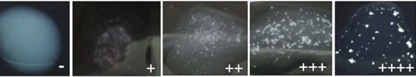
For RALT, bacterial isolates were grown on DMEM-agar and the test was done using latex sensitized with anti-EspB MAb. Figure 3 presents typical negative and semi-quantitative positive reactions. From the total of the positive reactions by RALT, +
correspond to 44.8%;++to 26.4%;+++to 11.2% and++++to
14.4% of the isolates. By this assay only four LEE-positive isolates (one aEPEC and three tEPEC) did not react with anti-EspB MAb and one false positive occurred (Proteus mirabilis) (Table 1). Considering the LEE-positive and -negative isolates, the test exhibited 97% sensitivity, 98% specificity and 97% efficiency.
Discussion
A fast and inexpensive diagnosis for EPEC/EHEC infections is highly required considering their global prevalence, the severity of
Figure 2. EspB production in different culture media.LEE-positive and LEE-negative isolates were cultivated in DMEM or T or DMEM-PC. The supernatants were tested by indirect ELISA for EspB detection using anti-EspB IgG-enriched fraction (30mg/mL) (A) and anti-EspB MAb (10mg/mL) (B). The mean optical densities for LEE-negative and LEE-positive isolates were determined. The cut-off obtained by the ROC curve for anti-EspB MAb was 0.027 for DMEM and 0.0145 for DMEM-T and DMEM-PC. For anti-EspB PAb was 0.152 for DMEM, 0.135 for DMEM-T and 0.001 for DMEM-PC.
doi:10.1371/journal.pntd.0003150.g002
Figure 3. Typical of agglutination latex assay: negative and a semi-quantitative positive (from+to++++) agglutination pattern with anti-EspB MAb coated beads.The test control with lysis buffer (B-PER) showed the same pattern as LEE-negative isolates.
the diseases associated with them, and the fact that the use of antibiotics to treat EHEC infections can be harmful. One appropriate approach for their rapid detection may utilize the
secreted proteins EspA and/or EspB, since the espA and espB
genes are present in LEE positive isolates and they are the major secreted proteins by both pathogens [4]. Thus, the aim of the present study was to develop and define sensitivity and specificity of EspA and EspB antibodies, determine the ideal target antigen, and design a simple and rapid test for the diagnosis of both emerging pathogens worldwide.
Production and secretion of virulence factors in pathogenic bacteria are tightly and coordinately regulated. Growth phase and environmental conditions characteristic of the host, including temperature and partial O2pressure, are the stimulus for virulence
factor expression in various gram-negative pathogens [48,49]. Additionally, in our experience, the production of virulence factors is a critical point for diarrheagenicE. colidiagnosis [50–52].
Thus, initially, one group of isolates (including tEPEC, aEPEC and EHEC) was cultivated in different media: LB broth, DMEM,
E. coli broth and Minimum medium. Besides, other culture conditions were tested, including pH (7.2 and 5.5), CO2presence,
and growth time period (6, 18 and 24 h). Our results showed that in general DMEM favored the production of secreted proteins after 6-h growth culture, but with individual variation (data not shown). Some reports describe that the use of preconditioned DMEM (DMEM-PC) provides signals from epithelial cells affecting virulence factors expression [47]. Also the secretion of
plasmid-encoded toxin (Pet) by enteroaggregative E. coli is
dependent on the addition of tryptone to DMEM (DMEM-T) [53]. Considering this, the bacterial isolates from our collection were cultivated in DMEM, DMEM-T and DMEM-PC, but EspB production and secretion was enhanced when bacterial isolates were cultivated in DMEM without enrichment.
Another important point of the present work was the evaluation of the four antibodies raised. We expected that EspA would be a biomarker for diagnosis and anti-EspA antibodies a detecting tool, since this protein is the major component of a transiently expressed surface organelle, which forms a direct link between the bacterium and the host cell [7]. However, our data pointed out EspB as the target antigen, and MAb anti-EspB the best antibody for defining LEE-positive isolates. Nakasone et al. [42,54] also defined EspB as the target antigen for identifying LEE-positive strains. In fact, EspA filaments exhibit antigenic polymorphisms [55].
The indirect ELISA using anti-EspB MAb showed 90.4% and 96.4%, sensitivity and specificity, respectively, indicating its
possible use in routine diagnostic laboratories. However, this methodology requires specific laboratory instrumentation, making it difficult to be performed in low-income country settings. Therefore, we standardized here a rapid agglutination test using latex beads coated with anti-EspB MAb (RALT), which has the sensitivity and specificity required for high impact diagnosis of neglected diseases in the developing world [56]. Two other assays have been described for LEE-positive isolates based on EspB detection; the 16–18 h reversed passive latex agglutination test (RPLA) [41] and a 10 min
immuno-chromatographic test (IC) [42]. Although more time
consuming, the RPLA test was more sensitive than the IC test [42].
Serotyping-based diagnosis is the only methodology available in limited-resources settings, employing either commercial or in-house antisera [28]. The standardized RALT for detection of EPEC and EHEC will have a remarkable impact in the diagnosis of these pathotypes, demonstrated by 97% sensitivity, 98% specificity and 97% efficiency in EspB detection. Also, no
cross-reaction was observed with other DEC pathotypes and E. coli
negative for DEC virulence factors. Among the enterobacteria species only oneProteus mirabiliswas recognized by MAb
anti-EspB. However, P mirabilis can be easily differentiated from
EPEC/EHEC by biochemical methods employed for species identification [57], a step necessary prior to the performance of our RALT. Thus the established agglutination latex in the present study is a simple, rapid (5 min) and easy to perform test, which can be employed in less equipped laboratories in low-income countries.
Supporting Information
Checklist S1 STARD checklist. (DOC)
Acknowledgments
The authors thank Camila B. P. Pereira for technical support. Dr. A. Leyva helped with English editing of the manuscript.
Author Contributions
Conceived and designed the experiments: RMFP LBR ARRS DDM DEL. Performed the experiments: LBR ARRS DDM LTAC DEL FBA. Analyzed the data: LBR ARRS DDM LTAC DEL DSPQH WPE RMFP. Contributed reagents/materials/analysis tools: RMFP. Wrote the paper: RMFP LBR ARRS DDM DEL DSPQH WPE.
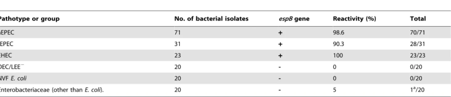
Table 1.Rapid agglutination latex test reactivity (%) with bacterial isolates.
Pathotype or group No. of bacterial isolates espBgene Reactivity (%) Total
aEPEC 71 + 98.6 70/71
tEPEC 31 + 90.3 28/31
EHEC 23 + 100 23/23
DEC/LEE2 20 - 0 0/20
NVFE. coli 20 - 0 0/20
Enterobacteriaceae (other thanE. coli). 20 - 5 1a/20
tEPEC (typical enteropathogenicE. coli); aEPEC (atypical enteropathogenicE. coli); EHEC (enterohemorrhagicE. coli); DEC/LEE2(LEE-negative diarrheagenicE. coli); NVFE.
coli(fecalE. colinegative for DEC virulence factors). aProteus mirabilis.
References
1. World Health Organization. (2013) Diarrhoeal disease. Fact Sheet 330. http:// www.who.int/mediacentre/factsheets/fs330/en/.
2. Kotloff KL, Nataro JP, Blackwelder WC, Nasrin D, Farag TH, et al. (2013) Burden and aetiology of diarrhoeal disease in infants and young children in developing countries (the Global Enteric Multicenter Study, GEMS): a prospective, case-control study. Lancet 382: 209–222.
3. Hill DR, Beeching NJ. (2010) Travelers’ diarrhea. Curr Opin Infect Dis 23: 481–487.
4. Croxen MA, Law RJ, Scholz R, Keeney KM, Wlodarska M, et al. (2013) Recent advances in understanding enteric pathogenicEscherichia coli. Clin Microbiol Rev 26: 822–880.
5. Hartland EL, Leong JM, (2013). Enteropathogenic and enterohemorrhagicE. coli: ecology, pathogenesis, and evolution. Front Cell Infect Microbiol 3: 1–3. 6. Knutton S, Lloyd DR, McNeish AS. (1987) Adhesion of enteropathogenic
Escherichia colito human intestinal enterocytes and cultured human intestinal mucosa. Infect Immun 55: 69–77.
7. Knutton S, Rosenshine I, Pallen MJ, Nisan I, Bain C, et al. (1998) A novel EspA-associated surface organelle of enteropathogenicEscherichia coliinvolved in protein translocation into epithelial cells. The EMBO J 17: 2166–2176. 8. Ide T, Laarmann S, Greune L, Schillers H, Oberleithner H, et al. (2001)
Characterization of translocation pores inserted into plasma membranes by type III-secreted Esp proteins of enteropathogenicEscherichia coli. Cell Microbiol 3: 669–679.
9. Kenny B, DeVinney R, Stein M, Reinscheid DJ, Frey EA, et al. (1997) EnteropathogenicE.coli(EPEC) transfers its receptor for intimate adherence into mammalian cells. Cell 91: 511–520.
10. Deibel C, Kra¨mer S, Chakraborty T, Ebel F. (1998) EspE a novel secreted protein of attaching and effacing bacteria, is directly translocated into infected host cells, where it appears as a tyrosine-phosphorylated 90 kDa protein. Mol Microbiol 28: 463–474.
11. Tarr PI, Gordon CA, Chandler WL. (2005) Shiga-toxin-producingEscherichia coliand haemolytic uraemic syndrome. Lancet 365: 1073–1086.
12. Nataro JP, Baldini MM, Kaper JB, Black RE, Bravo N, et al. (1985) Detection of an adherence factor of enteropathogenicEscherichia coliwith a DNA probe. J Infect Dis 152: 560–565.
13. Sohel I, Puente JL, Murray WJ, Vuopio-Varkila J, Schoolnik GK. (1993) Cloning and characterization of the bundle-forming pilin gene of enteropatho-genicEscherichia coliand its distribution in Salmonella serotypes. Mol Microbiol 7: 563–575.
14. Kaper JB. (1996) Defining EPEC. Rev Microbiol 27: 130–133.
15. Trabulsi LR, Keller R, Gomes TAT. (2002) Typical and atypical enteropatho-genicEscherichia coli. Emerg Infect Dis 8: 508513.
16. Hernandes RT, Elias WP, Vieira AM, Gomes TAT. (2009) An overview of atypical enteropathogenicEscherichia coli. FEMS Microbiol Lett 297: 137–149. 17. Abe CM, Trabulsi LR, Blanco J, Blanco M, Dahbi G, et al. (2009) Virulence features of atypical enteropathogenicEscherichia coliidentified by the eae(+) EAF-negative stx(-) genetic profile. Diagn Microbiol Infect Dis 64: 357–365. 18. Nara JM, Cianciarullo AM, Culler HF, Bueris V, Horton DS, et al. (2010)
Differentiation of typical and atypical enteropathogenicEscherichia coliusing colony immunoblot for detection of bundle-forming pilus expression. J Appl Microbiol 109: 35–43.
19. Moreno ACR, Fernandes-Filho A, Gomes TAT, Ramos STS, Montemor LPG, et al. (2008) Etiology of childhood diarrhea in the northeast of Brazil: significant emergent diarrheal pathogens. Diagn Microbiol Infect Dis 66: 50–57. 20. Ochoa TJ, Barletta F, Contreras C, Mercado E. (2008) New insights into the
epidemiology of enteropathogenicEscherichia coliinfection. Trans R Soc Trop Med Hyg 102: 852–856.
21. Viljanen M, Peltola T, Junnila S, Olkkonen L, Jarvinen H, et al. (1990) Outbreak of diarrhoea dueEscherichia coliO11:B4 in schoolchildren and adults: association of Vi antigen-like reactivity.Lancet336: 381–384.
22. Hedberg C, Savarino S, Besser J, Paulus C, Thelen V, et al. (1997) An outbreak of foodborne illness caused byEscherichia coliO39:NM, an agent not fitting into the existing scheme for classifying diarrheogenicE. coli. J Infect Dis 176: 1625– 1628.
23. Afset JE, Bergh K, Bevanger L. (2003) High prevalence of atypical enteropathogenic Escherichia coli (EPEC) in Norwegian children with diarrhoea. J Med Microbiol 11: 1015–1019.
24. Gomes TAT, Irino K, Gira˜o DM, Gira˜o VB, Guth BE, et al. (2004) Emerging enteropathogenicEscherichia colistrains? Emerg Infect Dis 10: 1851–1855. 25. Franzolin MR, Alves RCB, Keller R, Gomes TAT, Beutin L, et al. (2005)
Prevalence of diarrheagenic Escherichia coli in children with diarrhea in Salvador, Bahia, Brazil. Mem Inst Oswaldo Cruz 100: 359–363.
26. Araujo JM, Tabarelli GF, Aranda KR, Fabbricotti SH, Fagundes-Neto U, et al. (2007) Typical enteroaggregative and atypical enteropathogenic types of Escherichia coliare the most prevalent diarrhea-associated pathotypes among Brazilian children. J Clin Microbiol 10: 3396–3399.
27. Bueris V, Sircili MP, Taddei CR, Santos MF, Franzolin MR, et al. (2007) Detection of diarrheagenicEscherichia colifrom children with and without diarrhea in Salvador, Bahia, Brazil. Mem Inst Oswaldo Cruz 6: 839–844. 28. Piazza RMF, Abe CM, Horton DSPQ, Miliwebsky E, Chinen I, et al. (2010)
Detection and subtyping methods of diarrheagenicEscherichia colistrains. In:
Alfredo Torres, editor. PathogenicEscherichia coliin Latin America. Dubai: Bentham Science Publishers. pp. 95–115.
29. Adu-Bobie J, Trabulsi LR, Carneiro-Sampaio MM, Dougan G, Frankel G. (1998) Identification of immunodominant regions within the C-terminal cell binding domain of intimin alpha and intimin beta from enteropathogenic Escherichia coli. Infect Immun 66: 5643–5649.
30. Batchelor M, Knutton S, Caprioli A, Huter V, Zanial M, et al. (1999) Development of a universal intimin antiserum and PCR primers. J Clin Microbiol 37: 3822–3827.
31. Koga PCM, Menezes CA, Lima FA, Nara JM, Magalha˜es CA, et al. (2003) Polyclonal anti-intimin antibody: immunological characterization and its use in EPEC and EHEC diagnosis. Braz J Microbiol 34: 5–7.
32. Menezes MA, Rocha LB, Koga PC, Fernandes I, Nara JM, et al. (2010) Identification of enteropathogenic and enterohaemorrhagic Escherichia coli strains by immunoserological detection of intimin. J Appl Microbiol 108: 878– 887.
33. Menezes MA, Aires KA, Ozaki CY, Ruiz RM, Pereira MC, et al. (2011) Cloning approach and functional analysis of anti-intimin single-chain variable fragment (scFv). BMC Res Notes 4: 30.
34. Caravelli A, Luz DE, Andrade FB, Moraes CT, Maranha˜o AQ, et al. (2013) Sensitive and specific detection of enteropathogenic and enterohemorrhagic Escherichia coli using recombinant anti-intimin antibody by immunofluores-cence assay. Diagn Microbiol Infect Dis 77: 301–303.
35. Haigh R, Baldwin T, Knutton S, Williams PH. (1995) Carbon dioxide regulated secretion of the EaeB protein of enteropathogenic Escherichia coli. FEMS Microbiol Lett 129: 63–67.
36. Abe H, Tatsuno I, Tobe T, Okutani A, Sasakawa C. (2002) Bicarbonate ion stimulates the expression of locus of enterocyte effacement-encoded genes in enterohemorrhagicEscherichia coliO157:H7. Infect Immun 70: 3500–3509. 37. Beltrametti F, Kresse AU, Guzma´n CA. (1999) Transcriptional regulation of the
esp genes of enterohemorrhagicEscherichia coli. J Bacteriol 181: 3409–3418. 38. Ide T, Michgehl S, Knappstein S, Heusipp G, Schmidt MA. (2003) Differential
modulation by Ca2+of type III secretion of diffusely adhering enteropathogenic Escherichia coli. Infect Immun 71: 1725–1732.
39. Yu S, Gu J, Wang HG, Wang QX, Luo P, et al. (2011) Identification of a novel linear epitope on EspA from enterohemorrhagicE. coliusing a neutralizing and protective monoclonal antibody. Clin Immunol 138: 77–84.
40. Ebel F, Deibel C, Kresse AU, Guzma´n CA, Chakraborty T. (1996) Temperature- and medium-dependent secretion of proteins by Shiga toxin-producingEscherichia coli. Infect Immun 64: 4472–4479.
41. Lu Y, Toma C, Honma Y, Iwanaga M. (2002) Detection of EspB using reversed passive latex agglutination: application to determination of enteropathogenic Escherichia coli. Diagn Microbiol Infect Dis 43: 7–12.
42. Nakasone N, Toma C, Lu Y, Iwanaga M. (2007) Development of a rapid immunochromatographic test to identify enteropathogenic and enterohemor-rhagicEscherichia coliby detecting EspB. Diagn Microbiol Infect Dis 57: 21–25. 43. Donnenberg MS, Finlay B. (2013) Combating enteropathogenicEscherichia coli
(EPEC) infections: the way forward. Trend in Microbiol 21: 317–319. 44. Rocha LB, Luz DE, Moraes CT, Caravelli A, Fernandes I, et al. (2012)
Interaction between Shiga toxin and monoclonal antibodies: binding charac-teristics and in vitro neutralizing abilities. Toxins 4: 729–747.
45. Levine MM, Nataro JP, Karch H, Baldini MM, Kaper JB, et al. (1985) The diarrheal response of humans to some classic serotypes of enteropathogenic Escherichia coli is dependent on a plasmid encoding an enteroadhesiveness factor. J Infect Dis 152: 550–559.
46. Kohler G, Milstein C. (1975) Continuous cultures of fused cells secreting antibody of predefined specificity. Nature 256: 495–497.
47. Giro´n JA, Torres AG, Freer E, Kaper JB. (2002) The flagella of enteropatho-genicEscherichia colimediate adherence to epithelial cells. Mol Microbiol 44: 361–379.
48. Lee CA, Falkow S. (1990) The ability of Salmonella to enter mammalian cells is affected by bacterial growth state. Proc Natl Acad Sci USA 87: 4304–4308. 49. Dorman CJ, Bhriain NN. (1993) DNA topology and bacterial virulence gene
regulation. Trends Microbiol 1: 92–99.
50. Vilhena-Costa AB, Piazza RM, Nara JM, Trabulsi LR, Martinez MB. (2006) Slot blot immunoassay as a tool for plasmid-encoded toxin detection in enteroaggregativeEscherichia coliculture supernatants. Diagn Microbiol Infect Dis 55: 101–106.
51. Rocha LB, Piazza RM. (2007) Production of Shiga toxin by Shiga toxin-expressing Escherichia coli (STEC) in broth media: from divergence to definition. Lett Appl Microbiol 45: 411–417.
52. Rocha LB, Ozaki CY, Horton DS, Menezes CA, Silva A, et al. (2013) Different assay conditions for detecting the production and release of labile and heat-stable toxins in enterotoxigenicEscherichia coliisolates. Toxins 5: 2384–2402. 53. Betancourt-Sanchez M, Navarro-Garcia F. (2009) Pet secretion, internalization
and induction of cell death during infection of epithelial cells by enteroag-gregativeEscherichia coli. Microbiology 155: 2895–2906.
55. Neves BC, Shaw RK, Frankel G, Knutton S. (2003) Polymorphisms within EspA filaments of enteropathogenic and enterohemorrhagicEscherichia coli. Infect Immun 71: 2262–2265.
56. Urdea M, Penny LA, Olmsted SS, Giovanni MY, Kaspar P, et al. (2006) Requirements for high impact diagnostics in the developing world. Nature 444: 73–79.
57. Farmer JJ, III (2003) Enterobacteriaceae: introduction and identification. In: Murray PR, Baron EJ, Jorgensen JH, Pfaller MA, Yolken RH, editors. Manual of Clinical Microbiology.8th